Rare Gastric Diverticulum Mimicking Adrenal Abscess on Computed Tomography
Alexander Maraveyas

TL;DR
A rare case of a gastric diverticulum was mistaken for an adrenal abscess on CT scans, highlighting the need for accurate diagnostic methods.
Contribution
This case report highlights the diagnostic challenge of gastric diverticula mimicking adrenal abscess on CT.
Findings
Gastric diverticula can mimic adrenal masses on CT due to their anatomical proximity.
The case showed apparent infection spread to the lung, complicating the diagnosis.
Upper gastrointestinal series or endoscopy are more reliable for diagnosing gastric diverticula.
Abstract
Gastric diverticula are a rare phenomenon that is typically asymptomatic and encountered incidentally. Due to the relative proximity between the gastric diverticula and the left adrenal gland, they may mimic adrenal masses on computed tomography (CT). For this reason, the preferred diagnostic methods for gastric diverticula are upper gastrointestinal series or direct visualization on endoscopy. The present report describes an unusual case of a gastric diverticulum mimicking an abscess of the adrenal gland with the apparent spread of infection to the left lower lobe of the lung.
Genes, proteins, chemicals, diseases, species, mutations and cell lines named across the full text — each resolved to its canonical identifier and authoritative record.
Click any figure to enlarge with its caption.
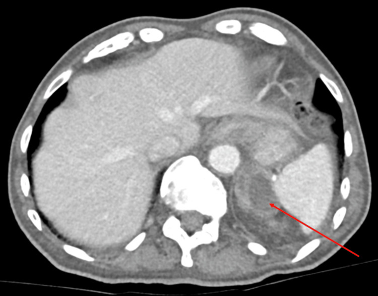 Figure 1
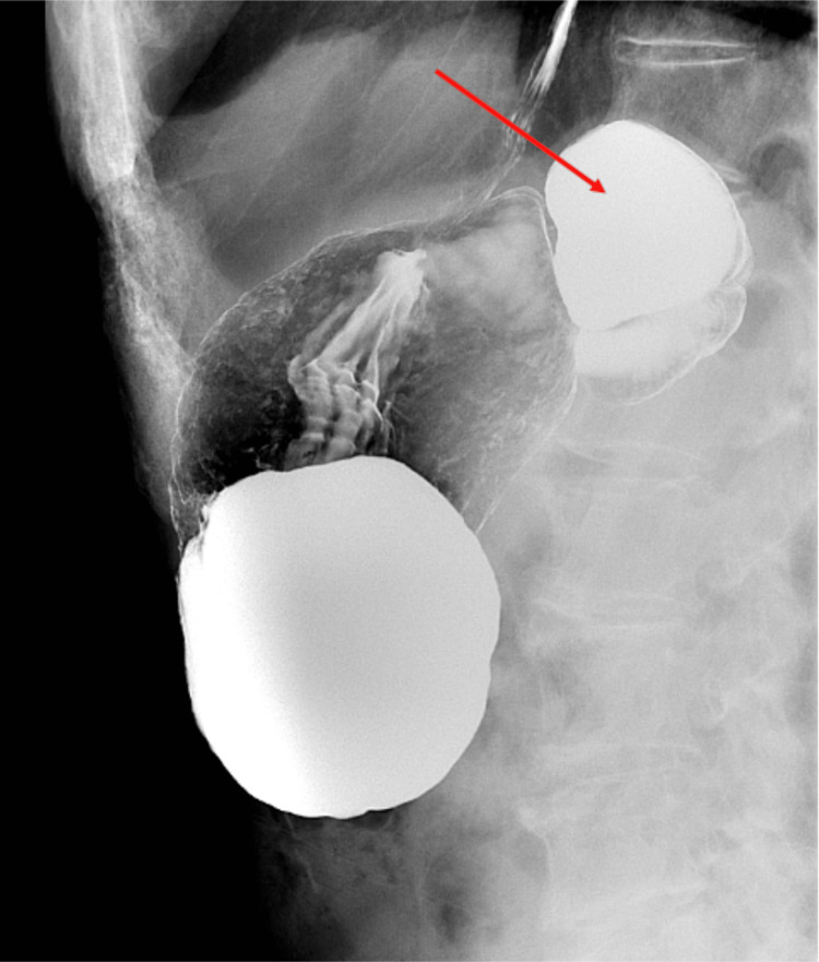 Figure 2
Peer Reviews
No public reviews on file for this paper yet. If you reviewed it on a platform where reviews are public (OpenReview, ICLR, NeurIPS, ICML), you can paste yours below so the community can read it here.
Videos
No videos yet. Explain this paper in a talk, walkthrough, or lecture? Add one.
Taxonomy
TopicsGastrointestinal disorders and treatments · Biliary and Gastrointestinal Fistulas · Eosinophilic Esophagitis
Introduction
Gastric diverticula are exceptionally rare, with a reported prevalence of 0.02% at autopsy and 0.04% in the upper gastrointestinal series [1]. Due to the relative proximity of gastric diverticula to the left adrenal gland, they have been recognized as a rare mimic of masses of the left adrenal gland on computed tomography (CT) [2]. Consequently, the recommended diagnostic methods for gastric diverticula are upper gastrointestinal series or direct visualization on endoscopy [3,4]. This case is described as an unusual case of a gastric diverticulum mimicking an abscess of the adrenal gland with the apparent spread of infection to the left lower lobe of the lung.
Case presentation
A 78-year-old male with a history of colonic diverticulosis presented to the emergency department complaining of left flank discomfort and no other symptoms. Blood tests revealed mild anemia and leukopenia but were otherwise unremarkable; urinalysis was also unremarkable. A chest X-ray showed a left lower lobe opacity, possibly suggestive of pneumonia. Contrast-enhanced CT of the abdomen and pelvis demonstrated an apparent subdiaphragmatic abscess arising from the left adrenal gland (Figure 1), with the apparent spread of infection to the adjacent left lower lung.
CT abdomen and pelvis with contrastAxial view demonstrating an apparent abscess arising from the left adrenal gland (red arrow); in reality, a gastric diverticulum arising from the posterior fundus of the stomachCT, computed tomography
The patient was started on intravenous piperacillin-tazobactam. Interventional radiology and surgery services were consulted to evaluate the abscess. At this point, collateral information was received from a relative of the patient that this possible abscess had been present on scans performed 10 years ago at an outside hospital system; the family had been informed at the time that it was gastric in origin. On receipt of this information, the CT images were reevaluated by radiology, and gastric diverticulum was determined to be the most likely cause for both the appearance on the CT and the patient’s presentation. The patient, who had remained stable and afebrile throughout this admission, was discharged to follow-up outpatient gastroenterology. An upper gastrointestinal series was performed as an outpatient, which confirmed the diagnosis: gastric diverticulum arising from the posterior fundus of the stomach (Figure 2).
Upper gastrointestinal seriesGastric diverticulum arising from the posterior fundus of the stomach (red arrow)
Discussion
Gastric diverticula are a rare phenomenon, typically asymptomatic, and most commonly encountered incidentally. They are divided into two main types: congenital (or true) diverticula, which contain all layers of the abdominal wall, and acquired (or false) diverticula, which do not [5]. The congenital type is the more common, comprising an estimated 75% of cases [3]. The most common location for congenital diverticula is the posterior wall of the stomach, approximately 2 cm below the gastroesophageal junction and 3 cm from the lesser curve of the stomach [4].
Although gastric diverticula are a principally asymptomatic phenomenon, when symptomatic, the clinical picture is usually nonspecific. The most common symptom is the vague sensation of fullness or epigastric discomfort, although patients may also present with serious complications of gastric diverticula, including gastrointestinal hemorrhage [4]. In our case, the presenting symptom of left flank discomfort is broadly consistent with a typical symptomatic presentation.
The preferred techniques for diagnosing gastric diverticula are upper gastrointestinal series or direct visualization on endoscopy. When seen on CT, the typical appearance is a thin-walled cystic lesion located in the left paravertebral region [6], although, as this case illustrates, CT is not a preferred modality because of the potential for misdiagnosis. A 2015 review of the literature identified seven reported cases of gastric diverticula misdiagnosed as adrenal masses; of these, six had initially used CT to visualize the lesion [7]. Misdiagnosis can have deleterious effects on investigation and management and has been documented to have led to unnecessary endocrinological investigation [5], exploratory laparotomy [1], and even adrenalectomy [8].
The first-line treatment for gastric diverticula is medical management with either proton pump inhibitor therapy, H2 receptor antagonist therapy, or antacids [5]. Surgical resection is the treatment of choice when gastric diverticula are particularly large, symptomatic, or complicated by bleeding, perforation, or malignancy [9].
Conclusions
This case demonstrates the importance of exercising caution when interpreting apparent adrenal masses as visualized on a CT scan. Additionally, the case highlights the importance of obtaining correlative studies, such as upper gastrointestinal series, when there is doubt regarding the provenance of an adrenal mass. We hope this case contributes to the literature on this uncommon phenomenon and raises the index of suspicion for gastric diverticula, in particular, as a cause of adrenal pseudotumor.
The reference list from the paper itself. Each links out to its DOI / PubMed record.
- 1Gastric diverticulum simulating a left adrenal tumor Surgery Chasse E Buggenhout A Zalcman M Jeanmart J Gelin M El Nakadi I 44744813320031271736610.1067/msy.2003.47 · doi ↗ · pubmed ↗
- 2Commonly encountered adrenal pseudotumours on CT Br J Radiol Gokan T Ohgiya Y Nobusawa H Munechika H 1701747820051568133410.1259/bjr/18362306 · doi ↗ · pubmed ↗
- 3Gastric diverticulum: "a wayside house of ill fame" with a laparoscopic solution JSLS Du Bois B Powell B Voeller G 4734771620122331807710.4293/108680812 X 13462882736330 PMC 3535786 · doi ↗ · pubmed ↗
- 4A review on gastric diverticulum World J Emerg Surg Rashid F Aber A Iftikhar SY 1720122225743110.1186/1749-7922-7-1PMC 3287132 · doi ↗ · pubmed ↗
- 5Gastric diverticulum: a comprehensive review Inflamm Intest Dis Shah J Patel K Sunkara T Papafragkakis C Shahidullah A 161166320193111103110.1159/000495463 PMC 6501548 · doi ↗ · pubmed ↗
- 6Imaging findings of gastric diverticula Scientific World Journal Schramm D Bach AG Zipprich A Surov A 923098201420142540116010.1155/2014/923098 PMC 4225827 · doi ↗ · pubmed ↗
- 7Gastric diverticulum simulating a left adrenal mass: a case report and review of the literature Oncol Lett Feng YE Zhang Z 247724801020152662287410.3892/ol.2015.3559 PMC 4579981 · doi ↗ · pubmed ↗
- 8Misdiagnosed gastric diverticulum as a left adrenal lesion on imaging Clin Case Rep Zhang JY Qian C Pan CW 012202410.1002/ccr 3.8414 PMC 1079270038235412 · doi ↗ · pubmed ↗