Mutational Signature Changes in Patients With Metastatic Squamous Cell Carcinoma of the Anal Canal
Michael LaPelusa, Christopher Cann, Kristen K Ciombor, Cathy Eng

TL;DR
This study shows that mutations in cancer DNA circulating in the blood can change after treatment in anal canal cancer patients, suggesting ctDNA can track tumor evolution.
Contribution
The study demonstrates temporal mutational heterogeneity in metastatic SCCA and highlights ctDNA's potential for real-time tracking of tumor evolution during therapy.
Findings
34.5% of posttreatment ctDNA mutations were also found in pretreatment tumor tissue.
47.6% of pretreatment tumor tissue mutations were detected in posttreatment ctDNA.
Seven patients had new mutations in ctDNA not present in pretreatment tissue.
Abstract
We examined the concordance of genetic mutations between pretreatment tumor tissue and posttreatment circulating tumor DNA (ctDNA) in patients with metastatic squamous cell carcinoma of the anal canal (SCCA) and assessed the impact of therapy on this concordance. We analyzed next-generation sequencing reports from pretreatment tumor tissue and posttreatment ctDNA in 11 patients with metastatic SCCA treated at Vanderbilt University Medical Center between 2017 and 2021. Among the mutations identified in posttreatment ctDNA, 34.5% were also found in pretreatment tumor tissue, while 47.6% of pretreatment tumor tissue mutations were found in posttreatment ctDNA. Four patients had preservation of potentially actionable mutations in both pretreatment tissue and posttreatment ctDNA, while 7 patients had newly identified mutations in posttreatment ctDNA that were not present in pretreatment…
Genes, proteins, chemicals, diseases, species, mutations and cell lines named across the full text — each resolved to its canonical identifier and authoritative record.
Click any figure to enlarge with its caption.
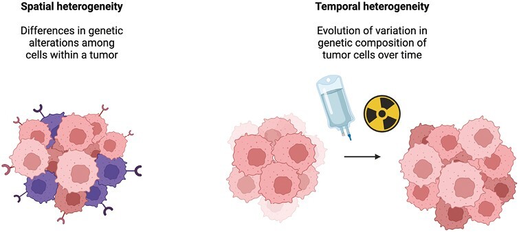 Figure 1
| Patient | Sample | HPV status | Stage at Time of Sample Collection | Collection interval (days) | Interval Therapy (cycles) | Somatic—potentially actionable (VAF), if reported) | Somatic—biologically relevant (VAF, if reported) | Variants of unknown significance (VAF, if reported) |
|---|---|---|---|---|---|---|---|---|
| 1 | Anus | Negative | IIA (T2N0M0) | T | -5-FU + mitomycin-C + radiation |
| - | -MTHFR (53.1%)* |
| ctDNA | IV | T + 643 |
| - | -ATM (0.4%)** | |||
| 2 | Anus | p16+ | IIIC (T4N1M0) | T | -5-FU + mitomycin-C + radiation | - | -ARID2 (15.1%)* | -SMAD3 (17.4%)* |
| ctDNA | IV | T + 414 | - | None | -KDR (50.4%)** | |||
| 3 | Anus | Negative | IIIA (T2N1M0) | T | -5-FU + mitomycin-C + radiation | None | -FAT1 (22.2%)* | -SYNE1 (22.2%)* |
| ctDNA | IV | T + 484 | None | -MYC** | -HNF1A (57.4%)** | |||
| 4 | Lung | p16+ | IV | T | -carboplatin + paclitaxel (7) | -PIK3CA (37.0%) | -CYLD (64.1%)* | -KIF1B (35.0%)* |
| ctDNA | IV | T + 1057 | None | None | None | |||
| 5 | Pelvic lymph node | p16+ | IV | T | -nivolumab (1) | -PIK3CA (18.6%) | -STK11 (38.0%) | - |
| ctDNA | IV | T + 763 | None | None | - | |||
| 6 | Brain | p16+ | IV | T | -irinotecan + cetuximab (1) | None | -CYLD (83.4%)* | -SETD2 (84.2%)* |
| ctDNA | IV | T + 763 | None | -SDHA (1.3%)** | -FGFR2 (49.7%)** | |||
| 7 | Anus | p16+ | IIIC (T4N1M0) | T | -5-FU + mitomycin-C + radiation | None | -APC | -APOB (25.1%)* |
| ctDNA | IV | T + 369 | None | None | None | |||
| 8 | Anus | p16+ | IIA (T2N0M0) | T | -5-FU + mitomycin-C + radiation | - | -KMT2D (43.5%)* | -PHOX2B (85.3%)* |
| ctDNA | IV | T + 205 | - | None | -PBRM1 (85.1%)** | |||
| 9 | Anus | p16+ | IIB (T3N0M0) | T | -5-FU + mitomycin-C + radiation | None | -CYLD (23.0%)* | -UBC (26.1%)* |
| ctDNA | IV | T + 989 | None | None | - | |||
| 10 | Anus | p16+ | IV | T | -carboplatin + paclitaxel (3) | - | -CDKN2B | -SDHC (18.8%)* |
| ctDNA | IV | T + 763 | - | -FBXW7 (28.7%)** | -ERBB2 (28.2%)** | |||
| 11 | Anus | p16+ | IIIC (T3N1M0) | T | -5-FU + mitomycin-C + radiation | -KMT2C/MLLC (18.8%)* | -EP300 (32.8%)* | -NSD1 (16.7%)* |
| ctDNA | IV | T + 1713 | None | None | None |
| Patient | Sample | HPV status | Stage at Time of Sample Collection | Collection interval (days) | Interval Therapy (cycles) | Somatic—potentially actionable | Somatic—biologically relevant | Variants of unknown significance | ||||||
|---|---|---|---|---|---|---|---|---|---|---|---|---|---|---|
| Gene | Mutation | VAF | Gene | Mutation | VAF | Gene | Mutation | VAF | ||||||
| 1 | Anus | Negative | IIA (T2N0M0) | T | -5-FU + mitomycin-C + radiation |
|
|
|
|
|
| MTHFR* | c.1462A > G p.I448V MV | 53.1% |
|
|
|
| FGF19* | CNG | WEE1* | c.107A > T p.E36V MV | 34.5% | |||||||
| FGF3* | CNG | |||||||||||||
| CCND1 | CNG | FGF4* | CNG | CBLB* | c.2401G > A p.D801N MV | 7.9% | ||||||||
| ctDNA | IV | T + 643 |
|
|
|
|
|
| ATM ** | c.8495G > A p.R2832H MV | 0.4% | |||
|
|
|
| ||||||||||||
| 2 | Anus | p16+ | IIIC (T4N1M0) | T | -5-FU + mitomycin-C + radiation |
|
|
| ARID2* | p.R274 SG—LOF | 15.1% | SMAD3* | c.733G?A p.G245R MV | 17.4% |
|
|
|
| ||||||||||||
| HIST1H1E* | c.343G > A p.E115K MV | 15.8% | ||||||||||||
| EPHA2* | c.274G > C p.E92Q MV | 15.4% | ||||||||||||
| FANCA* | p.W183 SG | 11.2% | LMNA* | c.307C > T p.Q103 SG | 14.4% | |||||||||
| FANCA* | c.604G > A p.D202N MV | 11.1% | ||||||||||||
| DYNC2H1* | c.2692C > T p.R898 SG | 7.3% | ||||||||||||
| SETD2* | c.1271G > A p.R424Q MV | 6.9% | ||||||||||||
| ctDNA | IV | T + 414 |
|
|
| None | -KDR** | c.1379G > T p.W460L MV | 50.4% | |||||
| SDHA** | c.1663 + 3G > C SRV | 46.8% | ||||||||||||
|
|
|
| ||||||||||||
| 3 | Anus | Negative | IIIA (T2N1M0) | T | -5-FU + mitomycin-C + radiation | None | FAT1* | p.E1027fs frameshift—LOF | 22.2% | SYNE1* | c.718G > A p.E240K MV | 22.2% | ||
| CASP8* | CNL | |||||||||||||
| ERCC3* | CNL | |||||||||||||
| FHIT* | CNL | PTPN11 | c.22C > T p.H8Y MV | 18.1% | ||||||||||
| FOXP1* | CNL | |||||||||||||
| LRP1B* | CNL | |||||||||||||
| ctDNA | IV | T + 484 | None | MYC** | CNG | -HNF1A** | c.1336G > A p.V446M MV | 57.4% | ||||||
| 4 | Lung | p16+ | IV | T | -carboplatin + paclitaxel (7) | PIK3CA | p.E726K MV | 37.0% | -CYLD* | p.V487fs frameshift—LOF | 64.1% | KIF1B* | c.2980G p.D994N MV | 35.0% |
| CREBBP* | p.R386 SG—LOF | 21.7% | ||||||||||||
| EBF1* | CNL | CREBBP* | c.1042C > T p.P3485 MV | 25.7% | ||||||||||
| ctDNA | IV | T + 1057 | None | None | None | |||||||||
| 5 | Pelvic lymph node | p16+ | IV | T | -nivolumab (1) | PIK3CA | p.E545K MV | 18.6% | STK11 | p.Q100 SG—LOF | 38.0% |
|
|
|
| TPM1* | c.116G > A p.R39K MV | 33.7% | ||||||||||||
| AMER1* | c.2036G > A p.R679Q MV | 24.1% | ||||||||||||
| BCL11B* | CNL | CTRC* | c.718C > T p.R240W MV | 22.3% | ||||||||||
| RAD51B* | c.234C > G p.F78L MV | 19.7% | ||||||||||||
| SLIT2* | CNL | ETS2* | c.1466T > A p.I489K MV | 18.7% | ||||||||||
| GATA2* | c.142T > A p.F481 MV | |||||||||||||
| ctDNA | IV | T + 763 | None | None | - |
|
| |||||||
| 6 | Brain | p16+ | IV | T | -irinotecan + cetuximab (1) | None | CYLD* | p.Q224 SG—LOF | 83.4% | SETD2* | c.6230G > A p.R2077Q MV | 84.2% | ||
| SEC63* | c.1666del p.V556fs frameshift | 45.6% | ||||||||||||
| AGO1* | c.431c > G p.A144G MV | 40.6% | ||||||||||||
| ATRX* | c.1718G > C p.G573A MV | 34.7% | ||||||||||||
| ctDNA | IV | T + 763 | None | SDHA** | c.456 + 2T > C SRV—LOF | 1.3% | FGFR2** | c.1364C > T p.T455M MV | 49.7% | |||||
| MAP2K1** | c.124C > T p.L42F MV | 0.9% | ||||||||||||
| 7 | Anus | p16+ | IIIC (T4N1M0) | T | -5-FU + mitomycin-C + radiation | None | APC | CNL | APOB* | c.4250C > T p.T1417M MV | 25.1% | |||
| ROS1 | c.274G > A p.E92K MV | 21.8% | ||||||||||||
| GRM3* | c.1296G > C p.K432N MV | 20.1% | ||||||||||||
| MYH11* | c.5125G > C p.E1709Q MV | 20.0% | ||||||||||||
| PMS2 | c.1688G > A p.R563Q MV | 19.4% | ||||||||||||
| ctDNA | IV | T + 369 | None | None | None | |||||||||
| 8 | Anus | p16+ | (T2N0M0) | T | -5-FU + mitomycin-C + radiation |
|
|
| KMT2D* | p.E1391 SG—LOF | 43.5% | PHOX2B* | c.191C > A p.S64Y MV | 85.3% |
| B2M* | CNL | HSD3B1* | c.503C > T p.A168V MV | 38.8% | ||||||||||
| ctDNA | IV | T + 205 |
|
|
| None | PBRM1** | c.1409A > G p.Y470C MV | 85.1% | |||||
| DDR2** | c.749T > A p.V250E MV | 49.9% | ||||||||||||
| MYCN** | c.1090C > T p.P364S MV | 48.6% | ||||||||||||
| APC** | c.3426T > A p.N1142K MV | 12.1% | ||||||||||||
| SDHA** | c.1915C > G p.L639V MV | 0.9% | ||||||||||||
| CDKN2A** | c.319C > T p.R107C MV | 0.2% | ||||||||||||
| 9 | Anus | p16+ | IIB (T3N0M0) | T | -5-FU + mitomycin-C + radiation | MUTYH* | c.1187G > A p.S344 SG—LOF | CYLD* | p.S344 SG—LOF | 23.0% | UBC* | c.910G > A p.G304R MV | 26.1% | |
|
|
|
| ||||||||||||
|
|
|
| ||||||||||||
| ctDNA | IV | T + 989 | None | None |
|
|
| |||||||
|
|
|
| ||||||||||||
| MTOR** | c.5714 + 1G > C SRV | 13.9% | ||||||||||||
| PMS2** | c.1427G > C p.S476T MV | 5.7% | ||||||||||||
| BRCA1** | c.2029_2030delinsTT p.G677L MV | 4.8% | ||||||||||||
| 10 | Anus | p16+ | IV | T | -carboplatin + paclitaxel (3) |
|
|
|
|
|
| SDHC* | c.485C > T p.S162F MV | 18.8% |
| EPHA2* | c.1987G > A p.E663K MV | 14.9% | ||||||||||||
| CDKN2A | CNL | CDKN2B* | CNL | ABL1* | c.323G > A p.R108Q MV | 13.9% | ||||||||
| CD70* | c.380C > A p.T127N MV | 12.3% | ||||||||||||
| SOX2* | CNG | SMARCA1* | c.514G > C p.E172Q MV | 11.7% | ||||||||||
| ctDNA | IV | T + 763 |
|
|
|
|
|
| ERBB2** | c.234G > T p.Q78H MV | 28.2% | |||
| 11 | Anus | p16+ | IIIC (T3N1M0) | T | -5-FU + mitomycin-C + radiation | KMT2C* | c.4661-1G > T SRV—LOF | 18.8% | EP300* | c.4617 + 1G > T SRV—LOF | 32.8% | NSD1* | C2230_2231 delinSGT p.S744V MV | 16.7% |
| PIK3CA | CNG | CREBBP* | p.S1074 SG—LOF | 28.4% | LRP1B* | c.13343T > C p.V448A MV | 13.7% | |||||||
| ASXL1* | p.G646fs frameshift—LOF | 7.0% | DNM2* | c.1782-5del SRV | 7.1% | |||||||||
| FH* | c.1431_1433dup p.K477dip inframe insertion | BCL6* | CNG | KLHL6* | c.1061C > T p.P354L MV | 6.1% | ||||||||
| MSH3 | c.316C > G p.Q106E MV | MCL1* | CNG | ERBB4* | c.1618G > T p.D540Y MV | 5.7% | ||||||||
| SOX2* | CNG | |||||||||||||
| ctDNA | IV | T + 1713 | None | None | None | |||||||||
| Mean number of mutations in tissue | 8.4 mutations per patient |
| Mean number of mutations in ctDNA | 2.6 mutations per patient |
| Percentage of mutations in ctDNA found in tissue | 34.5% |
| Percentage of mutations in tissue found in ctDNA* | 47.6% |
Peer Reviews
No public reviews on file for this paper yet. If you reviewed it on a platform where reviews are public (OpenReview, ICLR, NeurIPS, ICML), you can paste yours below so the community can read it here.
Videos
No videos yet. Explain this paper in a talk, walkthrough, or lecture? Add one.
Taxonomy
TopicsColorectal and Anal Carcinomas · Cholangiocarcinoma and Gallbladder Cancer Studies · Pancreatic and Hepatic Oncology Research
Introduction
In 2023, there will be an estimated 9760 patients diagnosed with anal cancer and 1870 deaths attributable to anal cancer in the US.^1^ The incidence of squamous cell carcinoma of the anal canal (SCCA) in the US increased by 2.7% per year, and anal cancer–related deaths increased by 3.1% per year, from 2001 to 2015.^2^ Several factors that increase the risk of developing SCCA have been identified, including more than 10 sexual partners, receptive anal intercourse before the age of 30, chronic immunosuppressed states such as solid-organ transplant or infection with human immunodeficiency virus, active tobacco use, and being older than 50 years old.^3,4^ Further, human papillomavirus (HPV) infection is associated with up to 95% of cases of SCCA.^5^ Chemoradiation is typically administered to patients with SCCA with locoregional disease, while patients with metastatic disease are treated with combination chemotherapy.^6-10^ Immunotherapy has also shown efficacy in patients with refractory metastatic disease and is being studied, in combination with chemotherapy for use in the first-line setting for patients with metastatic disease.^11,12^ Achieving disease control in patients with metastatic SCCA is a challenge, with one analysis finding that patients with metastatic SCCA treated with combination chemotherapy experience a median progression-free survival of 7 months and median overall survival of 22 months.^8^
Circulating tumor DNA (ctDNA) has been used effectively, in conjunction with imaging, to measure therapeutic response to chemoradiation in patients with locoregional anal cancer.^13^ Detection of HPV ctDNA is an emerging tool to detect minimal residual disease in patients with low disease burden, particularly following chemoradiation, and has been shown to be a mechanism to identify patients at high risk of recurrence in this setting.^14^
However, ctDNA has not been well studied in patients with SCCA as a means to understand spatial mutational heterogeneity (within a tumor or across sites of disease within one patient). This heterogeneity can lead to incomplete genomic information obtained via tumor biopsy.^15,16^ Temporal mutational heterogeneity, or the evolution of variation in the genetic composition of tumor cells over time that can occur as a response to treatment and ultimately leads to resistance to a particular therapy, represents another challenge to cancer treatment.^16^ A way to confront these challenges may be to serially assess a tumor’s mutational profile dynamically during treatment with peripheral blood measurements of ctDNA, particularly when patients exhibit disease progression, to identify newly acquired mutations that could serve as a target for therapy and allow for personalized changes in treatment while forgoing an invasive tissue biopsy (Fig. 1).
Spatial and temporal tumor heterogeneity.
The relatively low incidence and prevalence of SCCA pose a unique challenge to characterizing detailed tumoral genomic information in this patient population. Few analyses of the genomic profile of metastatic SCCA have been performed.^17,18^ The concordance, or lack thereof, of genomic alterations between tumor tissue and ctDNA has been reported in many tumor types, including EGFR in non-small cell lung cancer (NSCLC), KRAS in pancreatic cancer and metastatic colorectal cancer, and BRAF V600E in melanoma, but not in SCCA.^19-23^ In this study, we sought to describe how tissue-based and ctDNA-based mutational concordance, as identified by next-generation sequencing (NGS), is impacted by chemoradiation and systemic therapy in the context of patients’ unique clinicodemographic features.
Methods
We identified 11 patients with histologically confirmed metastatic SCCA treated at Vanderbilt University Medical Center between 2017 and 2021 with tissue-based TEMPUS 648 gene panel reports and ctDNA-based 105 gene panel reports available for analysis. Clinicodemographic features included in our study were: HPV status, clinical stage, the time interval between tissue biopsy and ctDNA collection, the type and amount of therapy administered during the interval between tumor tissue and ctDNA collection, and the mutations identified in each sample. Mutations were classified into predetermined categories on tissue-based TEMPUS 648 gene panel reports and ctDNA-based TEMPUS 105 gene panel reports (“Somatic—potentially actionable,” “Somatic—biologically relevant,” and “Variants of unknown significance”). We reported variant allele frequencies (VAF) if they were included in the sample’s reports.
Results
Table 1 displays by patient, the source of tissue, HPV status, clinical stage at the time of sample collection, collection Interval, interval therapy, and genes in which mutations were identified. Table 2 displays the same information but includes the specific mutations and genetic aberrations identified by NGS. Eight patients had tissue obtained from their primary tumor in the anal canal, while 3 patients had tissue obtained from sites of metastasis (lung [patient 4], pelvic lymph node [patient 5], and brain [patient 6]). Nine patients were HPV positive, as indicated by p16+ status. Four (1, 2, 8, and 10) patients with mutations in tumor tissue categorized as “somatic—potentially actionable” had the same mutations in ctDNA. In 3 (1, 2, and 10) of these 4 patients, the VAF was lower in ctDNA than in tumor tissue. Two^1,10^ patients with mutations in tumor tissue categorized as “somatic—biologically relevant” had the same mutations in ctDNA. In one of these patients, the VAF was lower in ctDNA than in tumor tissue. Two patients with mutations in tumor tissue categorized as “variants of unknown significance” had the same mutations in ctDNA. In one^2^ of the 3 patients, the VAF was lower in ctDNA than in tumor tissue.
Table 3 displays the mean number of mutations in tissue and ctDNA, in addition to the percentage of tumor tissue mutations and ctDNA mutations found in ctDNA and tissue, respectively. Among the 11 patients in our study, there were a total of 92 mutations found in tissue and 21 mutations found in ctDNA. The mean number of mutations in tissue was 8.4 (92 mutations/11 patients = 8.4 mutations per patient), and the mean number of mutations in ctDNA was 2.6 (29 mutations/11 patients = 2.6 mutations/patient). The percentage of mutations in ctDNA found in tissue was 34.5% (10 concordant mutations between tissue and ctDNA/29 mutations in ctDNA). Importantly, only 21 of the 92 mutations in tissue were in genes that were also analyzed in the ctDNA-based TEMPUS 105 gene panel reports. Therefore, to calculate the percentage of mutations in tissue found in ctDNA, only mutations in genes that were assessed both by the tissue-based TEMPUS 648 gene panel report and ctDNA-based TEMPUS 105 gene panel report were included. Therefore, the percentage of mutations in tissue found in ctDNA was 47.6% (10 concordant mutations between tissue and ctDNA/21 mutations in tissue).
Discussion
Our analysis outlines the impact of therapy on the mutational concordance between tumor tissue and ctDNA in patients with metastatic SCCA. Importantly, our real-world cohort of patients was similar to other cohorts described in the literature and the US, with regard to rates of HPV-positivity (9 patients; 81.8%) and mutations in PIK3CA (5 patients; 45.4%) which is one of the most commonly mutated genes in SCCA.^17,18,24^ The most frequently mutated “somatic—potentially actionable” and “somatic—biologically relevant” genes in tumor tissue among patients in our cohort were PIK3CA (4, 5, 9, 10, and 11) and CYLD (4, 6, and 7), respectively. Notably, one prior study suggested that mutations in TP53, PIK3CA, KMT2C, KMT2D, RB1, FAT4, NF1, CDKN2A, and CTNNB1 are driver mutations of anal cancer development.^25^ We identified several patients with tumor tissue containing mutations in these genes. A TP53 mutation was found in one patient’s^1^ tumor tissue. PIK3CA mutations were found in 5 patients’ (4, 5, 8, 10, and 11) tumor tissue. A KMT2C mutation and KMT2D mutation were found in 2 separate patients’ tumor tissues (11 and 8, respectively). CDKN2A mutations were found in 2 patients’ (1 and 10) tumor tissue.
Effect of Therapy on Concordance and VAF
VAF is a surrogate measure of the percentage of DNA in a sample that carries mutations.^26^ A higher VAF, as measured by ctDNA, is a marker of tumor burden and is associated with worse survival.^27^ Notably, genomic alterations with a VAF of less than 1% may not actually represent DNA shed from a patient’s tumor but rather clonal hematopoiesis of indeterminate potential (CHIP).^28^ It has been shown that VAF measured by ctDNA is lower than that of tissue among samples collected before the initiation of therapy in patients with non-small cell lung cancer.^29^ VAF, when measured serially in patients receiving systemic therapy, has been shown to decrease over time in patients who experience a response (determined by imaging) to treatment.^30^ In our analysis, we found that VAF was lower in ctDNA compared to tissue in the majority of concordant mutations (except PI3KCA in patient 8 and RAD51C and JAK2 in patient 9). However, it is unclear if this difference was related to the fact that tissue VAF is higher in tissue, generally, or whether this was representative of response to therapy.
Acquisition of Previously Absent Mutations
When patients with metastatic cancer experience disease progression, it is not uncommon for clinicians to obtain tissue from a new site of disease and assess for emerging actionable mutations with NGS. The viability of utilizing ctDNA for this purpose, as well as to understand whether certain changes in a tumor’s mutational profile are indicative of response (or lack thereof) to therapy, is currently being investigated in several clinical trials in patients with melanoma and NSCLC (NCT04166487, NCT04966676, NCT04093167). Seven (1, 2, 3, 6, 8, 9, and 10) patients had newly identified mutations in ctDNA that were not present in tumor tissue.
Three patients (6, 9, and 10) had potentially actionable mutations in ctDNA that were not previously present in tumor tissue. Patient 6 was found to have a mutation in FGFR2 (c.1364C>T p.T455M MV; VAF = 49.7%) in ctDNA that was not previously present in tumor tissue, for which several agents have been shown to demonstrate activity in advanced, refractory tumors harboring aberrations in FGFR 1-3.^31-33^ This patient was treated with pemigatinib, an FGFR2 inhibitor, and achieved a durable, objective response.^33^ Patient 9 was found to have a mutation in BRCA1 (c.2029_2030delinsTT p.G677L MV; VAF = 4.8%) in ctDNA that was not previously present in tumor tissue. In a meta-analysis that examined the activity of polyadenosine diphosphate-ribose polymerase (PARP) inhibitors, which have efficacy in specific patient populations with tumors containing BRCA1/2 mutations, 24 of 43 patients with tumors containing somatic BRCA mutations treated with PARP inhibitors had a response to therapy across 8 trials.^34^ Patient 10 was found to have a mutation in ERBB2 (.234G>T p.Q78H MV; VAF = 28.2%) in ctDNA that was not previously present in tumor tissue. Genetic alterations in ERBB2 have been established as a biomarker for targeted anti-human epidermal growth factor receptor 2 (HER2) therapy in patients with multiple types of solid tumors, including gastrointestinal malignancies such as gastroesophageal, gastric, and colorectal cancers.^35,36^
Four patients (2, 3, 6, and 8) had mutations with a VAF greater than 45% in ctDNA that were not previously present in tumor tissue, which may indicate they were drivers of resistance to therapy and metastasis. These mutations were in KDR (c.1379G>T p.W460L MV; VAF = 50.4%), SDHA (c.1663 + 3G>C SRV; VAF = 46.8%), HNF1A (c.1336G>A p.V446M MV; VAF = 57.4%), FGFR2 (c.1364C>T p.T455M MV; VAF = 49.7%), PBRM1 (c.1409A>G p.Y470C MV; VAF = 85.1%), DDR2 (c.749T>A p.V250E MV; VAF = 49.9%), and MYCN (c.1090C>T p.P364S MV; VAF = 48.6%). The function of the proteins these genes encode varies. According to the National Center for Biotechnology Information’s Gene database, KDR encodes vascular endothelial growth factor receptor 2, SDHA encodes a major catalytic subunit of a complex in the mitochondrial respiratory chain, HNF1A encodes a transcription factor required for the expression of some liver-specific genes, FGFR2 encodes fibroblast growth receptor 2, PBRM1 encodes a subunit of a chromatin remodeling complex integral for nuclear hormone-receptor mediated transcriptional activation, DDR2 encodes a receptor subclass of the receptor tyrosine kinase family, and MYCN encodes a nuclear protein involved in the regulation of transcription.^25^
Limitations
First, our ability to ascertain the true concordance of mutations between tissue and ctDNA was hampered by the fact that only 105 genes were analyzed on the ctDNA-based TEMPUS 105 gene panel reports, while 648 genes were analyzed on the tissue-based TEMPUS 648 gene panel reports. As annotated with a single asterisk (*) in Tables 1 and 2, 10 patients had mutations identified in tissue-based TEMPUS 648 gene panel reports categorized as “somatic—biologically relevant,” and all 11 patients had tissue with mutations categorized as “Variants of unknown significance” that were not tested for in ctDNA-based TEMPUS 105 gene panel reports. It is possible that if ctDNA-based TEMPUS 105 gene panel reports captured mutations in the same 648 genes as the tissue-based TEMPUS 648 gene panel reports, we would have observed a higher mutational concordance between tumor tissue and ctDNA.
Additionally, as seen in the third column of Tables 1 and 2, tumor tissue and ctDNA samples were obtained at separate, discrete points in time. Had our analysis been solely focused on the concordance of tissue and ctDNA mutational signatures rather than also trying to capture the impact of therapy on changes in SCCA mutational signatures, it would have been imperative that both tissue and ctDNA be collected simultaneously either before the start of treatment or at a discrete point in time during the course of therapy.
Lastly, the relatively small number of patients included in our analysis may limit the generalizability of our findings. This highlights the need for multi-institutional registries to study and understand detailed genomic information in uncommon tumor types, such as SCCA.
Conclusions
Among all solid tumor types, including SCCA, NGS of biopsy-obtained tumor tissue is crucial for identifying genomic alterations that could potentially serve as therapeutic targets. While tissue biopsies have primarily served as the gold standard for identifying mutations in patients with a new cancer diagnosis, noninvasive measures to identify genomic alterations are also being studied as a noninvasive and complementary approach to predict and identify dynamic responses to therapy in the neoadjuvant, adjuvant, and metastatic setting. Our analysis showed a relatively high degree of mutational temporal heterogeneity in patients with SCCA who received locoregional and systemic therapy, supporting the hypothesis that ctDNA may serve as a mechanism to track the real-time evolution of the molecular underpinnings of solid tumors subjected to systemic therapy, which can allow for the identification of mutations that, when present, allow patients with refractory SCCA the opportunity to participate in a clinical trial or receive off-label targeted therapy. As targeted therapies are developed and approved for this rare cancer, the utility of this technique will only become more relevant.
Our study demonstrated some degree of concordance, particularly in commonly mutated genes in SCCA, such as PI3KCA, between tumor tissue and ctDNA in patients with SCCA. The absence of evidence for a high mutational concordance between tissue and ctDNA limits the ability to rely solely on ctDNA-based mutations when administering targeted therapy in most tumor types, including SCCA.
Furthermore, integrative models of the tumoral microenvironment and more advanced sequencing technologies of peripheral blood, should continue to be investigated. Another critical area that should be explored is whether dynamic changes in a tumor’s genetic profile, and that of the ctDNA tumor shedding may be correlated to radiologically confirmed response or disease progression and if this information can be used to guide future decisions related to the continuation, de-escalation, or revision of treatment options.
The reference list from the paper itself. Each links out to its DOI / PubMed record.
- 1Siegel RL , Miller KD, Fuchs HE, Jemal A. Cancer statistics, 2022. CA Cancer J Clin. 2022;72(1):7-33.35020204 10.3322/caac.21708 · doi ↗ · pubmed ↗
- 2Deshmukh AA , Suk R, Shiels MS, et al. Recent trends in squamous cell carcinoma of the anus incidence and mortality in the united states, 2001-2015. J Natl Cancer Inst. 2020;112(8):829-838. 10.1093/jnci/djz 21931742639 PMC 7825484 · doi ↗ · pubmed ↗
- 3Daling JR , Madeleine MM, Johnson LG, et al. Human papillomavirus, smoking, and sexual practices in the etiology of anal cancer. Cancer. 2004;101(2):270-280.15241823 10.1002/cncr.20365 · doi ↗ · pubmed ↗
- 4Johnson LG , Madeleine MM, Newcomer LM, Schwartz SM, Daling JR. Anal cancer incidence and survival: the surveilance, epidemiology, and end results experience, 1973-2000. Cancer. 2004;101(2):281-288. 10.1002/cncr.2036415241824 · doi ↗ · pubmed ↗
- 5Baricevic I , He X, Chakrabarty B, et al. High-sensitivity human papilloma virus genotyping reveals near universal positivity in anal squamous cell carcinoma: different implications for vaccine prevention and prognosis. Eur J Cancer. 2015;51(6):776-785. 10.1016/j.ejca.2015.01.05825702585 · doi ↗ · pubmed ↗
- 6Ajani JA , Winter KA, Gunderson LL, et al. Fluorouracil, mitomycin, and radiotherapy vs fluorouracil, cisplatin, and radiotherapy for carcinoma of the anal canal: a randomized controlled trial. JAMA. 2008;299(16):1914-1921. 10.1001/jama.299.16.191418430910 · doi ↗ · pubmed ↗
- 7James RD , Glynne-Jones R, Meadows HM, et al. Mitomycin or cisplatin chemoradiation with or without maintenance chemotherapy for treatment of squamous-cell carcinoma of the anus (ACT II): a randomised, phase 3, open-label, 2 × 2 factorial trial. Lancet Oncol. 2013;14(6):516-524. 10.1016/S 1470-2045(13)70086-X 23578724 · doi ↗ · pubmed ↗
- 8Eng C , Chang GJ, You YN, et al. The role of systemic chemotherapy and multidisciplinary management in improving the overall survival of patients with metastatic squamous cell carcinoma of the anal canal. Oncotarget. 2014;5(22):11133-11142. 10.18632/oncotarget.256325373735 PMC 4294384 · doi ↗ · pubmed ↗