Implementation of a Robotic Hand Glove in the Physiotherapy Treatment of Carpal Tunnel Syndrome Secondary to Volar Barton Fracture: A Case Report
Vaishnavi M Thakre, Pratik Phansopkar

TL;DR
This case report explores the use of a robotic hand glove in physiotherapy to treat carpal tunnel syndrome following a wrist fracture, showing improved recovery and daily function.
Contribution
The novel use of a robotic glove in post-operative rehabilitation for carpal tunnel syndrome secondary to a volar Barton fracture is highlighted.
Findings
A tailored physiotherapy regimen using a robotic glove improved wrist mobility and grip strength.
Early functional recovery and enhanced activities of daily living were observed in the patient.
Cryotherapy and range of motion exercises complemented the robotic glove therapy effectively.
Abstract
A volar Barton fracture is a compression injury that usually results from a fall onto an outstretched, pronated wrist. It is characterized by an intraarticular marginal volar shearing fracture of the distal radius. Despite the prevalence of distal radius fractures, consensus regarding optimal treatment remains challenging. To achieve adequate alignment, a variety of treatment techniques are available, including open reduction with plate and screw fixation, external fixation, and closed reduction with pinning. Regardless of the method of treatment, carpal tunnel syndrome (CTS) is the most prevalent complication that is commonly seen in distal radius fractures. Establishing an effective post-operative rehabilitation regimen, primarily comprising functional physiotherapy interventions, is vital to managing this condition. This case report discusses the management of acute CTS secondary to…
Genes, proteins, chemicals, diseases, species, mutations and cell lines named across the full text — each resolved to its canonical identifier and authoritative record.
Click any figure to enlarge with its caption.
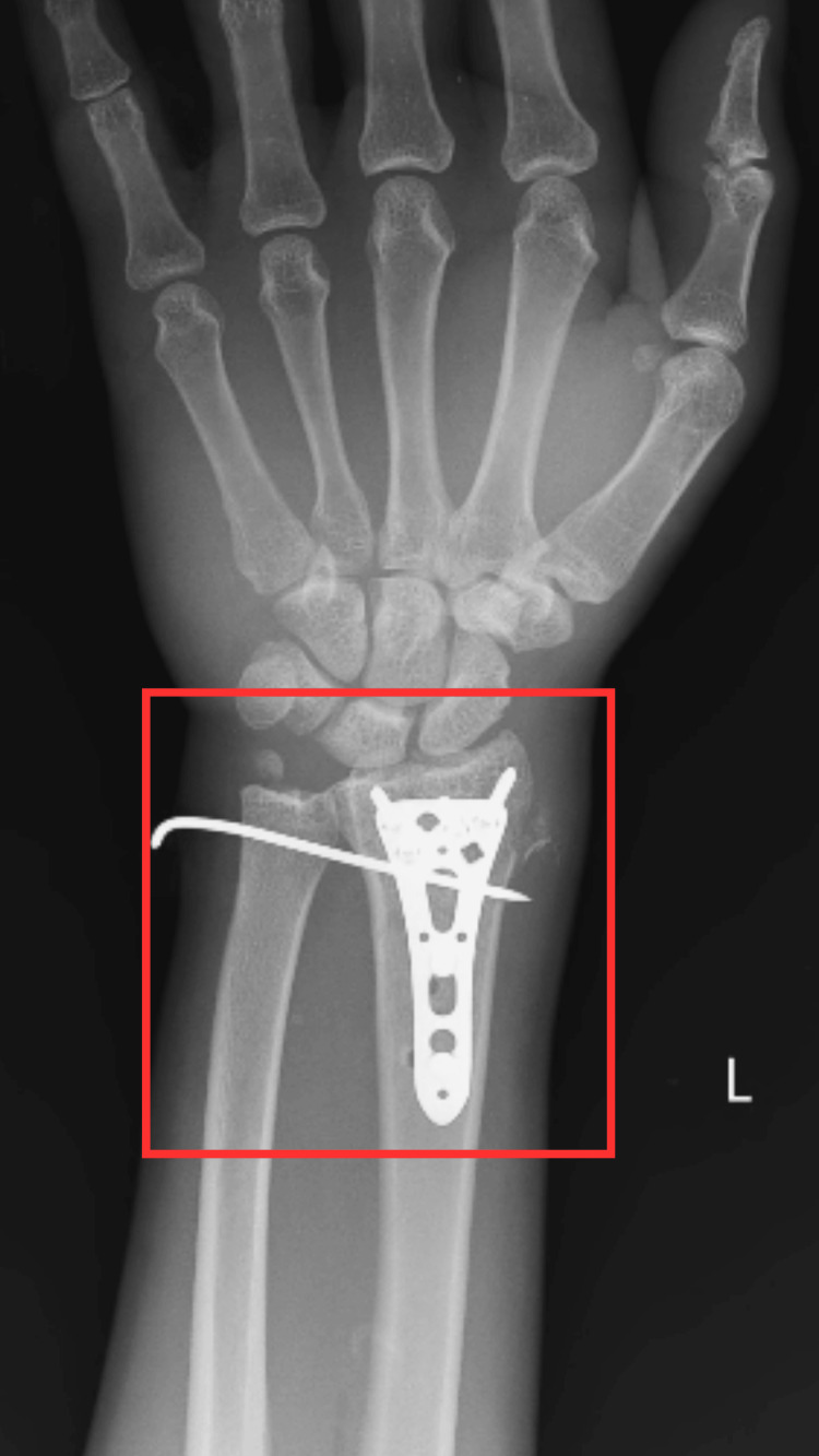 Figure 1
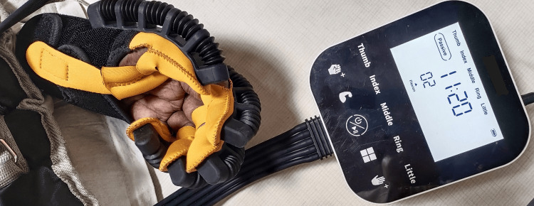 Figure 2
| Week | Goals | Intervention | Repetitions | Rationale |
| Weeks 1-2 (during plaster cast) | To prevent edema | Hand elevation above heart level | Twenty reps × one set (two times per day) | Mobility will be restored by lowering edema and swelling |
| To maintain the strength of the intrinsic muscles of the hand and wrist musculature | Isometrics: intrinsic muscles of the hand (week 1) and wrist flexors and extensors (week 2) | Ten reps × one set (two times per day) | Prevents muscle wasting | |
| To maintain the strength and ROM of the unaffected limb | AROM exercises for unaffected limb | Ten reps × one set (10-second hold) | Prevents joint stiffness | |
| Weeks 3-12 (after removal of the plaster cast) | To reduce pain | Cryotherapy | Duration: seven minutes twice daily | Efficient pain control facilitates patient mobility and engagement in physical activities, thereby promoting the healing process |
| To improve strength for wrist and hand muscles | Gentle resistive exercises for the digits and wrist (weeks 3-7) | Ten reps × one set (two times per day) | Contractions of the muscles against resistance cause an “overload” phenomenon, which stimulates the muscular system to adapt and gain training effects | |
| Progressive resistive exercises to the wrist, digits, and all groups of muscles (weeks 8-12) | ||||
| To improve ROM, wrist joint, and grip strength | Isometric exercises for wrist flexors, extensors, and radial and ulnar deviators | Ten reps × one set (10-second hold) | By gradually increasing their flexibility and ROM, patients can improve their physical function and progress toward the restoration of their autonomy | |
| Supination and pronation are encouraged. Active ulnar deviation and radial deviation using a robotic glove | ||||
| Pain-free PROM, AAROM, and AROM exercises for the wrist joint using a robotic glove | Ten reps × one set (two times per day) | |||
| Neural mobilization |
| Muscle group | Pre-treatment MMT | Post-treatment MMT |
| Wrist flexors | 2 | 4 |
| Wrist extensors | 2 | 4 |
| Pronators | 2 | 4 |
| Supinators | 2 | 4 |
| Ulnar deviator | 2 | 4 |
| Radial deviators | 2 | 4 |
| Movement | Pre-treatment active ROM | Post-treatment active ROM |
| Wrist flexion | 30o | 80o |
| Wrist extension | 20o | 75o |
| Pronation | 10o | 75o |
| Supination | 15o | 80o |
| Radial deviation | 5o | 25o |
| Ulnar deviation | 8o | 30o |
| Outcome measures | Pre-intervention | Post-intervention |
| UEFI score | 42 | 72 |
| Boston Carpal Tunnel Syndrome Questionnaire | Symptom severity: 3 | Symptom severity: 0 |
| Functional status: 4 | Functional status: 1 | |
| NPRS | On activity: 6/10 | On activity: 3/10 |
| On rest: 2/10 | On rest: 1/10 |
Peer Reviews
No public reviews on file for this paper yet. If you reviewed it on a platform where reviews are public (OpenReview, ICLR, NeurIPS, ICML), you can paste yours below so the community can read it here.
Videos
No videos yet. Explain this paper in a talk, walkthrough, or lecture? Add one.
Taxonomy
TopicsOrthopedic Surgery and Rehabilitation · Peripheral Nerve Disorders · Elbow and Forearm Trauma Treatment
Introduction
There are a variety of distal end fractures of the radius, depending on the bone quality, age, and mechanism of injury. These fractures include variations such as the dorsal and volar Barton, Smith, and Colles fractures [1]. Since they account for 75% of forearm fractures and 17% of all fractures, fractures around the lower end of the radius are the most common upper extremity fractures encountered in clinical practice. The range of incidence rates per 10,000 people per year is 5.7-124.6 people [2]. John Rhea Barton, an orthopedic surgeon in Philadelphia, was the first to describe the Barton fracture. It is characterized by a distal radius fracture that passes through the dorsal portion of the articular surface; the radiocarpal joint is frequently dislocated as a result [3]. The Barton fracture is distinctive among distal radius fractures and dislocations due to the fact that it maintains intact contact between the radius and carpus as the radiocarpal ligament is not disrupted [4]. According to the Arbeitsgemeinschaft Osteosynthese (AO) classification system, distal radius fractures resulting from shearing force are categorized as type B fractures. Specifically, B3 fractures denote volar Barton fractures. These B3 fractures are then subcategorized based on the size of the volar fragment into B3.1 for small fragment fractures, B3.2 for large fragment fractures, and B3.3 for comminuted fractures [5].
Comminuted distal radius fractures are often caused by a fall onto the outstretched hand from a standing height. However, it is crucial to emphasize that a minority of patients may experience substantial energy-related damage as a result of this mode of injury [6]. Individuals with Barton fractures frequently seek medical attention in urgent care when presenting with deformity, edema, and acute wrist pain as a result of recent trauma. Tenderness, ecchymosis, and joint swelling are common findings during a physical examination of the wrist. Pain typically results in a limited range of motion (ROM) for the wrist joint [7]. The initial assessment of a Barton fracture commences with wrist radiographs, typically comprising lateral and frontal views. Additionally, oblique views of the wrist may be acquired to aid in diagnosis. Anteroposterior radiographs are utilized to determine radial height inclination, articular step, and ulnar variance. Meanwhile, lateral radiographs allow detection of comminution, coronal split, and volar tilt [8]. Nonoperative management typically yields unsatisfactory outcomes and is accompanied by potential complications, including deformity, early-onset osteoarthritis, instability, and subluxation [9]. Several surgical approaches have been reported in the medical literature for the management of volar Barton fractures. These include percutaneous Kirschner pinning and closed reduction with external fixation, as well as open reduction with volar buttress plating, which is currently recommended for the management of such fractures. These procedures often produce favorable realignment and immediate stability. Additionally, they enable expedited wrist mobilization, potentially mitigating the risk of wrist stiffness [10].
Post-traumatic osteoarthritis, weakened grip strength and endurance, carpal instability, and restricted motion are all sequelae of articular malalignment after distal radius fractures. Carpal tunnel syndrome (CTS) is the most frequent nerve-related complication, and it occurs often in distal radius fractures regardless of treatment mode [11]. Internal fixation for distal radius fractures is often justified by the possibility of improved wrist motion and function from early mobilization and grip strengthening, similar to outcomes observed with other periarticular fractures. [12]. Another innovative way to support rehabilitation therapy is the development of robotic gloves for hand rehabilitation therapy. A robotic glove prosthesis specifically designed for hand rehabilitation in patients with grip disorders is presented in this scholarly publication [13]. In order to restore hand strength and movement, repetitive hand movements are frequently employed as a rehabilitation strategy. A wearable hand rehabilitation gadget can improve the quality of rehabilitation activities and allow physiotherapists to extend their training [14]. Our aim was to evaluate the effect of utilizing a robotic glove in order to achieve early wrist movement and grip strength to gain early fine movement in the case of acute CTS secondary to a volar Barton fracture.
Case presentation
Patient information
We report the case of a 45-year-old male who was apparently alright two months ago when he had a road traffic accident and fell from a two-wheeler, after which he experienced pain and swelling of the left wrist. Pain was sudden in onset, gradually progressive, sharp shooting in nature, constant throughout, increased in intensity on movement, and decreased on rest and immobilization. Immediately after the trauma, the patient came to our tertiary care hospital, where dressing was done, and he had undergone an X-ray investigation that revealed a B3.3 (according to the AO classification) volar Barton fracture on the left side. He underwent open reduction and internal fixation (ORIF) with plate osteosynthesis for a volar Barton fracture (Figure 1). After surgical repair, the patient developed acute CTS and pain at the wrist joint, reduced strength and range of the wrist joint, and a loss of grip strength, for which a tailor-made physiotherapy regimen was started.
X-ray of the wrist jointThe square shows ORIF with plate osteosynthesis for a volar Barton fracture.ORIF, open reduction and internal fixation
Physiotherapy assessment
Prior to initiating the examination, the patient provided informed consent. He was cooperative, conscious, and oriented to place, person, and time. Upon examination, he was hemodynamically stable. The patient had a mesomorphic build with a BMI of 29 kg/m^2^. Visible swelling was noted near the wrist joint. Palpation revealed Grade 2 tenderness based on the tenderness grading system. The patient reported dull pain, rated 8/10 during activity and 3/10 at rest on the Numerical Pain Rating Scale. The ROM and strength were notably diminished at the wrist joint of the left upper limb.
Physiotherapy intervention
Based on the patient’s clinical state, the physiotherapist designed tailored exercise sessions. The treatment duration was four weeks. Table 1 and Figure 2 depict the physiotherapy protocol.
Patient performing passive ROM exercises for the wrist joint using a robotic gloveROM, range of motion
Follow-up and outcome measures
For four weeks, the patient underwent a structured physical therapy regimen, followed by a subsequent follow-up evaluation. The findings of the outcome measure are shown in Tables 2-4.
Discussion
Even though volar Barton fractures are not uncommon, the optimal treatment approaches for them are still up for debate. The closed reduction with percutaneous Kirschner wire fixation and external fixation or open reduction with buttress plating has garnered significant support at this time [15]. In this study, we have discussed a case of a 45-year-old male diagnosed with acute CTS secondary to a volar Barton fracture and managed surgically with ORIF with plate osteosynthesis. The ORIF led to post-operative pain and loss of wrist proprioception, ROM, and strength, for which he had undergone physiotherapy management, including robotic gloves to regain early grip strength, which proved to be effective in early functional recovery.
We incorporated robotic gloves into our treatment, which helped in improving strength and ROM of the wrist and fingers, which led to early grip strengthening and regaining fine movements, which also led to an improved score on the upper extremity functional index and Boston Carpal Tunnel Syndrome Questionnaire. Biggar and Yao also stated that robotic exoskeletons are an emerging way of supporting physical therapy, owing to their capacity to provide a stable structure that improves patient posture. Furthermore, they provide a strong foundation for positioning the components that direct and support movements [16]. A wearable soft robotic device holds promise for enhancing the benefits of rehabilitative therapy by offering enhanced portability, improved affordability, simplified tailoring, reduced weight, grip-strengthening capabilities, safer interactions between humans and robots, expanded ROM, and the ability to conduct task-specific training or exercises simulating activities of daily living (ADLs) [17].
According to Kochar and Samal, for those with volar Barton fractures, physiotherapeutic therapies such as ROM exercises, cryotherapy, and muscle-strengthening exercises can be administered [18]. Additionally, exposure to cold decreases nerve transmission velocity and vasoconstriction, which, in turn, reduces blood flow and relieves pain and edema [19]. In this case, we have seen that the patient has undergone physiotherapy treatment that included isometric and active ROM exercises using a robotic glove, cryotherapy, strengthening exercises, and neural mobilization, which increased wrist grip strength and ROM, reduced pain, and regained fine movements, which led to improved ADLs and early functional recovery.
Conclusions
Dealing with a volar Barton fracture unveils significant challenges and can result in various secondary consequences, such as CTS. Physiotherapy plays a pivotal role in the rehabilitation of individuals with CTS secondary to volar Barton fractures. Wearable hand rehabilitation devices can aid physiotherapists in promoting early wrist mobility and grip strength, facilitating early fine motor movement, and improving the efficacy of rehabilitation exercises. Physiotherapeutic interventions, including cryotherapy, neural mobilization, ROM exercises using robotic gloves, and muscle strengthening exercises, are beneficial for individuals experiencing acute CTS secondary to volar barton fractures.
The reference list from the paper itself. Each links out to its DOI / PubMed record.
- 1Volar barton fracture: treatment comparison of plating and external fixation with krischner wires Ind Jour Orthop Surg Sharma J Upadhyaya P 61022016 https://www.ijos.co.in/journal-article-file/1604
- 2A prospective study in surgical management of distal end radius fracture with variable angle locking compression plate Int J Orthop Sci Shariff SM 70371272021
- 3Do variable locking plates provide better functional and radiological outcomes in volar Barton fractures?Cureus S JK Ethiraj P 014202210.7759/cureus.31427 PMC 974510136523730 · doi ↗ · pubmed ↗
- 4A study of surgical management of volar barton’s fractures using ellis plate J Evid Based Med Healthc Kolla R Aasupathri PK Rajaiah D Mallepogu RK Thadi RR Tej A 53453862019 https://www.jebmh.com/abstract/a-study-of-surgical-management-of-volar-bartons-fractures-using-ellis-plate-78251.html
- 5A classification and grading system for Barton fractures Int Orthop Lu Y Li S Wang M 172517344020162656663910.1007/s 00264-015-3034-x · doi ↗ · pubmed ↗
- 6Functional outcome of patients undergoing volar buttress plate for fixation in volar Barton’s fracture of the distal radius SAS J Surg Islam MA Kumar N Haque R 101482022
- 7Barton fracture Stat Pearls [Internet] Szymanski JA Reeves RA Taqi M Carter KR Treasure Island (FL)Stat Pearls Publishing 2024 https://pubmed.ncbi.nlm.nih.gov/29763081/29763081 · pubmed ↗
- 8Essential radiographic evaluation for distal radius fractures Hand Clin Medoff RJ 2792882120051603943910.1016/j.hcl.2005.02.008 · doi ↗ · pubmed ↗