A simple and rapid technique to achieving an airtight seal for negative pressure wound therapy in externally fixated lower limb open fractures
L Boyce, C Jordan, G Pafitanis

Abstract
Genes, proteins, chemicals, diseases, species, mutations and cell lines named across the full text — each resolved to its canonical identifier and authoritative record.
Click any figure to enlarge with its caption.
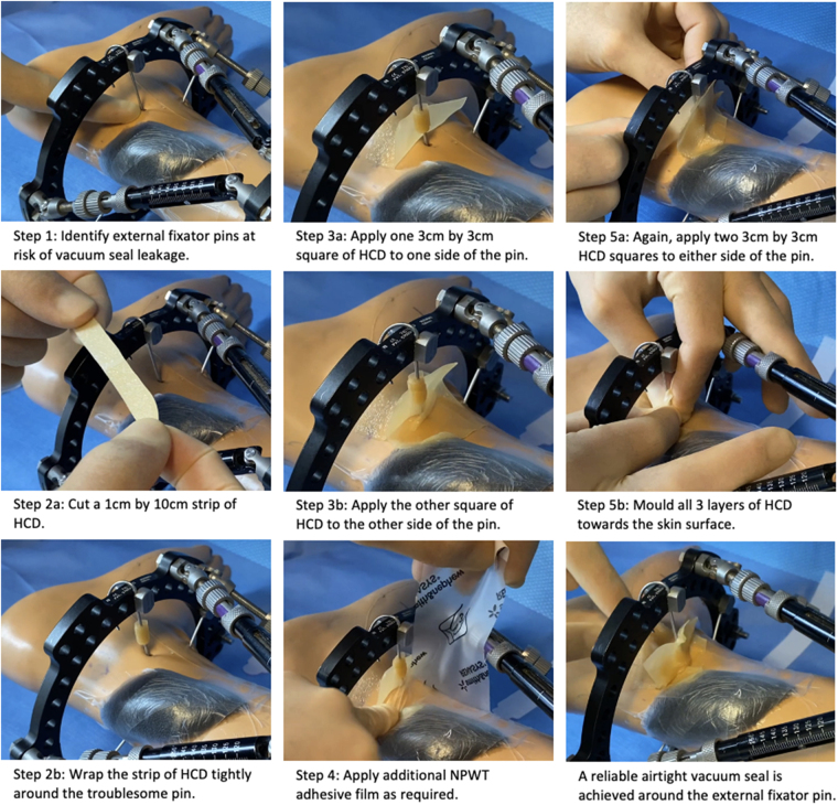 Figure 1
Peer Reviews
No public reviews on file for this paper yet. If you reviewed it on a platform where reviews are public (OpenReview, ICLR, NeurIPS, ICML), you can paste yours below so the community can read it here.
Videos
No videos yet. Explain this paper in a talk, walkthrough, or lecture? Add one.
Taxonomy
TopicsBone fractures and treatments · Surgical site infection prevention · Surgical Sutures and Adhesives
Background
External fixation and negative pressure wound therapy (NPWT) dressings are routinely used in the management of lower limb open fractures.^1^ Achieving an airtight seal for NPWT can be challenging when multiple pins are inserted close to the debrided wound. Once negative pressure is applied, tearing of the adhesive film on the fixation hardware results in loss of the vacuum seal.^2^
We developed a five-step technique that exploits the elastic, adhesive and mouldable properties of hydrocolloid dressings (HCDs) to enhance the vacuum seal around external fixator pins (Figure 1).
The simple five-step technique to achieving an airtight vacuum seal for negative pressure wound therapy in externally fixated open fractures
Technique
Our technique had the following steps:
- • Step 1: identify external fixator pins at risk of vacuum seal leakage – pins entering the skin within 2cm of the wound edge are candidates for this technique. Apply the NPWT foam and adhesive film to the wound.
- • Step 2: cut a 1 × 10cm strip of HCD and wrap it tightly around the troublesome pin.
- • Step 3: cut two identical 3 × 3cm squares of HCD and apply them to either side of the pin, overlying the adhesive film on the skin surface and sandwiching the HCD strip wrapped around the pin.
- • Step 4: apply additional adhesive film as required, overlying the HCD already applied.
- • Step 5: apply two further identical 3 × 3cm squares of HCD either side of pin and film. Mould all three layers of HCD towards the skin surface.
Discussion
Alternative solutions to this problem have previously been described, despite numerous flaws.^3–5^ HCDs are cheap, sterile, durable and easy to remove. Our technique provides a standardised method for ensuring an optimal, time-efficient vacuum seal using simple HCD.
The reference list from the paper itself. Each links out to its DOI / PubMed record.
- 1Jordan DJ, Malahias M, Khan W et al. The ortho-plastic approach to soft tissue management in trauma. Open Orthop J 2014; 8: 399–408.25408781 10.2174/1874325001408010399 PMC 4235068 · doi ↗ · pubmed ↗
- 2Lemmon JA, Ahmad J, Ghavami A et al. Vacuum-assisted closure over an external fixation device. Plast Reconstr Surg 2008; 121: 234e–235e.10.1097/01.prs.0000305394.80769.8b 18349619 · doi ↗ · pubmed ↗
- 3Ozer K, Smith W. A simple technique for applying vacuum-assisted closure therapy over the circular type external fixation device. Ann Plast Surg 2006; 56: 693–694.16721088 10.1097/01.sap.0000203997.70550.f 8 · doi ↗ · pubmed ↗
- 4Bulla A, Farace F, Uzel AP et al. Negative pressure wound therapy and external fixation device: a simple way to seal the dressing. J Orthop Trauma 2014; 28: e 176–7.24296597 10.1097/BOT.0000000000000013 · doi ↗ · pubmed ↗
- 5Fan W, Hou F, Xi K et al. A filled chocolates technique to seal negative-pressure wound therapy around external fixation devices: a randomized controlled trial. J Orthop Surg Res 2021; 16: 587.34641918 10.1186/s 13018-021-02747-1PMC 8507364 · doi ↗ · pubmed ↗