Response to commentary “The importance of assessing left ventricular longitudinal function in presence of increased afterload”
Hugues de Courson, Alexandre Loiseau, Grégoire Chadefaux, Matthieu Biais

Abstract
Genes, proteins, chemicals, diseases, species, mutations and cell lines named across the full text — each resolved to its canonical identifier and authoritative record.
Click any figure to enlarge with its caption.
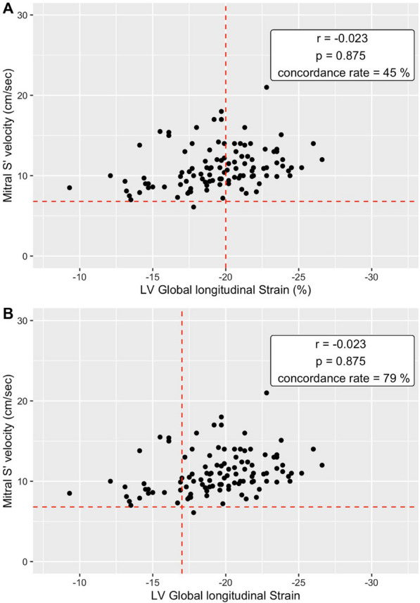 Figure 1
Peer Reviews
No public reviews on file for this paper yet. If you reviewed it on a platform where reviews are public (OpenReview, ICLR, NeurIPS, ICML), you can paste yours below so the community can read it here.
Videos
No videos yet. Explain this paper in a talk, walkthrough, or lecture? Add one.
Taxonomy
TopicsCardiovascular Function and Risk Factors · Cardiac Imaging and Diagnostics · Blood Pressure and Hypertension Studies
Dear Editor,
We very much appreciate Dr. Santonocito's thoughtful comments on our research, titled Myocardial dysfunction assessed by speckle-tracking in good-grade subarachnoid hemorrhage patients (WFNS 1–2): a prospective observational study [1].
The authors raise two valuable points:
Threshold for defining left ventricular damage We agree that the chosen threshold may have been too high for our specific population. We addressed this issue in our paper by presenting and discussing results for a lower threshold of − 17%. However, it's important to note that the research questioning this common threshold in critical care patients wasn't published at the time our protocol design and clinical trial registration (NCT03761654) were finalized.
Accounting for high afterload We agree that high afterload could influence the results. Note that the S' wave results presented were for the right ventricle. However, we were able to re-analyse the ultrasound images to obtain the value of the lateral S' wave at the mitral level. So, we performed, as asked, further analysis to assess the concordance and correlation between global longitudinal strain and mitral S' wave, providing a more comprehensive picture of left ventricular function in this context.The correlation between S' wave and SLG was very low and not statistically significant (r = − 0.023; p = 0.875). Using a threshold of 6.8 according to the work of *Park *et al. [2], the concordance rate between GLS and S' wave was 45% for a pathological GLS threshold of − 20% and 79% for a pathological GLS threshold of − 17% (Fig. 1).Fig. 1. Relationship between left ventricular ejection fraction (LVEF) and Mitral S’ velocity. A Strain threshold of − 20. B Strain threshold of − 17
These results therefore suggest that mitral S' wave analysis is not a better surrogate for GLS in this population.
The reference list from the paper itself. Each links out to its DOI / PubMed record.
- 1De Courson H Chadefaux G Loiseau A Georges D Biais M Myocardial dysfunction assessed by speckle-tracking in good-grade subarachnoid hemorrhage patients (WFNS 1–2): a prospective observational study Crit Care 202327145510.1186/s 13054-023-04738-637990276 PMC 10664298 · doi ↗ · pubmed ↗
- 2Park YS Park J-H Ahn KT Jang WI Park HS Kim JH Lee JH Choi SW Jeong JO Seong IW Usefulness of Mitral annular systolic velocity in the detection of left ventricular systolic dysfunction: comparison with three dimensional echocardiographic data J Cardiovasc Ultrasound 2010181110.4250/jcu.2010.18.1.120661328 PMC 2889389 · doi ↗ · pubmed ↗