Predicting central choroidal thickness from colour fundus photographs using deep learning
Yusuke Arai, Hidenori Takahashi, Takuya Takayama, Siamak Yousefi, Hironobu Tampo, Takehiro Yamashita, Tetsuya Hasegawa, Tomohiro Ohgami, Shozo Sonoda, Yoshiaki Tanaka, Satoru Inoda, Shinichi Sakamoto, Hidetoshi Kawashima, Yasuo Yanagi, Tatsuya Inoue, Tatsuya Inoue, Tatsuya Inoue

TL;DR
This paper introduces a deep learning method to estimate choroidal thickness from fundus images, which could aid in detecting eye diseases.
Contribution
The paper presents the first validated deep learning algorithm for estimating central choroidal thickness from color fundus photographs.
Findings
The standard deviation of 10-fold cross-validation was 73 μm.
Re-learning reduced the standard deviation in validation datasets from other institutions.
The algorithm can help identify choroidal thickening and thinning in clinical settings.
Abstract
The estimation of central choroidal thickness from colour fundus images can improve disease detection. We developed a deep learning method to estimate central choroidal thickness from colour fundus images at a single institution, using independent datasets from other institutions for validation. A total of 2,548 images from patients who underwent same-day optical coherence tomography examination and colour fundus imaging at the outpatient clinic of Jichi Medical University Hospital were retrospectively analysed. For validation, 393 images from three institutions were used. Patients with signs of subretinal haemorrhage, central serous detachment, retinal pigment epithelial detachment, and/or macular oedema were excluded. All other fundus photographs with a visible pigment epithelium were included. The main outcome measure was the standard deviation of 10-fold cross-validation. Validation…
Genes, proteins, chemicals, diseases, species, mutations and cell lines named across the full text — each resolved to its canonical identifier and authoritative record.
Click any figure to enlarge with its caption.
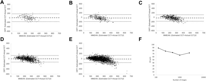 Figure 1
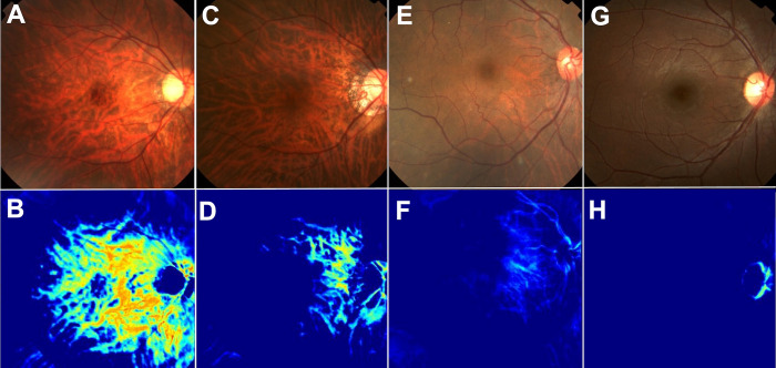 Figure 2
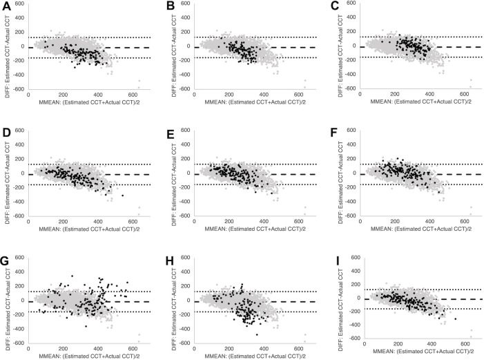 Figure 3
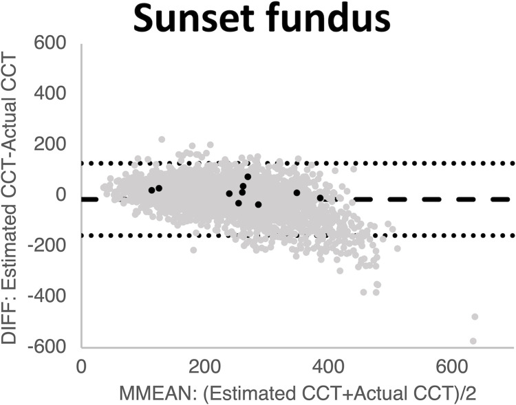 Figure 4
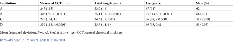 Figure 5
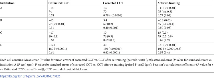 Figure 6
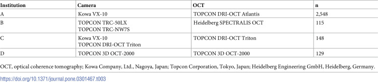 Figure 7
Peer Reviews
No public reviews on file for this paper yet. If you reviewed it on a platform where reviews are public (OpenReview, ICLR, NeurIPS, ICML), you can paste yours below so the community can read it here.
Videos
No videos yet. Explain this paper in a talk, walkthrough, or lecture? Add one.
Taxonomy
TopicsRetinal Imaging and Analysis · Retinal Diseases and Treatments · Retinal and Optic Conditions