Can white matter hyperintensities based Fazekas visual assessment scales inform about Alzheimer’s disease pathology in the population?
Aishwarya Pradeep, Sheelakumari Raghavan, Scott A. Przybelski, Gregory Preboske, Christopher G. Schwarz, Val J. Lowe, David S. Knopman, Ronald C. Petersen, Clifford R. Jack, Jonathan Graff-Radford, Petrice M. Cogswell, Prashanthi Vemuri

TL;DR
This study explores how white matter hyperintensities (WMH) relate to Alzheimer's disease pathology and cognitive decline, finding similar patterns for periventricular and deep WMH.
Contribution
The study introduces a harmonized approach to assess WMH spatial patterns and their reproducibility across MRI scanners in relation to Alzheimer's biomarkers.
Findings
Periventricular and deep WMH showed similar correlations with age, vascular risk, and cognition.
Minimal association was found between WMH and amyloidosis, with no link to tau burden.
High inter-scanner reproducibility of Fazekas ratings supports data combinability for future research.
Abstract
White matter hyperintensities (WMH) are considered hallmark features of cerebral small vessel disease and have recently been linked to Alzheimer’s disease pathology. Their distinct spatial distributions, namely periventricular versus deep WMH, may differ by underlying age-related and pathobiological processes contributing to cognitive decline. We aimed to identify the spatial patterns of WMH using the 4-scale Fazekas visual assessment and explore their differential association with age, vascular health, Alzheimer’s imaging markers, namely amyloid and tau burden, and cognition. Because our study consisted of scans from GE and Siemens scanners with different resolutions, we also investigated inter-scanner reproducibility and combinability of WMH measurements on imaging. We identified 1144 participants from the Mayo Clinic Study of Aging consisting of older adults from Olmsted County,…
Genes, proteins, chemicals, diseases, species, mutations and cell lines named across the full text — each resolved to its canonical identifier and authoritative record.
Click any figure to enlarge with its caption.
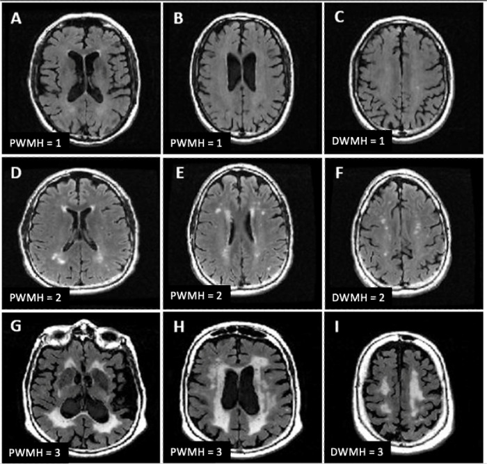 Figure 1
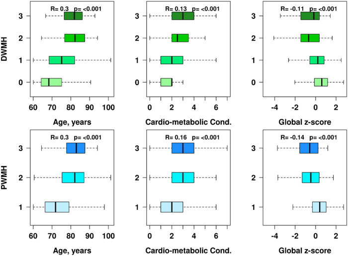 Figure 2
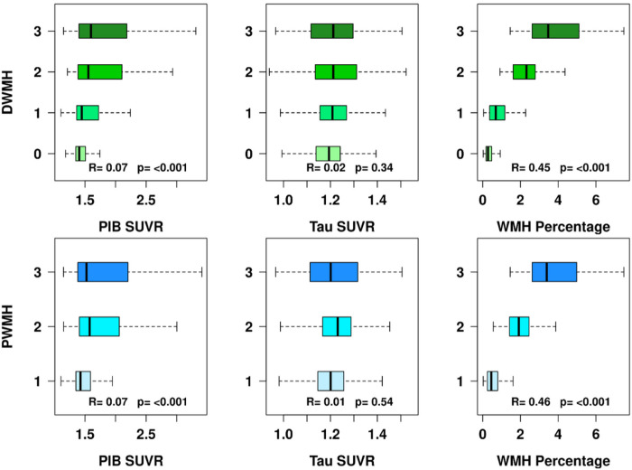 Figure 3
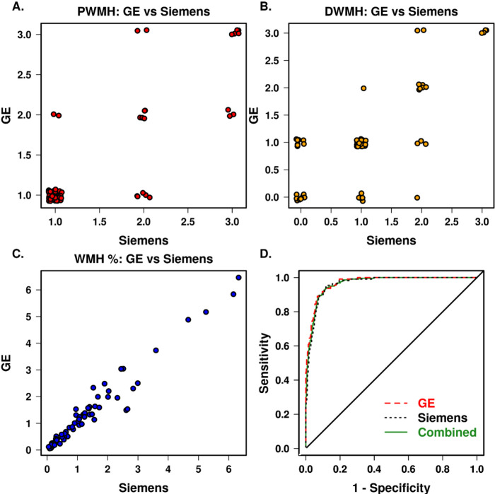 Figure 4
Peer Reviews
No public reviews on file for this paper yet. If you reviewed it on a platform where reviews are public (OpenReview, ICLR, NeurIPS, ICML), you can paste yours below so the community can read it here.
Videos
No videos yet. Explain this paper in a talk, walkthrough, or lecture? Add one.
Taxonomy
TopicsDementia and Cognitive Impairment Research · Retinal Imaging and Analysis · Neurological Disease Mechanisms and Treatments