Correction: Neural stem cells genetically-modified to express neprilysin reduce pathology in Alzheimer transgenic models
Mathew Blurton-Jones, Brian Spencer, Sara Michael, Nicholas A. Castello, Andranik A. Agazaryan, Joy L. Davis, Franz-Josef Müller, Jeanne F. Loring, Eliezer Masliah, Frank M. LaFerla

Abstract
Genes, proteins, chemicals, diseases, species, mutations and cell lines named across the full text — each resolved to its canonical identifier and authoritative record.
Click any figure to enlarge with its caption.
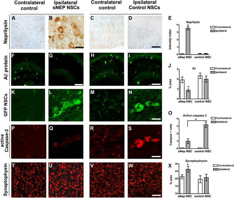 Figure 6
Peer Reviews
No public reviews on file for this paper yet. If you reviewed it on a platform where reviews are public (OpenReview, ICLR, NeurIPS, ICML), you can paste yours below so the community can read it here.
Videos
No videos yet. Explain this paper in a talk, walkthrough, or lecture? Add one.
Taxonomy
TopicsAnesthesia and Neurotoxicity Research · Pluripotent Stem Cells Research · Biomedical Ethics and Regulation
**Correction: Stem Cell Research & Therapy 2014, 5:46 ** 10.1186/scrt440
The original article contains an artifact in Fig. 6H that obscures the view of the lower-middle portion of the image. The correct original image for Fig. 6H can be viewed in this Correction article.
Fig. 6sNEP-NSCs reduce plaque pathology and resist degeneration in a second transgenic AD model. Neprilysin immunoreactivity in the contralateral (A) and ipsilateral (B) hippocampus of sNEP-NSC transplanted transgenic mice reveals high levels of NSC neprilysin expression in vivo. (C-D) Control NSCs, in contrast, produce little to no neprilysin following transplantation, quantified in (E). At 10 months of age, Thy1-APP mice exhibit considerable amyloidosis (6E10 labelling, green) within the hippocampus (F). However, transplantation of sNEP-NSCs significantly reduced Aβ pathology within the ipsilateral hippocampus (G). Control NSCs by comparison have no effect on Aβ levels (H-I), quantified in (J). GFP labelling (green) reveals examples of NSCs engrafted into the ipsilateral hippocampus (L, N), but not within the contralateral vehicle-injected side of the brain (K, M). In line with in vitro findings, caspase activation is reduced by expression of neprilysin (O). Little active caspase-3 immunoreactivity (red) is detected within the ipsilateral hippocampi of transgenic mice (P, R). However, caspase-3 activation (red) within sNEP-NSCs (Q) is significantly reduced versus control NSCs (S). Furthermore, levels of the presynaptic terminal marker synaptophysin (T-X) are significantly increased by sNEP-NSC transplantation (U), suggesting that neprilysin expression can reduce Aβ-induced synaptotoxicity. N = 6/group, error bars represent standard error of the mean (SEM). Scale Bar = 30 μm in A-D, 350 μm in F-I, 14 μm in K-S, 45 μm in T-W. Aβ, beta-amyloid; AD, Alzheimer’s disease; NSCs, neural stem cells; sNEP, secreted neprilysin.