MRI Findings Reflecting Ongoing Cardiac Arrest While Being on an MRI Scanner
Abdallah Q Al Khateeb, Pokhraj P Suthar, Sudeep Bhabad

TL;DR
This paper presents a rare case where MRI scans captured signs of ongoing cardiac arrest in a patient during an MRI scan.
Contribution
The paper reports the first known MRI findings of ongoing cardiac arrest due to pulseless electric activity during an MRI scan.
Findings
Abnormal brain MRI findings indicated ongoing cardiac arrest during the scan.
The imaging findings were unique and not commonly seen in routine practice.
The case highlights unusual MRI manifestations of pulseless electric activity.
Abstract
Sudden cardiac arrest (SCA) is the abrupt loss of cardiac function that results in acute cardiovascular collapse and subsequent decreased or loss of various organ perfusion. Here, we present an interesting case of a 58-year-old man who developed abnormal brain MRI findings reflecting ongoing cardiac arrest due to pulseless electric activity (PEA) during an MRI scan. To our knowledge, this is the first case describing the MRI findings of ongoing cardiac arrest due to PEA. Our case is unique in imaging findings, which are not routinely encountered in day-to-day practice. This case raised awareness among the readers.
Genes, proteins, chemicals, diseases, species, mutations and cell lines named across the full text — each resolved to its canonical identifier and authoritative record.
Click any figure to enlarge with its caption.
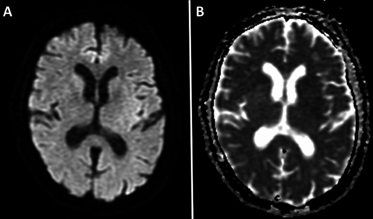 Figure 1
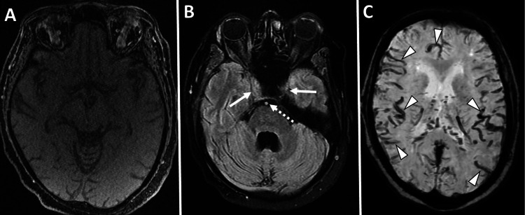 Figure 2
Peer Reviews
No public reviews on file for this paper yet. If you reviewed it on a platform where reviews are public (OpenReview, ICLR, NeurIPS, ICML), you can paste yours below so the community can read it here.
Videos
No videos yet. Explain this paper in a talk, walkthrough, or lecture? Add one.
Taxonomy
TopicsCardiac Arrest and Resuscitation · Traumatic Brain Injury and Neurovascular Disturbances · Advanced MRI Techniques and Applications