Phytophthora pluvialis maintenance, spore production and detached needle assays
Sophie Eccersall, Leann S. Vinson, Rebecca McDougal, Claudia-Nicole Meisrimler

TL;DR
This paper outlines a method to study how Phytophthora pluvialis infects pine trees, using spore production and detached needle assays to assess virulence and infection mechanisms.
Contribution
The paper introduces a reproducible protocol for spore production and detached needle assays to study P. pluvialis pathogenicity in pine needles.
Findings
Detached needle assays enable high-throughput screening of pine needles with different genetic backgrounds.
qPCR was used to assess gene expression of the candidate effector protein PpR01 during infection.
Combining assays with RNA sequencing and metabolomics can reveal infection mechanisms in detail.
Abstract
Phytophthora pluvialis is an oomycete that primarily infects Pinus radiata and Pseudotsuga menziesii causing the destructive foliar disease red needle cast (RNC). Recent observations show that P. pluvialis can also infect western hemlock inducing resinous cankers. High-throughput and reproducible infection assays are integral to find key information on tree health and oomycete pathogenicity. In this protocol, we describe the propagation and spore induction of P. pluvialis, followed by detached needle assays for verification and quantification of virulence of P. pluvialis in P. radiata needles. These needle assays can be employed for high-throughput screening of tree needles with diverse genetic backgrounds. In downstream analysis, Quantitative PCR (qPCR) was utilized to assess relative gene expression, as exemplified by candidate RxLR effector protein PpR01. Additional techniques like…
Genes, proteins, chemicals, diseases, species, mutations and cell lines named across the full text — each resolved to its canonical identifier and authoritative record.
Click any figure to enlarge with its caption.
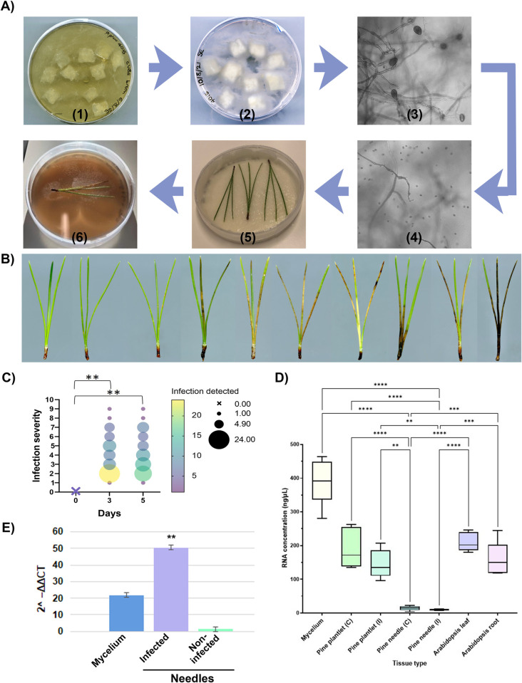 Fig 1
Fig 1- —Radiata Pine Breeding Company New Zealand
- —http://dx.doi.org/10.13039/100014111Bio-Protection Research Centre
Peer Reviews
No public reviews on file for this paper yet. If you reviewed it on a platform where reviews are public (OpenReview, ICLR, NeurIPS, ICML), you can paste yours below so the community can read it here.
Videos
No videos yet. Explain this paper in a talk, walkthrough, or lecture? Add one.
Taxonomy
TopicsPlant Pathogens and Resistance · Plant Pathogens and Fungal Diseases · Plant-Microbe Interactions and Immunity
Introduction
Phytophthora pluvialis is an oomycete that primarily infects Pinus radiata and Pseudotsuga menziesii causing the destructive foliar disease red needle cast (RNC) [1]. RNC causes accelerated needle senescence and premature defoliation of trees and can lead to loss of growth. Typical disease symptoms include a dark resinous band, which extends to discoloured khaki lesions on the needle, followed by premature needle shedding. First recovered in 2002 in Oregon, P. pluvialis is found throughout the pacific northwest of the USA and New Zealand, and most recently the United Kingdom, causing branch and stem cankers and crown die back on mature western hemlock (Tsuga heterophylla) [2].
A reliable and reproducible protocol was created for the maintenance and growth of P. pluvialis and detached needle assays in laboratory setting. Detached needle assays have been performed on both known and unknown hosts of P. pluvialis and provide insight into virulence for breeding and research purposes, otherwise difficult to access due to the tree hosts slow growth and reproduction [3]. Detached needle assays can be combined with DNA, RNA, protein, and metabolite analysis [4] from infected and control tissues for further investigation of molecular mechanisms of the infection process. High throughput and reproducible infection assays are integral to find key information around tree health and oomycete pathogenicity. Classical or conventional breeding is one of the principal ways to improve pest and disease resistance. In tree species, not only is this costly, but is substantially complicated by their long generation times [5]. An -omics based approach can generate large amounts of data on forest tree resistance mechanisms which can ultimately contribute to the improvement and protection of commercially important trees, such as Pinus radiata by providing insight on effector targets and recognition [6]. This information mitigates the need for blind breeding which becomes costly and time consuming.
In this protocol, we describe the propagation and spore induction of P. pluvialis, followed by detached needle assay for conducting the virulence assay of P. pluvialis on P. radiata needles. These needle assays can be used for semi-high throughput assays to screen hundreds of trees genotypes in a laboratory setting and allow for further downstream analysis as shown by Graham et al., 2018 [5]. Additionally, we performed qPCR on P. pluvialis mycelium and both infected and non-infected P. radiata pine needles to confirm the presence or absence of oomycete RNA as shown in S1 [7].
Phytophthora species require cultivation on the appropriate selective media for successful isolation and investigation in laboratory conditions [8]. Important characteristics to observe are mycelium growth habit- aerial or appressed; mycelium pattern- uniform, radiate, stellate, or petaloid; and the structures present in agar—sporangia, oospores, hyphal swellings and chlamydospores [9]. Five New Zealand strains of P. pluvialis received from Scion (New Zealand Forest Research Institute, Ltd),were propagated in the laboratory at the University of Canterbury. In contrast to the original protocol for maintenance [1], P. pluvialis was grown on cV8 agar in this study [9]. All five strains grew on this medium similar to the original carrot agar medium with appressed hyphae with an angular and petaloid growth pattern. This phenotype confirmed previous descriptions of growth patterns for these P. pluvialis strains [10].
Zoospore production is a crucial part of the infection process of Phytophthora species [1, 11]. For needle infection assay experiments, a previous zoospore induction protocol was tested without success in reliable zoospore induction. To circumvent this issue, two main points of the original protocol were modified: 1) the agar and broth media for P. pluvialis growth 2) the treatments used to induce zoospores. Carrot agar and the carrot broth were replaced by 20% cV8 agar and broth, supplemented with β-sitosterol [6]. The cV8 medium is well-known media for growing Phytophthora species [12] and supports development of sporangia, making it an ideal media to optimise P. pluvialis growth. Within seven days, all five P. pluvialis strains grew without issues on the new media type and followed the appropriate growth patterns [9] and were ready for use in experiments. Upon induction of zoospores following the new protocol, the number of zoospores released increased significantly. Following the initial release, the zoospore solution was collected and used for needle infection assays. Once the zoospore solution was siphoned off, the petri dish containing the sporangia was flooded a second time with cold sterile pond water and repeated shock treatments. Upon this second induction treatment, a second “wave” of released zoospores was observed. This second wave of zoospores was also used to perform needle infection assays. No change of virulence between first and second wave of zoospores was noted. Plates containing mature sporangia can be re-induced multiple times over successive days.
Material and methods
The protocol described in this peer-reviewed article is published on protocols.io dx.doi.org/10.17504/protocols.io.kqdg3p9d7l25/v1 and is included for printing as supporting information file 1 with this article.
Expected results
Maintenance and spore production of P. pluvialis is essential for experiments associated to downstream infection assays to test for resistance or sensitivity in Pinus radiata, Pseudotsuga menziesii, Tsuga heterophylla and other potential host species. For quantitative infection assays numbers of zoospores will need to be the same, allowing comparison between infection severity. Generally, this protocol will allow for a minimum amount of 2.5 x 10^3^ mL of zoospores from the first wave of zoospore solution. Similar yields can be expected from subsequent waves by refreshing the petri dish with 15 mL of sterile pond water. For detached needle assays, the suggested number of spores/μl 2.5 x 10^3^. Although currently the minimum number of zoospores for successful infection is unknown, the suggested number will allow qualitative and quantitative infection of needles showing symptoms within three days. If the infection is successful, it is expected that 100% of needles show signs of infection at day three and day five post infection. In the shown example (Fig 1C.) a two-way ANOVA analysis was performed and demonstrated statistically significant difference in infection between day zero and day three and five respectively (p-value = 0.0072); no statistically significant difference was observed between day three and five (p-value = >0.999).
A) Protocol at a glance: (1) agar cubes in v8 broth; (2) agar cubes in sterilised pond water; (3) formation of mature sporangia; (4) zoospore induction and release; (5) detached needle assays; new mycelium propagation. B) Severity of infected pine samples from left to right on a scale of 1–10. C) Graph depicting infection severity (visual quantification) from detached needle assays on a scale of 1–10; n = 65 needles. KEY: Colour scale and circle size indicating the number of replicates and the level of infection; × indicates the full set of replicates at day zero with no infection symptoms. D) Graph depicting RNA concentrations from various tissue samples. C = control; (I) = infected. E) Graph depicting expression of PpR01 normalised to P. pluvialis actin-1 calculating its 2^–ΔΔCT.
To demonstrate expected results for downstream analysis, RNA extraction was chosen. RNA extraction is essential for many applications, such as quantitative RT-PCR, RNA sequencing, Yeast-2-Hybrid cDNA library preparation. Analysis indicated no statistically significant difference between RNA concentrations extracted from control treated needles vs P. pluvialis infected pine needles (after three days); importantly, using the Plant/Fungi Total RNA purification kit (Norgen Biotek) to extract RNA from mycelium generally results in higher RNA amounts than pine samples. This needs to be considered during experimental planning. The pine needles used in the presented RNA extraction were ≤1 year old and harvested from trees 3 years of age or older. It can be stated that younger pine needles will result in higher amounts of RNA or protein than older pine needles (≥1 year). Additionally, for comparison, Arabidopsis leaves and roots have been extracted with the same RNA extraction protocol, indicating a significantly higher RNA concentration than for pine samples in general (Fig 1C and I; p-value≤0.0001). Furthermore, a statistically significant difference was observed for RNA extraction of pine plantlets vs needles for control and infected samples. These results are not unsurprising, as the phenolic compounds in mature pine needles can make RNA extraction difficult. Nevertheless, no effect is to be expected for RNA concentrations in dependence of the infection for day zero and day five. Importantly, longer infection time will lead to increased cell death resulting in reduced RNA and protein concentrations.
To confirm the presence of infection in the detached needle assay, quantitative RT-PCR was performed on three sample types. P. pluvialis mycelium, P. radiata needles infected with P. pluvialis zoospores and non-infected needles were tested for the expression of the candidate RxLR effector protein PpR01. No expression of PpR01 was detected in non-infected needles compared to high expression in mycelium and infected needles (Fig 1E).
Supporting information
S1 FileStep-by-step protocol, also available on protocols.io dx.doi.org/10.17504/protocols.io.kqdg3p9d7l25/v1.(PDF)
The reference list from the paper itself. Each links out to its DOI / PubMed record.
- 1Dick MA, Williams NM, Bader MK-F, Gardner JF, Bulman LS. Pathogenicity of Phytophthora pluvialis to Pinus radiata and its relation with red needle cast disease in New Zealand. New Zealand Journal of Forestry Science. 2014;44(1). doi: 10.1186/s 40490-014-0006-7 · doi ↗
- 2Pérez‐Sierra A, Chitty R, Eacock A, Jones B, Biddle M, Crampton M, et al. First report of Phytophthora pluvialis in Europe causing resinous cankers on Western Hemlock. New Disease Reports. 2022;45(1). doi: 10.1002/ndr 2.12064 · doi ↗
- 3Gomez-Gallego M, Gommers R, Bader MK-F, Williams NM. Modelling the key drivers of an aerial Phytophthora foliar disease epidemic, from the needles to the whole plant. PLOS ONE. 2019;14(5). doi: 10.1371/journal.pone.0216161 31136583 PMC 6538149 · doi ↗ · pubmed ↗
- 4Rolando C, Gaskin R, Horgan D, Williams N, Bader MK-F. The use of adjuvants to improve uptake of phosphorous acid applied to Pinus radiata needles for control of Foliar Phytophthora diseases. New Zealand Journal of Forestry Science. 2014;44(1). doi: 10.1186/s 40490-014-0008-5 · doi ↗
- 5Graham N, Suontama M, Pleasants T, Li Y, Bader M, KlápštěJ, et al. Assessing the genetic variation of tolerance to red needle cast in a Pinus radiata breeding population. Tree Genetics & Genomes. 2018;14(4). doi: 10.1007/s 11295-018-1266-9 · doi ↗
- 6Kovalchuk A, KeriöS, Oghenekaro AO, Jaber E, Raffaello T, Asiegbu FO. Antimicrobial defenses and resistance in forest trees: Challenges and perspectives in a genomic era. Annual Review of Phytopathology. 2013;51(1):221–44. doi: 10.1146/annurev-phyto-082712-102307 23682916 · doi ↗ · pubmed ↗
- 7Vinson LS. Effects of abiotic stress on the oomycete Phytophthora pluvialis. M Sc dissertation. University of Canterbury. 2022. 10.26021/12833 · doi ↗
- 8Sarker SR, Mc Comb J, Burgess TI, Hardy GE. Antimicrobials in Phytophthora isolation media and the growth of Phytophthora species. Plant Pathology. 2020;69(8):1426–36. doi: 10.1111/ppa.13224 · doi ↗