Optimizing surgical approaches for anterior communicating artery aneurysms: Development and internal validation of a novel surgical scoring system
Metin Orakdogen, Orkhan Mammadkhanli, Osman Simsek

TL;DR
This paper introduces a new scoring system to help surgeons choose the best approach for treating anterior communicating artery aneurysms, reducing complications.
Contribution
A novel surgical scoring system for ACoA aneurysms is developed and internally validated to guide optimal surgical approaches.
Findings
The scoring system showed a significant correlation between higher scores and lower complication rates.
A cut-off value of 12.5 points was established for predicting complications and outcomes.
The system supports personalized surgical planning using objective 3D imaging criteria.
Abstract
The objective was to provide comprehensive preoperative information on both the aneurysm orientation and the side and method of surgical approach for optimal preoperative information and safe clipping using 3D imaging modalities. This was achieved by making an objective risk assessment on the surgical side/method and evaluating its effectiveness with internal validation. Radiologic data of 61 ACoA aneurysm patients between 2012 and 2020 were retrospectively analyzed. A scoring system based on five criteria; ACoA aneurysm dome orientation, A1 symmetry/control, perforating artery control, A2 trace orientation, and A2 fork symmetry was developed. The system is designed to align with the most common surgical approaches in ACoA aneurysm surgery. The patients were categorized into three groups based on the scoring results to determine the most appropriate surgical method. Group I was…
Genes, proteins, chemicals, diseases, species, mutations and cell lines named across the full text — each resolved to its canonical identifier and authoritative record.
Click any figure to enlarge with its caption.
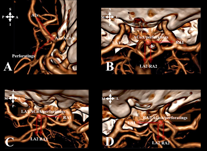 Figure 1
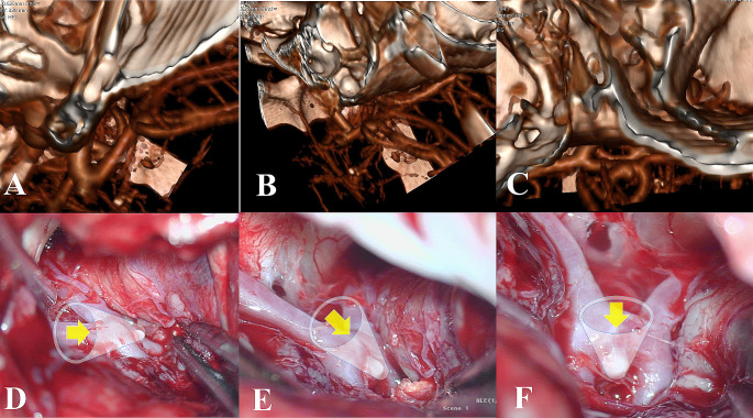 Figure 2
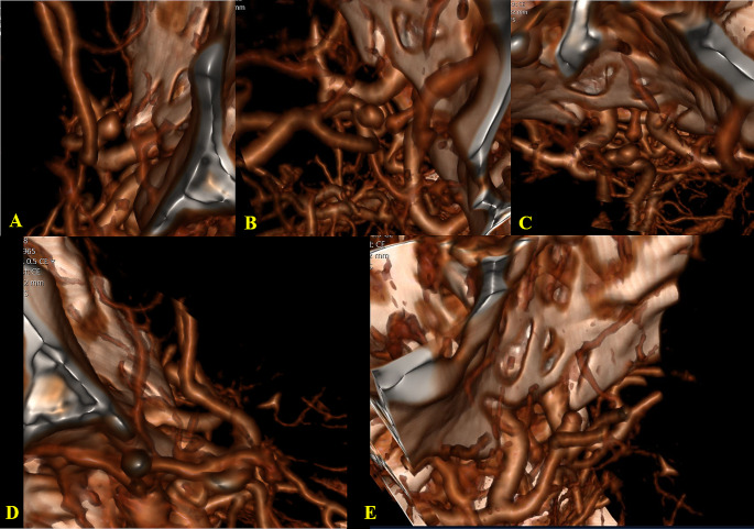 Figure 3
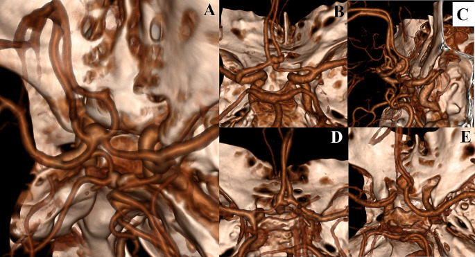 Figure 4
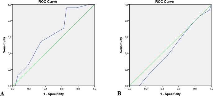 Figure 5
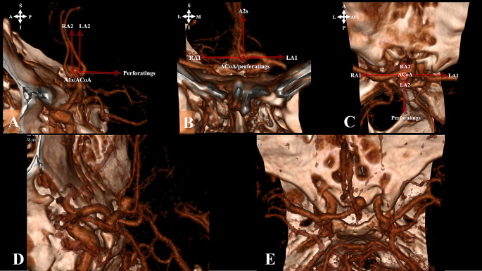 Figure 6
- —Trakya University
Peer Reviews
No public reviews on file for this paper yet. If you reviewed it on a platform where reviews are public (OpenReview, ICLR, NeurIPS, ICML), you can paste yours below so the community can read it here.
Videos
No videos yet. Explain this paper in a talk, walkthrough, or lecture? Add one.
Taxonomy
TopicsIntracranial Aneurysms: Treatment and Complications · Aortic Disease and Treatment Approaches · Vascular Malformations Diagnosis and Treatment
Introduction
Anterior communicating artery (ACoA) aneurysm projection is one of the important factors that determine the selection of the most appropriate surgical side, the difficulty level of the surgery, the precautions to be taken during surgical clipping, possible complications, and prognosis. Therefore, it is imperative to know the ACoA projection preoperatively [1–16].
Various factors have been taken into consideration with respect to the side of surgical approach to ACoA aneurysms. Aneurysm dome orientation, A1 dominance, A2 fork orientation, associated aneurysms and variations, surgeon’s experience, nondominant hemisphere/right-handed surgeon are among the most important factors [1, 3, 9, 13, 14, 17–22]. Furthermore, there are many surgical approaches to ACoA aneurysms such as transsylvian, subfrontal, lateral supraorbital, anterior interhemispheric, and orbitozygomatic [3, 9, 13, 14, 17–27].
In our study, we developed a comprehensive and reliable surgical scoring system for ACoA aneurysm clipping, considering essential factors including aneurysm dome orientation, A1 control, perforating artery control, A2 trace orientation, and A2 fork symmetry. This system provides neurosurgeons with valuable insights into safe and risky zones within the surgical field, facilitating precise decision-making during the procedure. Through internal validation, we established the reliability and applicability of our system, aiming to improve surgical outcomes for patients with ACoA aneurysms by providing objective preoperative information and guiding safe clipping procedures.
Materials and methods
Study population
After the approval of the local Institutional Committee (number TÜTF-GOBAEK 2023/255), we retrospectively analyzed the radiological data from a cohort of 96 patients with ACoA aneurysms with single/multiple aneurysms, with/without rupture, diagnosed with at least one of 3D CTA, MRI/MRA, or DSA at our University hospital between 2012 and 2020. Out of 96 patients, 61 cases underwent surgical intervention, and their data were employed for internal validation.
Defining the scoring system
The scoring system was based on the dome orientation of the aneurysm and the vascular variations in the anterior communicating complex, which would allow for the safest placement of the clip by reaching the neck of the aneurysm.
To determine the most appropriate surgical approach and method and to perform risk assessment, a scoring system based on five criteria was established. These criteria include aneurysm dome orientation, A1 control, perforating artery control, A2 trace orientation, and A2 fork symmetry .
- A.A1 control was based on A1 symmetry/control and direct vision without trying to dissect the opposite A1.
- B.Perforating artery control was evaluated as direct view and control, indirect view/control, and no view/no control. Direct vision and control imply direct control without aneurysm neck dissection. Indirect vision and control refer to control with aneurysm neck dissection, while no vision and control indicate no perforating control even with aneurysm dissection.
- C.Proximal A2/neck control was used to demonstrate the relationship between proximal A2s and aneurysm neck according to A2 trace orientation. It was evaluated as bilateral proximal A2/neck or unilateral proximal A2/neck control according to horizontal, vertical, and oblique orientations. In the horizontal orientation, the ACoA complex is positioned without lateral rotation, and both ACoA and its A2 segments are positioned in a coronal plane. In the vertical orientation, extreme rotation of the complex positions the ACoA and its A2 segments along a sagittal plane. An oblique orientation is considered if the position is between horizontal and vertical orientation.
- D.Aneurysm dome projection was classified as contralateral, midline, or ipsilateral based on approach side and technique.
- E.TheA2 fork symmetry criterion was based on the relationship between the ipsilateral A2 and aneurysm neck, and was classified as open, symmetrical, and closed.
The “open A2 fork” is characterized by the ipsilateral A2 being positioned more posteriorly than the contralateral A2 on the approach side. Vice versa in the case of the “closed A2 fork”.
As the most common surgical approaches in ACoA aneurysm surgery are pterional, lateral supraorbital, and basal anterior interhemispheric/subfrontal approaches, the scoring system was based on these (right and left). However, all possible surgical sides and methods were assessed on 3D cerebral angiography (Figs. 1 and 2).
Fig. 1. Illustration of the ACoA complex using schematics and angiography based on surgical approach. (S-superior, I - inferior, A - anterior, P - posterior, M- medial, L- lateral)A. ACoA complex: vertex down pterional approachB. ACoA complex: vertex down basal anterior interhemispheric/subfrontal approachC. ACoA complex: vertex down right lateral supraorbital approachD. ACoA complex: vertex down left lateral supraorbital approach
Fig. 23D CTA (A-C) and intraoperative views (D-F) of superior projection ACoA aneurysm in left pterional, left supraorbital and basal anterior interhemispheric/subfrontal approachesA. Left pterional viewB. Left supraorbital viewC. Basal anterior interhemispheric/subfrontal viewD. Intraoperative view of the left pterional approachE. Intraoperative view of left supraorbital approachF. Intraoperative view of basal anterior interhemispheric/subfrontal approach
Scoring was performed by considering alternatives for each criterion. The scoring system ranged from 5 to 16 points. The scoring system also considered the surgeon’s use of the dominant hand, the distance of the aneurysm from the skull base, and the complexity of the aneurysm (Table 1; Fig. 3).
Table 1. Surgical approach scoring system for ACoA aneurysmsCriteriaScore A. A1 control Bilaterally A14 Dominant A13 Symmetrical A12 Nondominant A11 B. Perforating artery control Direct view and control3 Indirect view/control2 No view/No control1 C. Proximal A2/neck control Bilaterally (horizontal or vertical)3 Bilaterally (oblique)2 Unilateral (horizontal or vertical)1 D. The aneurysm dome orientation Contralateral3 Midline2 Ipsilateral1 E. A2 fork symmetry Open A2 fork3 Symmetrical A2 fork2 Closed A2 fork1Note:For right-handed surgeons, 1 point is added to the total score for right-sided approaches and 1 point is subtracted for left-sided approaches. The opposite is done for left-handed surgeonsIn patients with a skull base–aneurysm distance > 10 mm, one point is added to the total score in the basal anterior interhemispheric/subfrontal approach and one point is subtracted in other approachesFor aneurysm diameter > 10 mm and complex aneurysms, 1 point is subtracted from the total score in all approaches
Fig. 3. Case example of surgical scale calculationACoA aneurysm with anterosuperior medial projection. Right A1 hypoplasia and left A2 closed fork present. Surgical approach scores: RPT = 9, RSO = 9, INT/SF = 13, LPT = 11, LSO = 10.A. Right pterional view (RPT)B. Right supraorbital view (RSO)C. Basal anterior interhemispheric/subfrontal view (INT/SF)D. Left pterional view (LPT)E. Left supraorbital view (LSO)
Internal validation group
Out of 61 patients, 56 cases were ruptured, and 5 unruptured. These patients were assessed as ruptured and all patients (ruptured and unruptured). The patients were categorized into three groups based on the surgical approach used;
Group 1 consists of cases where the intervention suggested by the surgical scoring system is the first option (highest score).
Group 2 consists of cases where the intervention suggested by the surgical scoring system is the second option (second highest score).
Group 3 consists of cases where the intervention suggested by the surgical scoring system is the other options (lowest score).
The surgeries were performed by more than one experienced surgeon. Each group may have multiple surgical approach options, including pterional, basal anterior interhemispheric/subfrontal, and lateral supraorbital, depending on the scores given according to our surgical scoring system.
The mean score differences between the groups for the approach recommended by the scoring system and the preferred approaches were analyzed.
Outcomes and complication rates (perforating and parent artery infarction) were investigated in these groups. Glasgow Outcome Scale (GOS) 5 − 4 was considered a favorable outcome and GOS 3 − 1 as an unfavorable outcome.
Statistical analyses
Statistical analyses were performed using Jamovi (version 2.3). The results were presented as mean ± standard deviation for parametric continuous variables and as percentage for categorical variables. Two-group comparisons were made using student’s t-test for parametric variables and χ² test and Fisher’s exact test for categorical variables. One-way analysis of variance and Kruskal–Wallis tests were used to compare the means or medians of a continuous variable (age) between different groups. Mantel–Haenszel trend test was used to determine whether there was a linear trend between two categorical variables. A p-value of ≤ 0.05 was considered significant. In addition, a receiver operating characteristic (ROC) curve was generated to evaluate the prediction of postoperative complications and GOS by the surgical scale.
Results
Among the 96 patients included in the study, 76 had only ACoA aneurysms, while 20 had multiple aneurysms. Demographically, there were 39 males and 22 females with a mean age of 54.54 ± 11.8, ranging from 29 to 82 years. Aneurysm diameters ranged between 2 and 13 mm, with an average 6.62 mm ± 2.43. A1 and A2 variations (A1 control/diameter symmetries, aneurysm dome orientations, A2 trace orientations, and A2 fork symmetries) for all cases (96 patients) are given in Table 2 (Fig. 4).
Table 2A1 and A2 variations (A1 diameter asymmetry, A1 symmetry / Aneurysm projection, A2 trace orientation, A2 fork asymmetry)Anterior cerebral artery A1 and A2 variationsNumber of case (%)A1 symmetry / Aneurysm projectionn = 96Asymmetricaln = 78 (81,25%)Medial47 (60.25)Lateral4 (5.12)Midline24 (30.77)Bilobed3 (3.84)Symmetricaln = 18 (18,75%)Medial9 (50)Lateral1 (5.55)Midline8 (44.44)Bilobed-A1 diameter asymmetryLeft A1n = 78Dominance41Prominent hypoplasia21Aplasia8Only asymmetric8Right A1n = 78Dominance37Prominent hypoplasia14Aplasia18Only asymmetric9A2 variationsA2 trace orientationn = 96Classic57 (59.37)Oblique24 (25)Vertical15 (15.62)A2 forkn = 96Symmetrical73 (76)Asymmetric (closed/open)23 (24)
Fig. 4 Types of A1 and A2 Variations A. ACoA aneurysm: Symmetric A1B. ACoA aneurysm: Asymmetric A1C. ACoA aneurysm: Closed A2 forkD. ACoA aneurysm: A2 oblique orientation and Asymmetric A1E. ACoA aneurysm: A2 vertical orientation and Asymmetric A1
A total of 61 patients underwent surgical treatment. Standard pterional approach was used as the surgical method. In the choice of the operation side, the dominant A1 side was preferred. In the presence of concomitant aneurysms, side choice was performed taking these aneurysms into account.
The aneurysm projection was evaluated using a 3D classification system [28]. The approach methods were then analyzed according to these projections (Table 3).
Table 3. Relationship between aneurysm projections and first approach option based on surgical scoring systemProjectionPterional (n = 16)Supraorbital (n = 10)Basal Anterior Interhemispheric//Subfrontal (n = 44)Anterior-median213Anterior-lateral1--Anterior-medial1-4Anterosuperior-medial5410Anterosuperior-lateral-11Anterosuperior-median1-5Anteroinferior-median112Anteroinferior--1Anteroinferior-medial1-3Superior-median--4Superior-medial1-2Posterior-medial114Posterosuperior-medial11-Posterosuperior-median1--Posteroinferior-medial--1Complex-14
Based on the surgical scoring, 44 patients were suitable for the basal anterior interhemispheric/subfrontal approach (INT/SF), while 16 patients were suitable for the pterional approach (10 on the right, 6 on the left), and 10 patients for the right supraorbital (RSO) approach as the first approach option (4 patients had 2 options and 3 patients had 3 options with the maximum score). The surgical approach used was the first option in 15 (24.6%) patients, the second option in 33 (54.1%) patients, and the third option in 13 (21.31%) patients. The mean score difference in the surgical approach was 2.71 in Group II and 4.62 in Group III compared to Group I.
In the 61 operated cases, the rate of favorable outcome was 13/15 (86.7%) in Group I, 24/33 (72.7%) in Group II, and 10/13 (76.9%) in Group III.
Complications
In Group 1, there were two caudate and one caudate + hypothalamic infarct (three patients), in Group 2, there were five caudate, two hypothalamic, one hypothalamic + caudate infarct, and one hypothalamic + caudate + parent artery (distal anterior cerebral artery (DACA)) infarct in a patient with prominent vasospasm (nine patients), and in Group 3, there were six caudate, one DACA, one bilateral hypothalamic + caudate infarct, and one hypothalamic + DACA infarct (nine patients). Totally, there were 21 patients with perforating and parent artery infarcts.
Information about the cases in the study context is summarized in Table 4.
Table 4. Summary of patients included in the studyCaseAge and genderRPTLPTRSOLSOINT/SFPrefered choiceChoiceComp.Outcome145, M1481249RPT 141NoFavorable239, M141011913RPT 141NoFavorable360, M1013101212LPT 131NoFavorable449, M12812712 RPT 12
1
Yes Favorable564, M14612711 RPT 14
1
Yes Favorable658, M81171010LPT 111NoFavorable761, F15813914RPT 151No Unfavorable 858, F1410141014RPT 141NoFavorable938, M15914715RPT 151NoFavorable1038, M14611712RPT 141NoFavorable1157, M14512714RPT 141No Unfavorable 1252, M91211911 LPT 12
1
Yes Favorable1370, M111310912LPT 131NoFavorable1466, M51561415LPT 151NoFavorable1559, M13913812RPT 131NoFavorable1634, M13715711RPT 132NoFavorable1764, F999911LPT 92NoFavorable1844, F11911913RPT 112No Unfavorable 1958, M686812 LPT 8
2
Yes Favorable2063, M12810714RPT 122NoFavorable2165, M9108713LPT 102NoFavorable2244, F7118912 LPT 11
2
Yes
Unfavorable 2367, M998814 LPT 9
2
Yes Favorable2437, M11810714RPT 112NoFavorable2550, F101291212 RPT 10
2
Yes Favorable2670, F11812710 RPT 11
2
Yes Favorable2746, F12714814 RPT 12
2
Yes Favorable2855, F71061011 LPT 10
2
Yes
Unfavorable 2967, M8107912 LPT 10
2
Yes Favorable3059, M11810714RPT 112NoFavorable3165, M1113111115LPT 132NoFavorable3278, F11911914RPT 112No Unfavorable 3352, M888813RPT 82NoFavorable3464, M10912410RPT 102NoFavorable3555, M11814711RPT 112NoFavorable3664, F11991113RPT 112NoFavorable3767, F12911914RPT 122No Unfavorable 3882, F998813RPT 92No Unfavorable 3932, M12811715RPT 122NoFavorable4058, F13913715RPT 132No Unfavorable 4161, M910101015LPT 102NoFavorable4260, M.91181013LPT 112NoFavorable4357, F.111110914 RPT 11
2
Yes
Unfavorable 4434, F.129101014RPT 122No Unfavorable 4541, M.101110815LPT 112NoFavorable4629, M.71091015LPT 102NoFavorable4742, M.13613714RPT 132NoFavorable4846, M.91081014LPT 102NoFavorable4944, M.1399914 LPT 9
3
Yes Favorable5066, F.10813712RPT 103NoFavorable5146, M.11912714 RPT 11
3
Yes Favorable5259, M.9108913 RPT 9
3
Yes Favorable5349, F.989714 LPT 8
3
Yes Favorable5474, F.10129914 RPT 10
3
Yes Favorable5552, F.101381214 RPT 10
3
Yes
Unfavorable 5647, M.1010101115RPT 103 Yes Favorable5744, F.10714711 RPT 10
3
Yes
Unfavorable 5868, M.910111215RPT 93No Unfavorable 5952, M.7117915LSO 93NoFavorable6047, M.118101010 LPT 8
3
Yes Favorable6155, M.57101016RPT 53NoFavorableAbbreviations: RPT - right pterional approach; LPT - left pterional approach; RSO - right supraorbital approach; LSO - left supraorbital approach; INT/SF - basal anterior interhemispheric/subfrontal. Comp. - complication related to perforating and parent artery infarct; Outcome - Favorable (GODS 4–5), unfavorable (GOS 1–3)
Results of statistical analysis
Statistical analyses were performed in terms of age, sex, World Federation of Neurological Surgeons (WFNS) grading, Fisher’s score, complication rate and favorable GOS, and recommended surgical options (ruptured and all (ruptured and unruptured) surgically treated group) (Table 5).
Table 5. Statistical analysis of ruptured and total (ruptured and unruptured) aneurysmsSurgical optionsP value1st Choice2nd Choice3rd Choicen = 12n = 15n = 31n = 33n = 13n = 13RupturedTotal1 21 21 31 32 32 3n = 56n = 61Age (p value)0.4820,9680.9330,9590.8580,9680.50.946WFNS (p value)0.5920.9990.8030.3110.3280.2130.3010.216Fisher (p value)0.6001.0000.7570.2710.1450.0890.155 0.112 Ruptured
Total
Ruptured
Total
Ruptured
Total GenderM10 (83,3%)13 (86.7%)17 (54.8%)19 (57,6%)8 (61,5%)8 (61,5%)0,2230,132F2 (16.7%)2 (13.3%)14 (45.2%)14 (42,4%)5 (38,5%)5 (38,5%)Favorable Outcome110 (83.3%)13 (86.7%)22 (71%)24 (72,7%)10 (76.9%)10 (76.9%)0,8430,65702 (16.7%)2 (13.3%)9 (29%)9 (27.3%)3 (23.1%)3 (23.1%)Complication13 (25%)3 (20%)9 (29%)9 (27.3%)9 (69.2%)9 (69.2%) 0,033
0,016 09 (75%)12 (80%)22 (71%)24 (72.7%)4 (30.8%)4 (30.8%)n = number of patients, m – male, f – female, Favorable Outcome 1 (GOS 4–5), 0 - (GOS 1-2-3), Complication 1 (if present), 0 (absent)
There was no statistically significant difference between the surgical option groups in terms of age (p = 0.5 and 0.946), sex (p = 0.223 and 0.132), WFNS grading (p = 0.301 and 0.216), and Fisher’s score (p = 0.155 and 0.112). The distribution of these factors was homogeneous among the groups.
Although there was no statistically significant relationship between favorable outcome and surgical option (p = 0.843 and 0.657), the percentage of favorable outcome was higher in Group I. There was a statistically significant correlation between surgical options and complication rates (p = 0.033 and 0.016). This association between the two variables was seen with a high degree of confidence (ruptured: χ² = 5.31, p = 0.021; total group: χ² = 6.98, p = 0.008).
ROC analysis was performed on the inverted score (15-score) of the selected approach to predict complications and unfavorable outcomes. The cut-off point for complications was determined to be 12.5, with a sensitivity of 95.8% and a specificity of 34.4%. The cut-off point for unfavorable outcomes was also found to be 12.5, with a sensitivity of 78.6% and a specificity of 21.4% (Fig. 5).
Fig. 5. Receiver operating characteristic (ROC) curves of surgical scoring system for complications (A) and outcome (B)
Discussion
There are challenges in ACoA aneurysm surgery owing to different factors. In our study, by considering A1 control, A2 trace orientation, A2 fork symmetry, aneurysm dome orientation, and perforating artery control, a scoring system was created to provide maximum preoperative information for safe clipping to enable a more objective risk assessment of the side/method of approach. The internal validation revealed that fewer complications were met when the approach option suggested by this system was used.
Advantages of 3D CTA and DSA
Advances in 3D technology offer enhanced accuracy in evaluating aneurysm morphology and projections. Unlike conventional DSA, 3D CTA and DSA provide a 360-degree global, coronal, sagittal, and axial view, enabling detailed assessments of ACoA aneurysm relationships with proximal A1 and A2 outlets. Variations in A2 tracing and fork symmetry are better visualized. The greatest advantage is the ability to preoperatively decide the most appropriate surgical approach by visualizing aneurysms and vascular structures from the desired surgical head position.
Variations of the ACoA complex
Variations of the ACoA complex are frequent and may affect the side and method of surgical approach. These include A1 asymmetry, A2 trace orientation, and A2 fork symmetry [3, 9, 11, 14, 17–20, 29, 30]. Therefore, each aneurysm should be evaluated individually. A1 asymmetry was found in 81.25% of our patients .
In the classical ACoA complex, the A2 segments and ACoA do not show lateral rotation, and they are situated in the coronal plane. However, in cases where there is excessive rotation of the complex, the ACoA and A2 segments are positioned in the sagittal plane [11].
In the presence of vertical A2 trace orientation, aneurysm projections are also displaced owing to the 90-degree shift of the classical orientation. The anterior–posterior projection is replaced by the medial–lateral and the medial–lateral projection by the anterior–posterior. In superior projection aneurysms, the classical A2 orientation is a disadvantage for aneurysm visualization and clipping with the pterional approach and an advantage for the basal interhemispheric/subfrontal approach, while the opposite is true for the vertical A2 orientation (Fig. 6). In our study, classical A2 tracing was detected in 59.37% of the patients. Vertical A2 trace orientation was seen in 15.62% and oblique A2 trace orientation in 25% of the cases.
Fig. 6Schematic and Angiographic Views of the ACoA Complex in Sagittal, Coronal and Axial Planes with Vertical A2 Orientation (A-C). CT angiography views of pterional and basal anterior interhemispheric/subfrontal approaches in the presence of vertical A2 orientation in superior projection ACoA aneurysm (D,E). (S-superior, I - inferior, A - anterior, P - posterior, M- medial, L- lateral)A. Vertical A2 orientation in the sagittal planeB. Vertical A2 orientation in the coronal planeC. Vertical A2 orientation in the axial planeD. Left pterional approachE. Basal anterior interhemispheric/subfrontal approach
The orientation of the A2 fork refers to the relationship between the plane of the A2 fork and the midsagittal plane, where one A2 segment is positioned in front and the other behind. The “closed A2 plane” is described as the situation where the ipsilateral A2 is located more anteriorly, causing the A1-A2 junction and the A2 to obscure the neck of the aneurysm [17, 18, 29]. Asymmetric (open/closed) A2 fork was found in 24% of our patients with or without superior projection aneurysm.
Choice of surgical side and approach in ACoA aneurysms
The surgical approach to ACoA aneurysms depends on many factors, such as presence of rupture, aneurysm size, dome orientation, A1 length, tracing and dominance, A2 fork orientation, height of the ACoA complex from the base of the skull, neurovascular variations, presence of concomitant aneurysm, presence and extension of atherosclerosis in the parent artery and aneurysm base, presence of intracerebral/intraventricular hemorrhage, existing neurological deficits, previous surgeries, experience of the surgeon, and nondominant hemisphere/right-handed surgeon [19].
The surgical approach methods include transsylvian, subfrontal, lateral supraorbital, unilateral/bilateral/basal anterior interhemispheric, cranioorbitozygomatic, transorbital, skull base approaches, and their keyhole modifications [3, 19, 20, 27]. The view of the ACoA complex and its aneurysm varies with the surgical approach [27].
Yasargil et al. [22] used the standard pterional craniotomy and recommended the right side for right-handed surgeons, with exceptions. According to Solomon et al. [25], standard pterional craniotomy is sufficient.
According to Hernesniemi et al. [19], the most important factor in the selection of surgical side is A1 dominance, hence the lateral supraorbital approach and dominant A1 side are used. The anterior interhemispheric approach is preferred in aneurysms with anterior/posterior projection and in the presence of high ACoA complex.
In the pterional approach, it has been reported that A1 dominance is more important in inferior projection aneurysms and the dominant A1 side should be used [1, 20]. In the posterior projection, a closed A2 plan should be preferred [1]. However, other authors have stated that the dominant A1 side is a better choice for approaching ACoA aneurysms, whether the A2 plan is open or closed [17].
Nondominant side or A1 dominance can be used for side choice. Particular attention should be paid to the superior projection and the vertical A2 trace [3]. Other authors have employed the supraorbital keyhole approach [17, 26]. In the inferior projection, the lateral approach is more appropriate than the supraorbital approach [24].
A2 fork symmetry is important in determining the surgical approach and complications [14, 18, 20, 29]. When dealing with aneurysms projecting towards the rear, Chen et al. [18] recommend selecting the side where the A2 segment is positioned in front, considering potential obstruction by the ACoA complex. In such cases, the A2 segment on the same side should be carefully dissected from a posterior approach, possibly involving the removal of the gyrus rectus to achieve extensive exposure of the aneurysm base.
The importance of A2 plan in superior projection has been emphasized in terms of necessity of gyrus rectus resection, aneurysm neck residual, and postop complications [14, 20]. In the presence of a closed A2 plan in dorsal projection and high positioned aneurysms, the use of skull base techniques or interhemispheric approach instead of normal pterional craniotomy will improve postop patient outcome [14]. Liu et al. [21] suggested that the pterional approach from the opposite side of A1 dominance can provide good proximal control and is more convenient, safe, and effective.
Sekhar et al. [13] preferred the dominant A1 side and the nondominant hemisphere side in the presence of symmetric A1 and routinely used the frontoorbital pterional approach and the combined subfrontal and interhemispheric approach in giant aneurysms. In accordance with the ‘A2 fork,’ Chen L et al. [18] routinely selected the non-dominant hemisphere’s side as the surgical approach if the A2 fork had a non-dominant direction, and both A1 segments were of equal diameter. Riina et al. [24] prefer the orbitozygomatic or modified orbitozygomatic approach, and Petraglia et al. [23] use the modified subfrontal approach with unilateral frontal craniotomy.
Starting from the mid-1970s, many institutions have adopted the interhemispheric approach for treating ACoA aneurysms, particularly when these aneurysms are situated deep within the interhemispheric fissure, especially if they extend in a superior direction. Furthermore, the interhemispheric approach offers advantages in terms of avoiding rectus gyrus resection, it provides adequate orientation of the ACoA complex structures and exposure of the aneurysm with less dissection, particularly in ACoA aneurysms with a skull base distance greater than 10 mm. According to our scoring system, 44 patients were found to be suitable for the basal anterior interhemispheric/subfrontal approach. While the pterional approach is the most utilized method in ACoA aneurysm surgery, neurosurgeons should not restrict themselves to it alone. They should have knowledge and awareness of the surgical risks associated with this approach and also be informed about alternative methods, such as the basal interhemispheric/subfrontal, lateral supraorbital, and even orbitozygomatic approaches, which can serve as alternatives.
In our study, in accordance with the above information, the basal anterior interhemispheric/subfrontal approach was predominantly recommended for aneurysms including anterior and superior projections. This supports the usefulness of our scoring system for projection-based surgical approach.
In our opinion, it is incorrect to generalize that all ACoA aneurysms should operate by the nondominant hemisphere or A1 dominance side or that only pterional, subfrontal, or orbitozygomatic approach may be sufficient. Choice of the surgical method based solely on aneurysm projection should consider the presence of a standard ACoA complex without variation. For example, if an aneurysm has a superior/posterior projection with vertical A2 orientation, the basal anterior interhemispheric/subfrontal approach may pose surgical difficulties. The diversity and frequency of ACoA complex variations, and the possibility of multiple coexisting variations, imply that relying on a single criterion while ignoring others may lead to surgical challenges and complications. Establishing a scoring system that considers the pros and cons of each potential surgical side and approach, evaluating objective criteria on a case-by-case basis, is essential. CTA offers advantages over 3D DSA and 3D MRA, providing clear visualization of the bone structure and ACoA complex, aligning with the planned surgical approach and desired angle.
In our study the predominant surgical approach applied in our patients was option two (54.1%). An average difference of 2.7 points was found between surgical approaches using the first and second options. In the unruptured group (five patients), first and second options were applied. All patients in this group had a good outcome and no perforating artery infarction was detected. In patients with ruptured aneurysms, perforating and parent artery infarction was found to be more common when moved away from the first option (p = 0.016) and the causal relationship (Mantel–Haenszel test) was also significant (p = 0.021). Moreover, we found that although the specificity of ROC analysis was low, the cutoff value for both complications and GOS was 12.5. These data suggest that the probability of complications and unfavorable outcomes may increase in cases with a score of 12 and below. Although there was no statistically significant difference, the favorable outcome was better among unruptured patients and those in Group I. Considering that caudate infarcts are mostly associated with vasospasm and hypothalamic infarcts with the surgical clipping method, the hypothalamic infarction, which was more common in Groups II and III, may be directly related to the surgical choice. Although the number of patients is small, complication rate and poorer outcome in patients with ruptured aneurysms compared with unruptured cases may be explained by the high surgical risk and complications related to vasospasm.
The experience of the surgeon is undoubtedly important in the selection of the side and the surgical method in ACoA aneurysm surgery. However, it is advisable for the surgeon not to limit themselves to a single approach method. Instead, it is recommended that the surgeon evaluate each case individually and use the method that will minimize the risk of complications. This is particularly crucial for less experienced surgeons.
Currently, there is a growing trend towards endovascular treatment for ACoA aneurysms due to advancements in endovascular technologies. However, surgical treatment remains crucial, particularly in underdeveloped and developing countries where access to endovascular treatment is limited and inadequate. Furthermore, it is imperative that every neurosurgeon is knowledgeable about surgical treatment methods and can apply them, when necessary, particularly in cases where endovascular treatment is not feasible, or complications may develop from endovascular treatment.
Limitations of the study
The limitations of our study include the fact that it was retrospective and consisted mostly of patients with ruptured aneurysms. There was more than one surgeon, and the number of patients was relatively small. It would be appropriate to evaluate the clinical implications of the study with a large case series to be created using the surgical approach scoring system proposed in this study.
Conclusion
The proposed surgical approach scoring system provides generalization of surgical approaches based on projection and may allow case-by-case evaluation by considering objective criteria that influence the choice of approach side and method. This allows for the prediction of potential surgical risks and complications associated with the surgical side and approach.
In addition, the following points are to be noted:
- The total score may indicate not only the approach with the least risk but also the level of difficulty if surgery other than the recommended approach is chosen,
- With the scoring system, it is possible to predict preoperatively which approach will be difficult for which criterion and the degree of this difficulty (the risks to be taken based on these criteria should be considered in the surgical approach to be applied),
- Combined approaches should also be considered in aneurysms whose score indicates high risk for all approaches, especially complex aneurysms.
In summary, our study has shed light on the complex nature of surgical approaches to ACoA aneurysms and has proposed a comprehensive and reliable scoring system to aid in surgical decision-making. Through the use of 3D technology, we have developed a new preoperative surgical scoring system that enhances the understanding of critical factors affecting the surgical approach.
The reference list from the paper itself. Each links out to its DOI / PubMed record.
- 1Liu P, Pan L, An X, Ma L, Yang M (2015) Effect of clipping anterior communicating artery aneurysms via pterional approach contralateral to supply of dominant blood: report of 15 patients. International Journal of Clinical and Experimental Medicine 8(2): 1912 PMC 440276725932120 · pubmed ↗
- 2Petraglia AL, Srinivasan V, Moravan MJ, Coriddi M, Jahromi BS, Vates GE, Maurer PK (2011) Unilateral subfrontal approach to anterior communicating artery aneurysms: a review of 28 patients. Surgical neurology international 210.4103/2152-7806.85056 PMC 320548822059119 · doi ↗ · pubmed ↗
- 3Orakdogen M, Mammadkhanli O, Chousein B, Simsek O (2024) Development of a comprehensive and clinically applicable novel projection classification system for anterior communicating artery aneurysms. Neurosurg Rev 47(39). 10.1007/s 10143-023-02275-y 10.1007/s 10143-023-02275-y PMC 1078186438200376 · doi ↗ · pubmed ↗