Alveolar adenoma with a single cyst: A case report
Hiroshi Matsui, Takahiro Utsumi, Natsumi Maru, Yohei Taniguchi, Tomohito Saito, Haruaki Hino, Kaori Ishida, Koji Tsuta, Tomohiro Murakawa

TL;DR
A 50-year-old man with a rare benign lung tumor called alveolar adenoma was diagnosed after a 3-year follow-up and surgery confirmed the condition.
Contribution
This case report adds to the limited clinical knowledge of alveolar adenoma by presenting a confirmed diagnosis and long-term follow-up.
Findings
Alveolar adenoma was confirmed via histopathological and immunohistochemical analyses after thoracoscopic wedge resection.
The patient showed no malignancy and had no recurrence after 46 months of follow-up.
Pre-operative diagnosis of alveolar adenoma is challenging due to its similarity to other lung tumors.
Abstract
Alveolar adenoma is a rare and benign pulmonary tumor, which originates from type II pneumocytes and is often incidentally identified on radiographic images. Alveolar adenoma presents as a peripleural, solitary and cystic nodule in the lung and may mimic other types of lung tumors, thus rendering its differential diagnosis difficult. Alveolar adenoma is diagnosed based on histopathological and immunohistochemical analyses. The present study describes the case of a 50-year-old male patient with alveolar adenoma. He visited a local doctor ~3 years prior due to left chest pain. A chest computed tomography scan revealed a cystic lesion in segment 8 of the left lung. A nodular shadow appeared in the cyst and gradually increased in size; the patient was thus referred to the authors' hospital. The nodule was well-defined, solitary and solid; thus, lung cancer or aspergilloma were suspected.…
Genes, proteins, chemicals, diseases, species, mutations and cell lines named across the full text — each resolved to its canonical identifier and authoritative record.
Click any figure to enlarge with its caption.
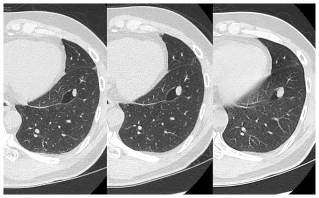 Figure 1
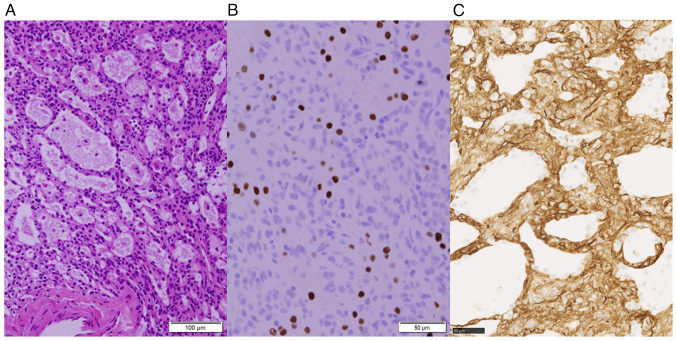 Figure 2
Peer Reviews
No public reviews on file for this paper yet. If you reviewed it on a platform where reviews are public (OpenReview, ICLR, NeurIPS, ICML), you can paste yours below so the community can read it here.
Videos
No videos yet. Explain this paper in a talk, walkthrough, or lecture? Add one.
Taxonomy
TopicsMedical Imaging and Pathology Studies · Lung Cancer Diagnosis and Treatment · Tracheal and airway disorders
Introduction
Alveolar adenoma (AA) is an exceedingly rare benign lung tumor that was first reported by Yousem et al in 1986(1). Patients with AA are usually asymptomatic and the tumor is incidentally detected on chest radiography as a clear solitary pulmonary nodule. When solid pulmonary nodules are >10 mm in size, there is an increased likelihood of surgical intervention due to a higher risk of malignancy and patient anxiety (2,3). A definitive diagnosis of AA depends on histopathology and immunohistochemistry findings as it is difficult to diagnose pre-operatively. Curative treatment consists of surgical resection, with no case of recurrence having been described to date in the literature, at least to the best of our knowledge.
The present study describes the case of a patient with alveolar adenoma and reports on the imaging and pathological differentiation of AA from other lung tumors.
Case report
A 50-year-old male patient presented to his previous doctor with a complaint of chest discomfort. A chest computed tomography (CT) scan revealed a 10-mm solitary nodule with a single cyst in the left lower lobe. The diameter of the pulmonary nodule increased to 14 mm within a span of 2 years (CT scan results shown in Fig. 1). He was referred to Kansai Medical University Hospital (Hirakata, Japan) due to the possibility of lung cancer. His medical history was notable for benign prostatic hyperplasia. A thoracoscopic wedge resection of the left lower lung was performed. The frozen sections were non-diagnostic, and the surgical procedure and postoperative course were uneventful, with no signs of recurrence 4 years post-operatively.
Overall, the tumor was well-defined and yellowish, measuring 10 mm in a subpleural cyst. Microscopically, the tumor consisted of polycystic structures resembling alveoli filled with pulmonary surfactant. The cyst lining cells were positive for thyroid transcription factor-1 (TTF-1) without atypia, corresponding to type II pneumocytes (Fig. 2). Additionally, the stroma lacked elastic fibers characteristic of alveoli and contained cluster of differentiation 34 (CD34)-positive cells with rounded nuclei and eosinophilic cytoplasm. These cells were negative for TTF-1, CD31, ERG, D2-40, SALL4, BRAF and STAT6. Immunohistochemical analysis was conducted on 4-µm-thick formalin-fixed paraffin-embedded tissue sections (details presented in Table I). Cultures were negative for fungi and tuberculosis, and no malignant cells were noted. Analyses were performed under a light microscope (Olympus Corporation) and whole slide images. Therefore, the final pathological diagnosis was that of AA.
Discussion
AA is a rare lung tumor with an incidence rate of <1% of all lung tumors, and is classified as an adenoma in the 2015 World Health Organization Classification of lung tumors (4). AA is often asymptomatic and is incidentally detected during imaging examinations, typically exhibiting no tendency to enlarge (5). The majority of patients are middle-aged to elderly, with a slight predominance in the female sex. There is no association between the occurrence of AA and a previous medical history or family history. AA commonly occurs in the middle and lower lobes of the lung and is pathologically characterized by multifocal cystic lesions resembling alveolar cavities, with the lumen lined by TTF-1-positive type II alveolar epithelium (6). In the present study, upon an examination of the patient, it was found that the tumor had similar histopathological features to the cases reported in the literature (6-11).
AAs are characterized by the presence of vacuoles within or around the tumor on imaging (7). It is speculated that alveoli rupture and fuse to create cavities, similar to the cavity formation mechanism observed in lung cancer. This occurs as tumor cells develop toward the bronchiole, forming a unidirectional check-valve system, which results in the accumulation of gas in the alveoli. This phenomenon may explain the cystic lesions observed in the pathology. Therefore, it is important to consider AA as a differential diagnosis in cases of pulmonary nodules with air images. In the case presented herein, the tumor increased in size with the presence of air images, highlighting the necessity to distinguish the tumor from lung cancer or pulmonary aspergilloma.
The diagnosis of AA is challenging when based on small biopsy tissue or frozen sections as it can resemble normal lung parenchyma or mimic malignancy with small glandular spaces lined by regular glandular epithelium (8). Additionally, there are other conditions in the differential diagnosis of AA, including papillary adenoma, sclerosing pneumocytoma and pulmonary hamartoma. Papillary adenoma is characterized by distinctive papillae covered by uniform cuboidal to columnar cells and a heterogeneous epithelial component. The presence of TTF-1 expression in AA can help distinguish it from sclerosing pneumocytoma. Pulmonary hamartoma consists primarily of benign cartilage mixed with a fibrovascular stroma and scattered bronchial glands (9).
The curative treatment for AA is surgical resection, typically performed to rule out malignancy and confirm the diagnosis through postoperative pathology. No recurrence has been reported following complete resection (10). In the case described herein, surgery was concluded with a wedge resection as the tumor was a peripheral lesion that could be completely resected, and the frozen section did not provide a definitive diagnosis. There is a risk of unnecessary extended resection, such as segmentectomy or lobectomy, if the patient is intraoperatively misdiagnosed with lung cancer based in the frozen section; hence, wedge resection is a viable option for small peripheral lesions (11).
In conclusion, AA is a rare, benign lung tumor. When encountering a well-defined solitary nodule with cystic spaces in the peripheral lung, an intraoperative diagnosis can be challenging. Therefore, it is critical to consider the possibility of AA, complete the surgery with a wedge resection and await the final pathological diagnosis.
The reference list from the paper itself. Each links out to its DOI / PubMed record.
- 1Yousem SA Hochholzer L Alveolar adenoma Hum Pathol 1710661071198610.1016/s 0046-8177(86)80092-23759064 · doi ↗ · pubmed ↗
- 2Klaveren RJ Oudkerk M Prokop M Scholten ET Nackaerts K Vernhout R Iersel CA Bergh KAM Westeinde SV Aalst C Management of lung nodules detected by volume CT scanning N Engl J Med 36122212229200910.1056/NEJ Moa 090608519955524 · doi ↗ · pubmed ↗
- 3Mac Mahon H Naidich DP Goo JM Lee KS Leung ANC Mayo JR Mehta AC Ohno Y Powell CA Prokop M Guidelines for management of incidental pulmonary nodules detected on CT Images: From the fleischner society 2017 Radiology 284228243201710.1148/radiol.201716165928240562 · doi ↗ · pubmed ↗
- 4Travis WD Brambilla E Burke AP Marx A Nicholson AG Introduction to the 2015 World Health Organization classification of tumors of the lung, pleura, thymus, and heart J Thorac Oncol 1012401242201510.1097/JTO.000000000000066326291007 · doi ↗ · pubmed ↗
- 5Bhavsar T Uppal G Travaline JM Gaughan C Huang Y Khurana JS An unusual case of a microscopic alveolar adenoma coexisting with lung carcinoma: A case report and review of the literature J Med Case Rep 5187201110.1186/1752-1947-5-18721592362 PMC 3113997 · doi ↗ · pubmed ↗
- 6Sak SD Koseoglu RD Demirag F Akbulut H Gungor A Alveolar adenoma of the lung. Immunohistochemical and flow cytometric characteristics of two new cases and a review of the literature APMIS 11514431449200710.1111/j.1600-0463.2007.00762.x 18184418 · doi ↗ · pubmed ↗
- 7Hsieh MS Tseng YH Hua SF Chou YH Cystic alveolar adenoma: An unusual clinical presentation of a rare lung neoplasm Pathology 477880201510.1097/PAT.000000000000020125474515 · doi ↗ · pubmed ↗
- 8Burke LM Rush WI Khoor A Mackay B Oliveira P Whitsett JA Singh G Turnicky R Fleming MV Koss MN Travis WD Alveolar adenoma: A histochemical, immunohistochemical, and ultrastructural analysis of 17 cases Hum Pathol 301581567199910.1016/s 0046-8177(99)90270-810029443 · doi ↗ · pubmed ↗