Successful removal of an intra-pericardic Kirschner wire via fluoroscopy-guided minimal invasive approach: a case report
Olivier Georges, Florence De Dominicis, Malek Ben Rahal, Jules Le Pessot, Pascal Berna, Osama Abou Arab, Christophe Beyls

TL;DR
A 65-year-old patient had a rare but dangerous Kirschner wire migration into the pericardium, successfully removed using a minimally invasive fluoroscopy-guided method.
Contribution
Demonstrates a safe, minimally invasive technique for removing intra-pericardial Kirschner wires without major surgery.
Findings
Multimodal imaging and fluoroscopy enabled safe wire extraction.
The minimally invasive approach avoided the need for median sternotomy.
The patient had an uncomplicated postoperative recovery.
Abstract
Kirschner wires are widely used in trauma surgery. Their migration into the pericardium is a rare but often fatal phenomenon, requiring urgent management. We describe the case of a 65-year-old patient who underwent Kirschner wire placement to treat a humeral head fracture. Three months after the operation, pleural and pericardial effusions with cardiac tamponade were observed, leading to the diagnosis of wire migration within the pericardium. A minimally invasive approach guided by fluoroscopy allowed emergency wire extraction without needing a median sternotomy. The postoperative clinical course was uncomplicated. The use of pre- and per-operative multimodal imaging allowed for the safe extraction of an intra-pericardial Kirschner wire through a minimally invasive approach. The online version contains supplementary material available at 10.1186/s40792-023-01803-8.
Genes, proteins, chemicals, diseases, species, mutations and cell lines named across the full text — each resolved to its canonical identifier and authoritative record.
Click any figure to enlarge with its caption.
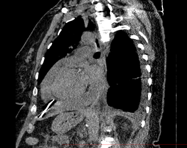 Figure 1
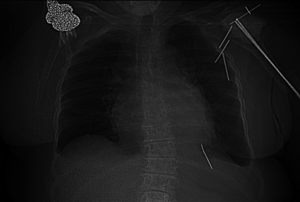 Figure 2
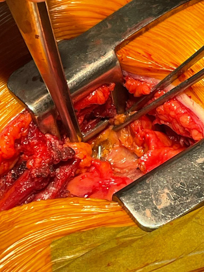 Figure 3
Peer Reviews
No public reviews on file for this paper yet. If you reviewed it on a platform where reviews are public (OpenReview, ICLR, NeurIPS, ICML), you can paste yours below so the community can read it here.
Videos
No videos yet. Explain this paper in a talk, walkthrough, or lecture? Add one.
Taxonomy
TopicsShoulder and Clavicle Injuries · Trauma Management and Diagnosis · Shoulder Injury and Treatment
Introduction
The use of orthopedic metal pins, known as Kirschner wires (K-wires), is a standard surgical treatment of humerus and clavicle fractures. Despite an initial intraosseous positioning, pin migration is a well-known complication, with many case reports already published. However, most of these pins migrations remain localized to the peri-fracture zone and do not constitute a life-threatening situation for the patient [1]. Rarely, due to their shape and size, the pins can migrate much further and cause severe lesions, especially in case of migration to the thoracic cavity or even to the pericardium.
Ethics
For the case we describe, we obtained study approval and informed written consent for publication from the patient, following the strengthening of reporting guidelines for case report studies CARE.
Case
A 65-year-old female patient with a history of diabetes and severe obesity (BMI 42.1) underwent open reduction and internal fixation of a proximal humerus fracture using five straight K-wires. In the following, a 1-month repeat X-ray showed 3 K-wires had started to migrate toward the thoracic cavity. In the third month, the patient presented to the emergency department with exertional dyspnea of progressive onset. Clinical examination revealed mild lower limb edema without other signs of right or left heart failure, except for dyspnea. The ECG showed atrial fibrillation with a rapid ventricular response at a rate of 150 beats per minute. The patient was hemodynamically stable (blood pressure: 110/75). At the transthoracic exam, the K-wire appeared intra-pericardial, associated with a circumferential pericardial effusion, a collapse of the right atrium, and a dilated inferior vena cava. The chest computed tomography (CT) confirmed the intra-pericardial migration of one of the K-wires (Figs. 1 and 2). However, the measurement of the fluid density favored a reactive, non-hemorrhagic effusion.Fig. 1. Sagittal view of a preoperative thoracic CT scan centered on the Kirschner wire without contrastFig. 2Scout view of preoperative thoracic CT scan
The pin appeared laterally positioned toward the left of the mediastinum. Its most superficial point measured 50 mm from the skin surface.
Upon presentation of cardiac tamponade, urgent surgical intervention was warranted. Subsequently, a comprehensive multidisciplinary discussion involving the anesthesiologist, cardiothoracic surgeon, and cardiologist was undertaken to establish the optimal surgical approach. Given the patient's specific characteristics of obesity and diabetes, a meticulously planned minimally invasive strategy was chosen, involving a short anterolateral thoracotomy with a primary focus on addressing the wire while deliberately avoiding the need for a median sternotomy. However, it was collectively agreed that in the event of sudden hemodynamic deterioration, myocardial injury, or uncontrolled bleeding, an emergency sternotomy would be swiftly performed to address the critical situation. Therefore, a dedicated team of cardiac perfusionists, as well as a cardiopulmonary bypass machine, was available. The two Scarpas underwent surgical cleansing and were placed within the sterile field.
Thus, the patient was positioned supine with the left arm at 90° and a bolster under the left flank. Due to the lack of reliable anatomical landmarks given the patient's body size, we utilized 2-plane orthogonal fluoroscopy (Siemens Cios Alpha device) to guide the skin incision (Additional file 1: Video S1). The initial step involved conducting an anteroposterior fluoroscopy to locate the position in a horizontal plane. This enabled us to make the incision centered on the pin, consisting in short left, 10 cm, anterior thoracotomy beneath the mammary fold. The initial step involved opening the left pleural cavity in the 6th intercostal space, resulting in the evacuation of 300 cc of reactive fluid. The dissection continued toward the anterior surface of the pericardium in the anterior mediastinum. With the assistance of fluoroscopy used in a lateral plane, the wire was quickly located. We proceeded with the opening of the pericardium, allowing for the evacuation of 250 cc of citrine fluid, and the wire was extracted (Additional file 2: Video S2, Fig. 3). No myocardial injury was found. The postoperative course was simple, allowing the removal of the pericardial and pleural drains on day 2. The patient was discharged on the 7th postoperative day.Fig. 3. Per-operative view of the K-wire after dissection
Discussion
This is the first case report describing the extraction of an intra-pericardial K-wire using a minimally invasive approach guided by fluoroscopy. The phenomenon of pin migration is well-known, and numerous case reports describe various migration sites, such as the inferior vena cava, lung, ascending aorta, and trachea [2]. There are multiple hypotheses proposed to explain these migrations. Factors such as muscular activity, breathing, and intra-thoracic negative pressure have been described. Insufficient fixation techniques have also been mentioned [3].
The site of K-wire implantation also determines the likelihood of migration. In a literature review, Freund found that most reported cases were associated with the sternoclavicular joint as the primary fixation location, followed by the clavicle, acromioclavicular joint, and proximal humerus [4]. Among approximately 50 reported cases in the literature, around 20 involved migration into the mediastinum, while 8 cases described an intra-cardiac. The exact migration trajectory remains uncertain. In our case, as in most articles found in the literature, the patient did not undergo any additional imaging assessments between the post-operative consultation and her urgent admission to our hospital.
In cases of intra-pericardial migration, the prognosis primarily depends on the presence of myocardial injury. In the case of heart or aortic injury, the outcome is invariably fatal [5].
Regardless, emergency sternotomy is considered the gold standard for diagnosis and emergent treatment. However, in our case, especially considering high-risk patients such as those with obesity and diabetes, our aim was to avoid this invasive approach in order to reduce postoperative complications (e.g., surgical site infection, osteitis, delayed wound healing).
The challenge was twofold: first, it was essential to ensure the absence of associated myocardial injury since the minimally invasive approach does not provide satisfactory control. Echocardiography, 3D reconstruction on thoracic CT scan, and measurement of pericardial fluid density assisted us in this regard.
Second, unlike the open and wide view provided by sternotomy, the approach through mini-thoracotomy limits the dissection. It was, therefore, crucial to locate the small-diameter pin perforating the pericardium within the adipose tissue of the anterior mediastinum. The use of continuous fluoroscopy in two orthogonal planes allowed us to guide the skin incision and progress the dissection centered on the pin. It is apparent that such technology is not universally available across all facilities. From our perspective, even single-plane fluoroscopy presents substantial advantages. While estimating pin depth might pose more challenges in anteroposterior fluoroscopy compared to biplanar methods, the procedure remains entirely viable.
Conclusion
Kirschner wire migration is a known phenomenon. Although rare, migration in the intrathoracic position is almost systematically associated with severe lesions. In the event of migration, rapid surgical management is essential. The use of multimodal perioperative imaging such as CT scanner, TTE, and intra-operative X-ray, allows safe extraction via a minimally invasive approach.
Supplementary Information
Additional file 1: Video S1. Continuous fluoroscopy centered on the thorax showing the K-wire.Additional file 2: Video S2. Intra operative view of K-Wire removal.
The reference list from the paper itself. Each links out to its DOI / PubMed record.
- 1Gallego Palmero Cde la Red Gallego MDLA Velez Garcia OM Gutiérrez Olivera N Migration of broken Kirschner wire to the brachial plexus after fixation of fracture dislocation of the acromioclavicular joint J Hand Surg Asian Pac 202227590791110.1142/S 242483552272044436178419 · doi ↗ · pubmed ↗
- 2Park SY Kang JW Yang DH Lim TH Intracardiac migration of a Kirschner wire: case report and literature review Int J Cardiovasc Imaging 201127 Suppl 1858810.1007/s 10554-011-9977-022101576 · doi ↗ · pubmed ↗
- 3Slongo T Technique and biomechanics of Kirschner wire osteosynthesis in children Oper Orthop Traumatol 202032650952910.1007/s 00064-020-00684-633237395 PMC 7688098 · doi ↗ · pubmed ↗
- 4Freund E Nachman R Gips H Hiss J Migration of a Kirschner wire used in the fixation of a subcapital humeral fracture, causing cardiac tamponade: case report and review of literature Am J Forensic Med Pathol 200728215515610.1097/PAF.0b 013e 31806195 a 117525569 · doi ↗ · pubmed ↗
- 5Tan L Sun DH Yu T Wang L Zhu D Li YH Death due to intra-aortic migration of Kirschner wire from the clavicle: a case report and review of the literature Medicine (Baltimore)20169521 e 374110.1097/MD.000000000000374127227938 PMC 4902362 · doi ↗ · pubmed ↗