Genome sequence of bacteriophage Djungelskog isolated from an Arthrobacter globiformis culture
Abigail M. Oliveros, Shelby A. McDougall, Miles A. Snyder, Sara K. Snowden, Joseph D. Richard, Christopher M. Rao, Marybeth Ponce, Christopher J. Pitonza, Mira Ozcelik, Sofia S. Mannina, Juliana R. Magna, Andrew S. Lopez, Linnea C. Gustafson, Brynn K. Glackin, Abigail E. Dolge

TL;DR
This paper reports the genome sequence of a new bacteriophage, Djungelskog, isolated from a bacterial culture in New York.
Contribution
The study provides a new genome sequence and classification for the bacteriophage Djungelskog.
Findings
Djungelskog's genome is 54,512 base pairs long and contains 86 putative protein-coding genes.
The phage has a siphovirus morphology and belongs to cluster AW based on gene content similarity.
Abstract
Actinobacteriophage Djungelskog was isolated from a sample of degraded organic material in Poughkeepsie, NY, using Arthrobacter globiformis B-2979. Its genome is 54,512 bp and encodes 86 putative protein-coding genes. Djungelskog has a siphovirus morphology and is assigned to cluster AW based on gene content similarity to actinobacteriophages.
Genes, proteins, chemicals, diseases, species, mutations and cell lines named across the full text — each resolved to its canonical identifier and authoritative record.
Click any figure to enlarge with its caption.
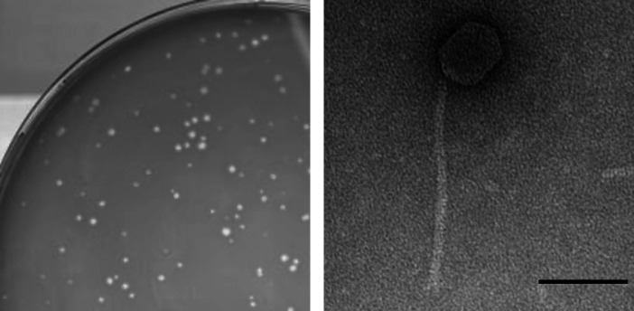 Fig 1
Fig 1Peer Reviews
No public reviews on file for this paper yet. If you reviewed it on a platform where reviews are public (OpenReview, ICLR, NeurIPS, ICML), you can paste yours below so the community can read it here.
Videos
No videos yet. Explain this paper in a talk, walkthrough, or lecture? Add one.
Taxonomy
TopicsBacteriophages and microbial interactions · Genomics and Phylogenetic Studies · Plant and Fungal Interactions Research
ANNOUNCEMENT
Bacteriophages are increasingly relevant in the field of biotechnology, agriculture, and medicine, particularly for controlling bacterial growth (1). Here, we report on bacteriophage Djungelskog that infects Arthrobacter, the latter capable of breaking down complex hydrocarbons and is therefore a potential use in bioremediation (2).
In August 2022, Djungelskog was isolated directly from an environmental sample consisting of degraded leaves and dirt, from Poughkeepsie, NY (GPS: 41.722735 N, 73.92931 W), using standard procedures (3). The sample was isolated in peptone-yeast calcium (PYCa) liquid media, then filtered through a 0.22-µm filter. The filtered sample was plated in PYCa top agar with Arthrobacter globiformis B-2979 and incubated at 30℃ for 48 h, yielding clear plaques that were ~0.7 mm in diameter (Fig. 1) after three rounds of purification. Using negative staining transmission electron microscopy, a siphovirus morphology with a prolate capsid (72–73 nm in length, 53–58 nm in width) and tail (length of 237–244 nm) was observed (n = 9, Fig. 1) (4).
Characterization of Actinobacteriophage Djungelskog. Image of plaques that were clear and approximately 0.7 mm in diameter (left) and transmission electron micrograph (right) of Djungelskog lysate negatively stained with UranylLess on a 300-mesh copper grid and imaged with a Hitachi HT7800 120 kV transmission electron microscope (scale bar = 100 nm).
Genomic DNA for Djungelskog was isolated from lysate using Promega Wizard DNA Clean-Up Kit, prepared for sequencing using NEB Ultra II Library Kit, and sequenced using Illumina MiSeq (v3 reagents), to yield 471,444,150-base single-end reads. The reads were assembled into a contig with 1,241× coverage using Newbler v2.9 and checked for accuracy using Consed v29 (5, 6). Default parameters were used for all software, unless otherwise stated. The resulting genome is 54,512 bp with a GC% of 51.70% and a 3′ single-stranded genome end (CGCCGACCT).
Djungelskog’s genome was automatically annotated using DNA Master v5.23.6 (https://phagesdb.org/DNAMaster/), embedded with Genemark v2.5p (7) and Glimmer v3.02 (8). The annotations were refined using Starterator v519 and Phamerator v528 (9) for comparison with similar phages. BLASTp (10) searches against the NCBI non-redundant and actinobacteriophage databases and HHPred (11) searches against the PDB_mmCIF70, Pfam-A, UniProt-SwissProt-viral70, and NCBI_Conserved_Domains (CD) databases were used to assign putative functions. Using ARAGORN (12) and tRNA scan-SE (13), it was found that this genome did not contain any tRNAs. SOSUI (14) and TMHMM (15) revealed six proteins with potential transmembrane regions. The annotation process revealed 86 genes, of which 21 genes could be assigned putative functions. Based on gene content similarity (GCS) of >35% to sequenced actinobacteriophages, using the GCS tool at the Actinobacteriophages database (https://phagesdb.org/), Djungelskog was assigned to the AW cluster (16, 17).
As with other cluster AW genomes, all genes are transcribed in the same direction, with virion structure and assembly genes clustered across one half of the genome and DNA metabolism genes clustered across the other half. Djungelskog encodes for a major capsid and protease protein, homologs of which are found in actinobacteriophage of various clusters and isolated on different bacteria, including cluster B isolated on Mycobacteria, cluster CC isolated on Rhodoccocus, cluster DJ isolated on Gordonia, cluster EL isolated on Microbacteria, and clusters AM, AU, FI, and FK isolated on Arthrobacter. No immunity repressor or integrase functions were identified, and clear plaque phenotype was observed, suggesting a lytic lifecycle.
The reference list from the paper itself. Each links out to its DOI / PubMed record.
- 1Hatfull GF. 2020. Actinobacteriophages: genomics, dynamics, and applications. Annu Rev Virol 7:37–61. doi:10.1146/annurev-virology-122019-07000932991269 PMC 8010332 · doi ↗ · pubmed ↗
- 2Bugayong MB, Cha A, Hamel CL, Johnson RR, Kim J, Kim JJ, Levy AS, Nguyen KD, Pham LH, Sapre A, Scanlan AC, Ye B, Bancroft C. 2022. Genome sequences of Arthrobacter globiformis B-2979 phages Globi Warming and Taylor Sipht. Microbiol Resour Announc 11:e 0092322. doi:10.1128/mra.00923-2236197292 PMC 9670964 · doi ↗ · pubmed ↗
- 3Poxleitner M, Pope W, Jacobs-Sera D, Sivanathan V, Hatfull G. 2018. Phage discovery guide. Howard Hughes Medical Institute, Chevy Chase, MD.
- 4Schneider CA, Rasband WS, Eliceiri KW. 2012. NIH image to Image J: 25 years of image analysis. Nat Methods 9:671–675. doi:10.1038/nmeth.208922930834 PMC 5554542 · doi ↗ · pubmed ↗
- 5Gordon D, Green P. 2013. Consed: a graphical editor for next-generation sequencing. Bioinformatics 29:2936–2937. doi:10.1093/bioinformatics/btt 51523995391 PMC 3810858 · doi ↗ · pubmed ↗
- 6Russell DA. 2018. Sequencing, assembling, and finishing complete bacteriophage genomes. Methods Mol Biol 1681:109–125. doi:10.1007/978-1-4939-7343-9_929134591 · doi ↗ · pubmed ↗
- 7Besemer J, Borodovsky M. 2005. Gene Mark: web software for gene finding in prokaryotes, eukaryotes and viruses. Nucleic Acids Res 33:W 451–W 454. doi:10.1093/nar/gki 48715980510 PMC 1160247 · doi ↗ · pubmed ↗
- 8Delcher AL, Harmon D, Kasif S, White O, Salzberg SL. 1999. Improved microbial gene identification with GLIMMER. Nucleic Acids Res 27:4636–4641. doi:10.1093/nar/27.23.463610556321 PMC 148753 · doi ↗ · pubmed ↗