Stanniocalcin 2 governs cancer cell adaptation to nutrient insufficiency through alleviation of oxidative stress
Shuo Qie, Haijuan Xiong, Yaqi Liu, Chenhui Yan, Yalei Wang, Lifeng Tian, Chenguang Wang, Nianli Sang

TL;DR
Stanniocalcin 2 (STC2) helps cancer cells survive when nutrients are scarce by reducing harmful oxidative stress, shifting their focus from growing to surviving.
Contribution
This study reveals that STC2 is induced by nutrient insufficiency and governs cancer cell adaptation through redox homeostasis.
Findings
STC2 is induced and secreted under nutrient insufficiency, promoting cell survival while reducing proliferation.
STC2 suppresses ROS production by downregulating MAOB, a mitochondrial enzyme that generates ROS.
Loss of STC2 increases apoptosis and necrosis in mouse xenografts, impairing tumor growth.
Abstract
Solid tumours often endure nutrient insufficiency during progression. How tumour cells adapt to temporal and spatial nutrient insufficiency remains unclear. We previously identified STC2 as one of the most upregulated genes in cells exposed to nutrient insufficiency by transcriptome screening, indicating the potential of STC2 in cellular adaptation to nutrient insufficiency. However, the molecular mechanisms underlying STC2 induction by nutrient insufficiency and subsequent adaptation remain elusive. Here, we report that STC2 protein is dramatically increased and secreted into the culture media by Gln-/Glc-deprivation. STC2 promoter contains cis-elements that are activated by ATF4 and p65/RelA, two transcription factors activated by a variety of cellular stress. Biologically, STC2 induction and secretion promote cell survival but attenuate cell proliferation during nutrient…
Genes, proteins, chemicals, diseases, species, mutations and cell lines named across the full text — each resolved to its canonical identifier and authoritative record.
Click any figure to enlarge with its caption.
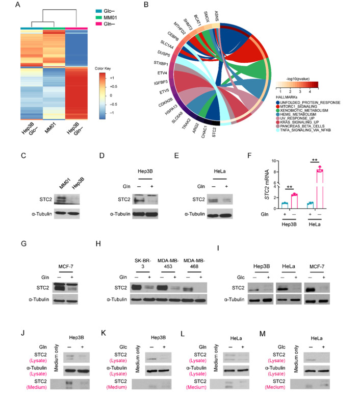 Figure 1
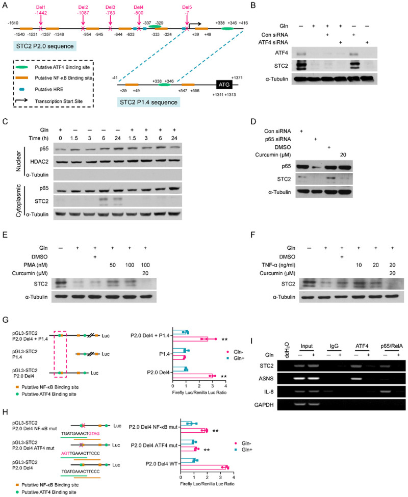 Figure 2
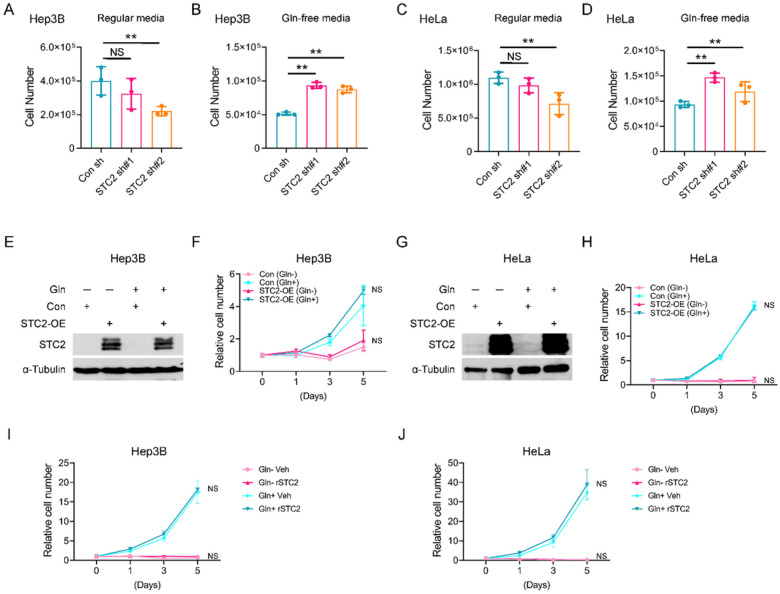 Figure 3
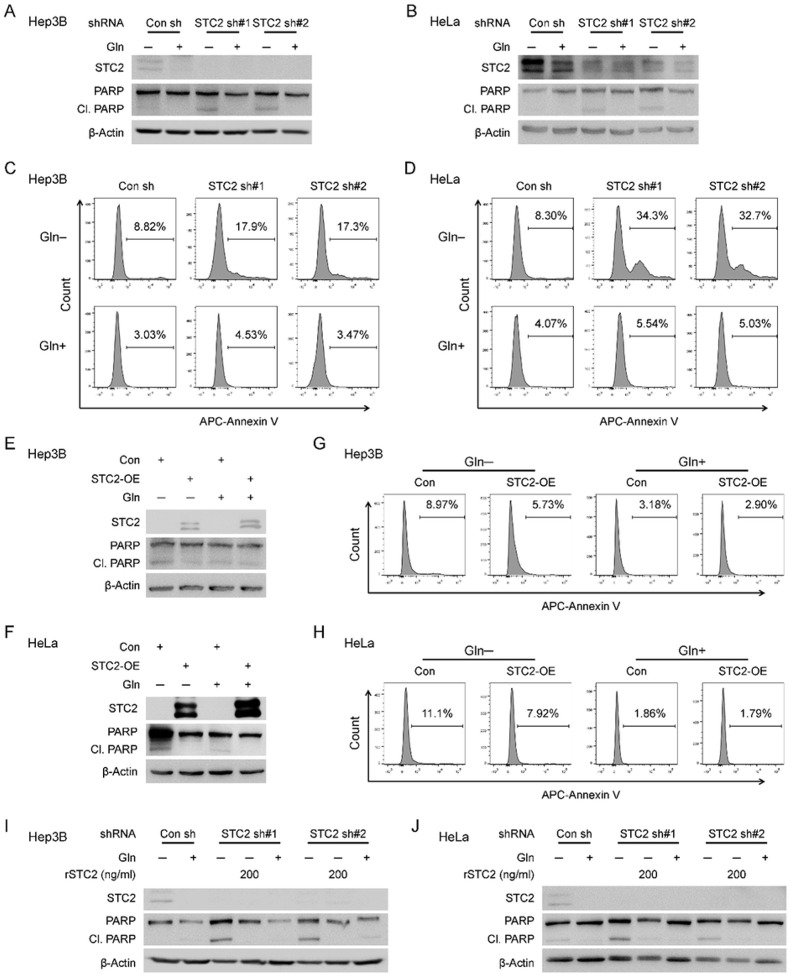 Figure 4
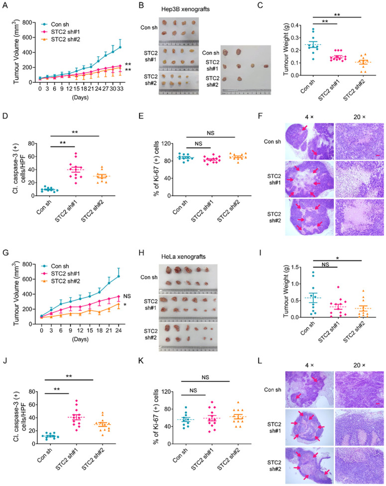 Figure 5
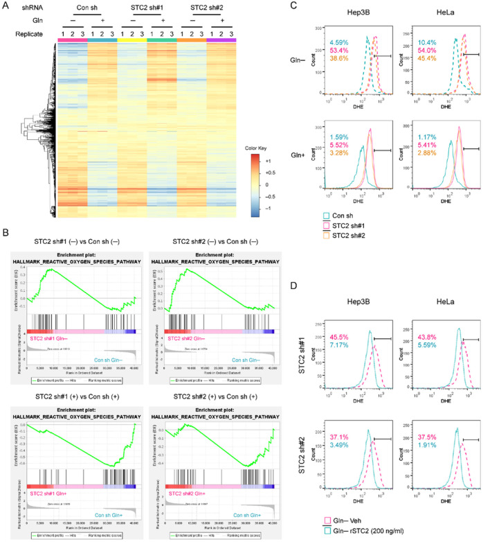 Figure 6
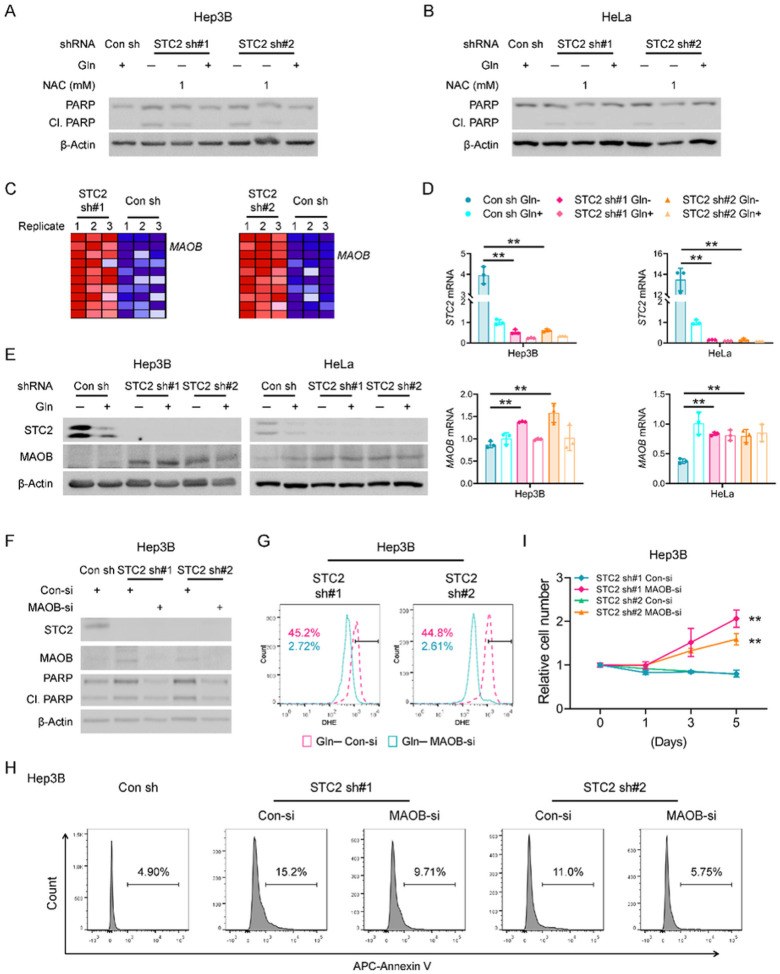 Figure 7
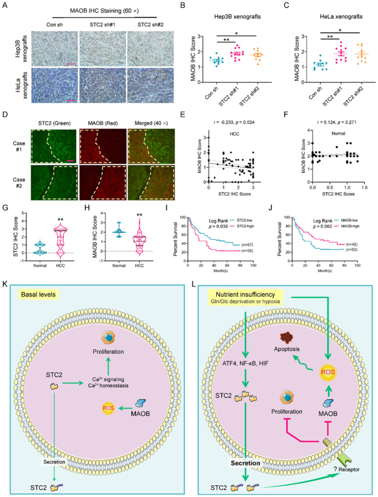 Figure 8
Peer Reviews
No public reviews on file for this paper yet. If you reviewed it on a platform where reviews are public (OpenReview, ICLR, NeurIPS, ICML), you can paste yours below so the community can read it here.
Videos
No videos yet. Explain this paper in a talk, walkthrough, or lecture? Add one.
Taxonomy
TopicsPhysiological and biochemical adaptations · Spaceflight effects on biology · Magnetic and Electromagnetic Effects
Introduction
The major characteristics of tumour metabolism include the Warburg effect, active glutaminolysis and hypoxia^1, 2, 3^; as such, tumour cells have increased demand of glucose (Glc), glutamine (Gln) and oxygen to keep cell survival and proliferation^4, 5, 6^. However, solid tumours often have poor circulation that leads to insufficient supply of Glc, Gln and oxygen; therefore, to investigate the mechanisms of how tumour cells respond to nutrient insufficiency could provide promising therapeutic targets. Our previous study identified Stanniocalcin 2 (STC2) as one of the most upregulated genes upon Gln-/Glc- deprivation and hypoxia exposure^1, 7^, suggesting a critical role of STC2 in cellular response to nutrient insufficiency.
STC2 belongs to the secreted glycoprotein hormone family that was reported to lower intracellular calcium concentrations by attenuating Ca^2+^ influx^8, 9^. Under physiological conditions, STC2 is highly expressed in skeletal muscle, heart, testis and pancreas^8, 10^; while under pathological conditions, STC2 is upregulated in a plethora of human tumours, including hepatocellular carcinoma (HCC), cervical cancer, nasopharyngeal carcinoma, colorectal cancer, gastric cancer, esophageal squamous cell carcinoma, prostate cancer, renal cell carcinoma, etc.^7, 8, 11, 12^. Increased STC2 levels are strongly correlated with tumour development, progression and poor prognosis for most human tumours except breast cancer^13^.
The expression of STC2 is regulated at both transcriptional and post-transcriptional levels, particularly under stress conditions including ER stress, hypoxia, nutrient insufficiency and radiation-mediated stress^1, 7, 8, 11, 14^. Biologically, STC2 is involved in many biological processes in human tumours, such as cell survival, proliferation, migration and immune escape^8, 15, 16, 17^. However, the precise mechanisms underlying the stress-induced STC2 induction remain unclear.
Previous reports reveal hydrogen peroxide (H_2_O_2_) induces STC2 expression, and loss of STC2 impairs the tolerability of cells to oxidative stress^18, 19, 20^, suggesting a potential mechanism that links nutrient insufficiency to STC2 induction, redox homeostasis and cellular adaption. Current understanding is that aberrant metabolic activities of tumour cells result in an elevation of reactive oxygen species (ROS)^21^. Low to moderate levels of ROS may serve as a signaling mechanism to maintain cellular physiology; increased levels of ROS facilitate tumorigenesis by causing genetic alteration and oncogenic transformation; however, long-term and/or excessive ROS may trigger apoptosis^22^. In general, the glutathione (GSH) and nicotinamide adenine dinucleotide phosphate (NADPH) systems work coordinatively to maintain intracellular redox homeostasis^23, 24^. As for tumour cell metabolism, Gln serves as the major source of glutamate that functions as an important substrate for de novo GSH biosynthesis; hence, Gln-deprivation results in decreased GSH levels and ROS accumulation^25, 26^. Glc insufficiency may trigger oxidative stress via impaired NADPH production, because Glc is the substrate for pentose phosphate pathway, the major pathway to generate NADPH^27^. Moreover, hypoxia impairs the electron transport chain (ETC) activity and also enhances ROS production^28, 29^. Therefore, a deprivation of each of these major nutrients may trigger oxidative stress, and cells must overcome the oxidative stress to maintain survival. A better understanding of how STC2 regulates the adaptation of tumour cells to nutrient insufficiency and associated oxidative stress may provide new therapeutic targets.
In this study, we first confirm that STC2 protein is induced and secreted into the culture media by nutrient insufficiency. We next present data to define the cis-elements in the STC2 promoter that physically interact with activating transcription factor 4 (ATF4) and nuclear factor-kappa B p65 (p65/RelA) during nutrient insufficiency. We show that STC2 induction inhibits apoptosis but attenuates tumour cell proliferation during nutrient insufficiency. In addition, loss of STC2 leads to elevated ROS levels accompanied by apoptosis during nutrient insufficiency. We finally show that STC2 induction suppresses the expression of monoamine oxidase B (MAOB), a mitochondrial membrane-associated enzyme that produces ROS, and the administration of recombinant human STC2 to culture media is sufficient to suppress MAOB expression. Taken together, our findings indicate that STC2 serves as a key regulator of redox homeostasis that governs the survival of tumour cells during nutrient insufficiency.
Materials and methods
Ethical approval
Animal studies were performed according to the protocol approved by the Laboratory Animal Ethic Committee at the Tianjin Medical University Cancer Institute & Hospital (TUMCIH) (#: AE-2022036). This study has complied with all relevant ethical regulations for animal experiments. All efforts were made to minimize the numbers and suffering of experimental animals.
Human HCC tissue microarray (TMA) was purchased from the Shanghai Outdo Biotechnology (China) with an Internal Review Board approval (YB M-05–02). The clinical samples (23 HCC specimens) were collected from the Department of Pathology at the TMUCIH from January 1st 2018 to December 31st 2018. All patients were provided with informed consent and the study was conducted according to a protocol approved by the Internal Review Board at the TMUCIH (bc2021308).
Cell culture
Hep3B, HeLa, MCF-7, SK-BR-3, MDA-MB-453, MDA-MB-468 and HEK293T (ATCC) cells were maintained in DMEM with 10% fetal bovine serum (FBS) supplemented with Pen/Strep. All cells were cultured in a humidified incubator at 37 °C with 5% CO_2_. MM01 cells were established through extended culture of Hep3B cells in Gln-free DMEM supplemented with 10% dialyzed FBS (Atlantic Biologicals, dialyzed 10 kDa), 0.8 mM ammonia and Pen/Strep^30^. The culture media for MM01 cells were changed every 2 days and the cells were passed once reaching ~ 90% confluency. All cells were tested routinely to ensure mycoplasma-free using Polymerase Chain Reaction (PCR) methods.
Tumour xenografts
The NTG and athymic nude, nu/nu mice were purchased from SPF Biotechnology Co. (Beijing, China). For Hep3B xenografts, 24 NTG mice (male, age: 4 weeks old) were randomly allocated into three groups (6 per group). For HeLa xenografts, 24 athymic nude, nu/nu mice (female, age: 4 weeks old) were randomly allocated into three groups (six per group). In general, 5×10^6^ Hep3B or HeLa cells were subcutaneously injected into each side of both flank regions. The xenografts were measured every three days to the completion of experiments. The growth curves were plotted with tumour sizes (calculated using the formula V_T_ =0.5 × L × W^2^, mm^3^). After euthanization, dissected xenografts were weighted and recorded for further analysis. The mice were euthanized if the xenograft size became ≥ 20 mm in the long diameter, and the mice died prior to the completion of experiments were excluded for analysis.
Statistical analysis
Statistical analyses were performed using SPSS 27.0 software (IBM SPSS Statistics). The results of qRT-PCR, tumour weight, IHC staining score, and luciferase analysis were presented as average ± standard deviation; student t test or one-way ANOVA was performed to compare these results. The curves for cell proliferation and xenograft growth were compared using two-way ANOVA. Linear correlation was assessed using Spearman’s correlation analysis. Survival analysis was performed using the Log Rank test to estimate the survival probability, and p-value less than 0.05 was set as statistically significant.
Results
Nutrient insufficiency triggers STC2 induction and secretion
In previous work, we cultured Hep3B cells for long term in Gln-free media supplemented with NH_4_^+^ as an alternative nitrogen source and established a long-term Gln-deprived cell line, MM01, which maintains a slow proliferation phenotype^30^. In addition, we cultured Hep3B cells in Gln-/Glc- free media for short terms that served as a model for acute nutrient insufficiency. cDNA microarray analysis revealed that STC2 was among the most upregulated 58 genes shared by cells enduring acute Gln-/Glc- deprivation as well as long-term Gln-deprived condition (Fig. 1A,B & S1A), indicating the potential of STC2 in mediating cellular adaptation to nutrient insufficiency. Further functional analyses suggested that most of these 58 genes were related to amino acid metabolism (Fig. S1B & Supplementary Table 1). Moreover, our prior study found STC2 was also upregulated by hypoxia^7^, suggesting STC2 induction is a common phenomenon when tumour cells exposed to nutrient insufficiency. In accordance, elevated STC2 protein levels were confirmed in MM01 cells relative to that in Hep3B cells (Fig. 1C), and STC2 was remarkably induced by acute Gln-deprivation (Fig. 1D–F & S1C), whereas re-supplementation with Gln rapidly suppressed STC2 expression (Fig. S1D). Similar results were observed when Gln was added back to MM01 cells (Fig. S1E). To address whether STC2 upregulation is a common phenomenon in different tumour types enduring nutrient insufficiency, we exposed breast cancer cells to Gln-deprivation as well. We observed that STC2 was similarly induced in MCF-7, SK-BR-3, MDA-MB-453 and MDA-MB-468 cells by Gln-free media (Fig. 1G,H). In consistent with the cDNA microarray, Glc-deprivation also increased STC2 protein levels (Fig. 1I).
To test whether STC2 is induced by the lack of Gln itself or by a lack of metabolic derivatives of Gln, we administered the glutaminase inhibitor, DON, to Hep3B cells. We observed that DON treatment efficiently induced STC2 expression in Gln-rich media (Fig. S1F,G), indicating that lack of Gln-derived metabolites is sufficient to trigger STC2 induction as well. As an inhibitor of protein secretion, BFA increased intracellular STC2 levels while decreased STC2 levels in the culture media (Fig. S1H), con rming STC2 as a secreted protein. To address whether increased STC2 levels in cells is the result of secretion inhibition, we analysed STC2 levels in cell lysate and their corresponding culture media. We observed that increased STC2 levels in both cell lysate and culture media in a proportional manner upon Gln-/Glc- deprivation, suggesting that increased intracellular STC2 levels are not a consequence of secretion inhibition (Fig. 1J–M). Taken together, these data suggest that nutrient insufficiency triggers STC2 induction and secretion, which represents a converging consequence in response to different nutrient insufficient conditions.
ATF4 and p65/RelA transcriptionally induce STC2 expression during nutrient insufficiency
Our previous studies indicated that STC2 mRNA levels were increased in cells under Gln-/Glc- deprived or hypoxic conditions^1, 8^, suggesting STC2 is transcriptionally upregulated by nutrient insufficiency. To further delineate the underlying mechanisms, we used ActD and CHX to block gene transcription and protein translation, respectively. ActD and CHX blocked STC2 expression at both mRNA and protein levels (Fig. S2A,B), demonstrating that STC2 transcription depends on de novo synthesis of a transcription factor. Additionally, to address whether Gln-deprivation affects STC2 protein stability, we performed CHX chase assay and found that after CHX treatment, STC2 showed a similar decay rate regardless of Gln levels (Fig. S2C,D), ruling out the possibility that Gln-deprivation stabilizes STC2 protein.
STC2 was reported to be induced by ER stress that is correlated with ATF4 activation. ATF4 is an important component of the integrated stress response system. Amino acid insufficiency leads to the accumulation of non-aminoacylated tRNAs, which promotes the activation of general control non-derepressible-2 (GCN2) and the phosphorylation of eIF2α, thereafter triggering the selective translation of ATF4^31, 32^. To determine whether STC2 gene is a direct transcription target of ATF4, we performed bioinformatic analysis and found STC2 promoter contains multiple copies of putative ATF4, NF-κB and HIF-1 binding sites (HRE) (Fig. 2A). Next, we confirmed that Gln-deprivation enhanced eIF2α phosphorylation and ATF4 induction in our experimental setting (Fig. S2E,F). In addition, ATF4 knockdown effectively abolished STC2 induction in cells cultured in Gln-free media (Fig. 2B). Indeed, ER stress inducers like MG132 which blocks proteosome-dependent degradation of proteins or BFA which suppresses protein secretory pathway also induced STC2 expression; while ActD or CHX effectively blocked this induction (Fig. S2G–J), suggesting ER stress triggered STC2 induction also depends on de novo transcription and translation. Finally, glutaminase inhibitor, DON or Glc-deprivation also induced co-upregulation of ATF4 and STC2 (Fig. S2K,L).
In consistence with the existence of putative NF-κB binding sites in the STC2 promoter, GSEA analyses highlighted the upregulation of NF-κB targets under Gln-insufficient conditions (Fig. S3A & Supplementary Table 2). To evaluate the role of NF-κB in STC2 induction, we performed cell fractionation analysis and observed that Gln-deprivation triggered p65/RelA accumulation in the nuclear fraction (Fig. 2C), demonstrating the activation of p65/RelA signaling in Gln-deprived cells. Next, we applied chemical inhibitors or activators to test the importance of p65/RelA in mediating STC2 expression upon Gln-deprivation. We observed that NF-κB inhibitors, BAY11–7082 or curcumin, efficiently blocked STC2 upregulation (Fig. S3B–G). Furthermore, genetically suppressing p65/RelA by siRNA or IκBαSR attenuated STC2 induction by Gln-deprivation (Fig. 2D & S3H). On the other hand, broadly used NF-κB activators, PMA or TNF-α, induced STC2 that could be blocked by curcumin (Fig. 2E,F). Taken together, these data demonstrate that p65/RelA functions as another transcription activator for STC2 induction.
To define the cis-elements that regulate STC2 expression in response to Gln-deprivation, we cloned two promoter fragments into the pGL3-Luciferase reporter with a length of 2026bp (P2.0) and 1412bp (P1.4), respectively (Fig. 2A). Guided by the identified putative binding sites, a series of deletions were constructed and cloned into the pGL3-Luciferase reporter. Luciferase assays indicated a DNA region included in STC2-P2.0 but not in STC2-P1.4 is responsive to Gln-/Glc- deprivation or hypoxia (Fig. S4A–C). Further deletion assays narrowed down to Deletion4 (Del4) region that is indispensable for the response of STC2 promoter to nutrient insufficiency (Fig. 2G & S4D). Mutation of either ATF4 or NF-κB binding sites in this region decreased the stress response (Fig. 2H & S4E), further confirming that these binding sites are functionally important in regulating STC2 induction. Finally, ChIP assays confirmed the direct binding of ATF4 and p65/RelA to this region of STC2 promoter (Fig. 2I).
STC2 induction compromises tumour cell proliferation under nutrient insufficient conditions
Our previous study revealed that Gln-/Glc- deprivation caused cell cycle arrest, hence attenuating cell proliferation^1^. Since STC2 is induced by nutrient insufficiency, it is intriguing to investigate the role of STC2 in regulating cell proliferation under such conditions. We next focused on Gln-deprivation as a model to investigate the role of STC2 in regulating cell proliferation. In regular media, STC2 knockdown impaired the optimal proliferation of Hep3B and HeLa cells (Fig. 3A,C), while in Gln-free media, STC2 knockdown cells showed higher proliferation rates (Fig. 3B,D), indicating that basal levels of STC2 are required to maintain the optimal proliferation when cells cultured in nutrient rich media, whereas under nutrient insufficient conditions, elevated STC2 levels can suppress cell proliferation. Interestingly, overexpression of STC2 did not affect cell proliferation of cells cultured in either regular media or nutrient insufficient media (Fig. 3E–H). Furthermore, supplementing exogenous recombinant STC2 (rSTC2) to media didn’t affect cell proliferation under either conditions (Fig. 3I,J). Taken together, these data demonstrate that basal levels of STC2 are required to support cell proliferation under nutrient rich conditions; while endogenous STC2 gene can be induced by nutrient insufficiency, thereby attenuating cell proliferation and facilitating cellular adaptation to stress conditions.
STC2 induction and secretion promote tumour cell survival during nutrient insufficiency
One common consequence of nutrient insufficiency and associated metabolic stress is apoptosis. We next focused on Gln-deprivation as a model to investigate whether STC2 induction played a role in maintaining cell survival during nutrient insufficiency. Western blot revealed STC2 knockdown increased PARP cleavage, a biomarker of apoptosis, under Gln-deprived conditions (Fig. 4A,B). In accordance, flow cytometry analysis also supported that cells with STC2 knockdown were more sensitive to apoptosis triggered by Gln-deprivation (Fig. 4C,D). On the other hand, overexpression of STC2 decreased apoptosis of cells exposed to Gln-free media (Fig. 4E–H). As a secretory protein, STC2 has been proposed to function as a signaling molecule or to function intracellularly as a calcium regulator^9^. To test whether STC2 protects cells from apoptosis triggered by Gln-deprivation in an autocrine/paracrine manner, we added rSTC2 to cell culture media and found that rSTC2 efficiently suppressed PARP cleavage as well as apoptosis of cells cultured in Gln-free media (Fig. 4I,J & S5). These data support that STC2 may function in an autocrine/paracrine manner to promote cell survival under nutrient insufficient conditions.
STC2 knockdown retards the growth of mouse xenografts
To evaluate the role of STC2 in a physiological context, we first run a large-scale Kaplan-Meier Plotter analyses and found that elevated STC2 levels were associated with poor prognosis in patients with a variety of tumours including HCC, cervical squamous cell carcinoma, thymoma, esophageal squamous cell carcinoma, renal papillary cell carcinoma, stomach adenocarcinoma, sarcoma, head and neck squamous cell carcinoma, bladder cancer and lung squamous cell carcinoma (Fig. S6). To confirm the role of STC2 induction in tumour progression in vivo, we established mouse xenografts by injecting 5×10^6^ Hep3B or HeLa cells transfected with STC2 knockdown or control shRNAs independently. We found that STC2 knockdown tumours grew slower than control ones (Fig. 5A,B,G,H). Moreover, STC2 knockdown xenografts overall had lighter tumour weights compared to control counterparts (Fig. 5C,I). To further determine the role of STC2 in cell survival and proliferation in vivo, we examined the expression of cleaved caspase-3 and Ki-67 in the dissected xenografts by IHC staining as markers of apoptosis and proliferation, respectively. We found that loss of STC2 led to increased caspase-3 cleavage, suggesting STC2 induction is a favourable factor for cell survival in vivo (Fig. 5D,J & S7A), while there was no obvious difference in cell proliferation rates evaluated by Ki-67 IHC staining (Fig. 5E,K & S7B). More importantly, larger necrotic areas were observed in STC2 knockdown xenografts compared to control ones (Fig. 5F,L), suggesting a failed adaptation to insufficient blood supply during tumour progression, which is consistent with a previous report that STC2 was involved in tumour angiogenesis^33^. These data highlight that STC2 plays a critical role in preventing cell death during tumour progression in vivo.
STC2 knockdown results in ROS elevation in cells under nutrient insufficient conditions
Next, we asked how STC2 protects tumour cells from nutrient insufficiency-induced apoptosis. To address this question, we first performed RNA-Seq analysis to search for alterations of signaling pathways in STC2 knockdown versus control cells cultured in Gln-free media (Fig. 6A). The analysis of the RNA-Seq data identified an alteration of genes related to “hallmark gene sets” (Fig. S8); particularly, both shRNAs targeting STC2 resulted in an elevation of genes related to redox homeostasis under Gln-deprived conditions (Fig. 6B). We next determined how STC2 status affected ROS levels in cells exposed to Gln-deprivation. Using dihydroethidium (DHE) staining combined with flow cytometry analyses, we observed that Gln-deprivation moderately increased ROS levels, and STC2 knockdown significantly intensified ROS accumulation (Fig. 6C). Moreover, the administration of rSTC2 to culture media obviously decreased ROS levels in STC2 knockdown cells (Fig. 6D). Taken together, these data indicate that STC2 induction and secretion are required to ameliorate ROS elevation triggered by nutrient insufficiency.
STC2 knockdown leads to MAOB dysregulation and redox imbalance during nutrient insufficiency
To confirm a causative relationship between ROS accumulation and apoptosis in our experimental model, we administered NAC, an ROS scavenger, to STC2 knockdown cells. Not surprisingly, NAC suppressed apoptosis of STC2 knockdown cells enduring Gln-deprivation (Fig. 7A,B & S5), indicating increased ROS is a bona fide mediator of apoptosis in our experimental setting. To explore the molecular mechanism underlying the protective effects of STC2 in response to nutrient insufficiency, we mined the RNA-Seq data and found MAOB, a mitochondrial membrane associated enzyme that directly participates in ROS production, was among the genes most upregulated by STC2 knockdown (Fig. 7C & Supplementary Table 3). Subsequent qRT-PCR and western blot assays further demonstrated that STC2 knockdown increased MAOB expression under Gln-deprived conditions (Fig. 7D,E & S9A), whereas we didn’t detect significant alteration of other genes related to ROS generation in two independent STC2 knockdown cell lines; particularly, no signal was detected for NADPH oxidase (NOX) family genes in both STC2 knockdown cell lines (Fig. S9B–E).
To further explore the regulatory relationship between STC2 and MAOB, we applied rSTC2 to cells with STC2 knockdown. We observed that rSTC2 effectively decreased MAOB expression in STC2 knockdown cells in Gln-free media (Fig. S10). To substantiate the biological importance of MAOB expression to ROS levels and cell survival, we used siRNA pool to knockdown MAOB, or specific inhibitors to suppress MAOB activity in STC2 knockdown cells. We observed that MAOB knockdown efficiently decreased ROS levels in STC2 knockdown cells cultured in Gln-free media (Fig. 7F,G & S11A,B). In addition, MAOB knockdown not only suppressed apoptosis but also rescued cell proliferation (Fig. 7H,I & S11C,D). To further evaluate whether MAOB is associated with STC2 loss, we employed two specific MAOB inhibitors, Rasagiline and Selegiline. Consistently, both inhibitors effectively attenuated ROS production and apoptosis in STC2 knockdown cells cultured in Gln-free media (Fig. S12A–D), thereby rescuing cell proliferation (Fig. S12E,F). Taken together, these findings indicate that under nutrient insufficient conditions, STC2 induction maintains redox homeostasis by suppressing MAOB expression, hence reducing intracellular ROS levels and maintaining cell survival.
The expression levels of STC2 and MAOB are negatively correlated, and elevated STC2 is associated with tumour progression in vivo
To address whether the observation in cell culture is relevant to tumour progression in vivo, we first performed IHC staining and confirmed the elevated expression of MAOB in STC2 knockdown xenografts (Fig. 8A–C). Furthermore, DepMap analyses suggested a reverse correlation between STC2 and MAOB expression in a series of human tumour cells (Fig. S13A). Other bioinformatic analyses also revealed a negative correlation between STC2 and MAOB expression in human tumours (Fig. S13B–F).
To investigate the potential of using the reciprocal STC2-MAOB expression as a prognostic indicator, we first performed immunofluorescent staining to examine their expression correlation in human HCC specimens. In consistency with cell culture findings, we confirmed an obviously negative correlation between STC2 and MAOB levels in the same HCC samples (Fig. 8D). IHC staining also verified a reverse correlation between STC2 and MAOB expression in human HCC tissues but not in normal counterparts (Fig. 8E–H & S14). Importantly, clinicopathological analyses revealed that elevated STC2 expression was significantly associated with poor prognosis in patients with HCC, whereas increased MAOB levels were likely correlated with a favourable prognosis with a log-rank p value of 0.062 (Fig. 8I,J). Taken together, these data strongly indicate that STC2 negatively regulates MAOB expression via an un-characterized mechanism (Fig. 8K,L), and a combined analysis of both STC2 and MAOB levels is a potential approach in assessing the prognosis of patients with HCC.
Discussion
Rapid proliferation of tumour cells demands increased supply of nutrients to support proliferation and active biosynthesis, while defective blood vessel system often fails to support solid tumour cells with sufficient blood supply, leading to nutrient insufficiency and subsequently oxidative stress during tumour progression. Our previous study revealed that STC2 is one of the most upregulated genes under Gln-/Glc-deprived and hypoxic conditions^1^. With these data in context, our study focuses on addressing how STC2 is upregulated by nutrient insufficiency, and the biological roles of STC2 in the adaptation of tumour cells to nutrient insufficiency.
We presented data to conclude that ATF4 and p65/RelA are activated by nutrient insufficiency that in turn transactivates STC2 gene. Previous studies reported that STC2 was upregulated upon ATF4 activation^8, 18^, but it was unclear whether ATF4 directly transactivates STC2 gene, or indirectly regulates its expression. Our current study identifies the cis-elements of the STC2 gene and demonstrates that these elements are indispensable for STC2 induction upon nutrient deprivation. It is well known that eIF2α-ATF4 axis represents the core of an integrated stress response mechanism^34^. Under various stress conditions, cells utilize multiple kinases to phosphorylate eIF2α. Phosphorylation of eIF2α results in a global translation inhibition but a selective translation of stress related genes exemplified by ATF4. As a stress responsive transcription factor, ATF4 transcribes several functional groups of genes including ER chaperones that are involved in protein folding, ER-associated degradation of terminally damaged proteins, and apoptosis. In addition to ATF4, NF-κB is another transcription factor ubiquitously involved in cellular stress response. It was suggested that Gln-deprivation activated NF-kB signaling^35^. Consistent with that report, our findings demonstrate that p65/RelA, a member of NF-κB family, directly transactivates STC2 gene by a physical interaction with identified cis-elements upon activation. Considering NF-κB is activated by various cell injury, pathogen infection and inflammatory signaling, we deduce that STC2 could also be induced by a variety of stress including cell injury and inflammatory factors.
Elevated ROS is a common result of many cellular stress and disease status, and is a major contributor to oxidative stress imbalance. ROS is mainly generated in the mitochondria as a byproduct of oxidative phosphorylation, or in the cytosol from reactions catalyzed by oxidoreductases such as NOX^36^. To some extent, ROS possesses beneficial effects for tumour cells, driving cell proliferation, genetic instability and metastasis^37^. However, excessive and/or long-term ROS may lead to the damage of macromolecules, and eventually cell death. From the nutrient point of view, Gln and Glc play critical roles in the intracellular counter-oxidative stress processes^25, 26^. Gln is the direct precursor of glutamate; both Gln and glutamate play central roles as a nitrogen donor in the biosynthesis of non-essential amino acids including cysteine and glycine. Glutamate, cysteine and glycine are the direct substrates for GSH biosynthesis. Indeed, we observed that Gln-/Glc- deprivation altered the amino acid metabolic pathway (Fig. S1B), reflecting a feedback response to global amino acid insufficiency that directly impair the biosynthesis of GSH and possibly other antioxidants. Our data revealed that tumour cells exposed to Gln-free media have elevated ROS levels, and STC2 knockdown further increases ROS levels, suggesting that STC2 plays a role in maintaining redox homeostasis. RNA-Seq analyses also revealed that the change of ROS metabolic signature is one of the most affected pathways in STC2 knockdown cells upon Gln-deprivation, further providing additional evidence to our hypothesis.
Previous studies indicated a connection between oxidative stress and STC2 induction. For example, H_2_O_2_ treatment leads to STC2 upregulation, and loss of STC2 causes cell death when challenged with oxidative stress^18, 19, 20^. We note that a variety of cell stress, including nutrient insufficiency, may trigger oxidative stress and ER stress. Accordingly, as a primary or a secondary alteration, oxidative stress itself may be sufficient to independently activate ATF4 and NF-kB, hence upregulating STC2.
Whereas STC1 has been reported to regulate redox homeostasis by promoting the expression of uncoupling protein 2, thus compromising the ETC-related ROS production^38, 39^, our findings in this study suggest that STC2 utilizes a novel mechanism to maintain the cellular redox homeostasis during nutrient insufficiency, i.e., STC2 induction by nutrient insufficiency suppresses MAOB expression to decrease ROS production. MAOB belongs to the family of monoamine oxidase (MAO) enzymes that localize in the outer mitochondrial membrane to catalyze the oxidation of various monoamines.^40^. MAOB is mainly expressed in liver, heart, duodenum, lung and brain^41^. Under physiological conditions, there is a clear increase of MAOB expression in aging or inflammatory brains^42, 43^. The reaction catalyzed by MAOB is regarded as a source of H_2_O_2_ that is involved in ROS-mediated cellular injury in neurological and cardiac disorders^44, 45, 46, 47^. In consistent with these literatures, our data indicate that STC2 suppresses MAOB expression, whereas loss of STC2 leads to MAOB induction, which coincides with elevated ROS levels and cell death upon Gln-deprivation, highlighting the role of MAOB as a redox regulator during nutrient insufficiency. Therefore, although MAOB is not a major ROS producer in regular metabolism, its activity in ROS generation may serve as a critical regulator of the redox balance in cells enduring stress.
Besides its functions in catalyzing monoamine, it remains unclear whether MAOB directly affects the activity of ETC, the major source of ROS production. A previous study reported that MAOA, another member of the MAO family, is involved in regulating mitochondrial bioenergetics, and MAOA specific inhibitor, clorgyline, enhances mitochondrial ROS production at high concentrations by increasing cellular respiration^48^.
Importantly, this study found the administration of rSTC2 can efficiently block Gln-deprivation-induced apoptosis, suggesting STC2 may perform its biological functions in an autocrine/paracrine manner. Till now, the majority of investigations have focused on utilizing the genetically overexpressing systems to investigate the biological functions of STC2 to establish its role in regulating cell proliferation, migration, invasion and apoptosis^8^. But as a secreted glycoprotein, overexpression of STC2 leads to increased secretion when it has its own signal peptide (our unpublished data). Therefore, it’s unreasonable to simply attribute the biological effects of STC2 overexpression to its intracellular functions^49^. Our study found rSTC2 efficiently blocks MAOB induction, resulting in reduced ROS production and apoptosis under Gln-deprived conditions. These findings strongly suggest STC2 works in an autocrine/paracrine manner to regulate cellular processes. A recent study found the administration of rSTC2 promotes epithelial-mesenchymal transition by inducing the expression of snail family transcription repressor 2 and matrix metalloproteinases^16^. Recently, several clinicopathological studies have identified serum STC2 levels are elevated in patients with HCC, gastric cancer and colorectal cancer, and particularly high serum STC2 levels are associated with poor prognosis^20, 50, 51, 52^, indicating secreted STC2 could perform its biological functions through an endocrine mechanism at organism levels. It has been reported that the cation-independent mannose-6-phosphate receptor/IGF2R interacts with STC1 and may serve as a receptor^53^. Whether STC2 shares the same receptor or uses a different surface protein as a receptor remains to be investigated.
Although a plethora of clinicopathological investigations indicate that STC2 expression is correlated with tumour size, disease stage and prognosis of various human tumours that is also demonstrated by the bioinformatic analyses of the TCGA data in our study^8^, it remains unclear how STC2 induction could biologically benefit tumour growth. This study provides new insights into our understanding of the role of STC2: serving as a critical regulator to coordinate cell survival and proliferation under nutrient insufficient conditions. Specifically, STC2 is required to support optimal cell proliferation under normal conditions, indicating basal levels of intracellular STC2 facilitate cell proliferation, which might be attributed to its function in maintaining intracellular calcium homeostasis (Fig. 8K,L). Under nutrient insufficient conditions, STC2 slows down cell proliferation that might be more beneficial for tumour cells by allocating limited resources to fulfill their basic demands, such as maintaining cell survival. A potential explanation might be that various types of cellular stress triggers STC2 induction and secretion into the culture media; and the secreted STC2 serves as a ligand to activate downstream signaling, which subsequently slows down cell proliferation (Fig. 8K,L). Therefore, STC2 serves as a switch of cell priority: under normal conditions, basal levels of STC2 facilitate maintaining normal cell biology and functions; under stress conditions, STC2 is upregulated and secreted, thereby triggering a stress response that shifts cellular priority to survival.
In summary, as a secreted glycoprotein, STC2 is quickly and remarkably transactivated by ATF4 and p65/RelA in cells enduring nutrient insufficiency. As a signaling molecule, secreted STC2 functions as a ligand in an autocrine/paracrine manner to promote cell survival by alleviating oxidative stress. One possible mechanism is that STC2 suppresses MAOB expression at transcriptional level, thus decreasing ROS production. Although the receptor of STC2 and its downstream signaling remain to be dissected, our current findings support a model that STC2 switches cancer cells’ priority from proliferation to survival, hence facilitating cellular adaptation to nutrient insufficiency. Therefore, STC2 can be regarded as a prognostic biomarker and a promising therapeutic target for cancer treatment.
The reference list from the paper itself. Each links out to its DOI / PubMed record.
- 1Qie S, Liang D, Yin C, Gu W, Meng M, Wang C, Glutamine depletion and glucose depletion trigger growth inhibition via distinctive gene expression reprogramming. Cell Cycle 2012, 11(19): 3679–3690.22935705 10.4161/cc.21944 PMC 3478318 · doi ↗ · pubmed ↗
- 2Qie S, Chu C, Li W, Wang C, Sang N. Erb B 2 activation upregulates glutaminase 1 expression which promotes breast cancer cell proliferation. J Cell Biochem 2014, 115(3): 498–509.24122876 10.1002/jcb.24684 PMC 4518873 · doi ↗ · pubmed ↗
- 3Qie S, Yoshida A, Parnham S, Oleinik N, Beeson GC, Beeson CC, Targeting glutamine-addiction and overcoming CDK 4/6 inhibitor resistance in human esophageal squamous cell carcinoma. Nat Commun 2019, 10(1): 1296.30899002 10.1038/s 41467-019-09179-w PMC 6428878 · doi ↗ · pubmed ↗
- 4Qie S, Diehl JA. Glutamine addiction: an Achilles heel in esophageal cancers with dysregulation of CDK 4/6. Mol Cell Oncol 2019, 6(4): 1610257.31211239 10.1080/23723556.2019.1610257 PMC 6548481 · doi ↗ · pubmed ↗
- 5Qie S, He D, Sang N. Overview of Glutamine Dependency and Metabolic Rescue Protocols. Methods Mol Biol 2019, 1928: 427–439.30725468 10.1007/978-1-4939-9027-6_22 · doi ↗ · pubmed ↗
- 6Wu S, Fukumoto T, Lin J, Nacarelli T, Wang Y, Ong D, Targeting glutamine dependence through GLS 1 inhibition suppresses ARID 1A-inactivated clear cell ovarian carcinoma. Nat Cancer 2021, 2(2): 189–200.34085048 10.1038/s 43018-020-00160-x PMC 8168620 · doi ↗ · pubmed ↗
- 7He H, Qie S, Guo Q, Chen S, Zou C, Lu T, Stanniocalcin 2 (STC 2) expression promotes post-radiation survival, migration and invasion of nasopharyngeal carcinoma cells. Cancer Manag Res 2019, 11: 6411–6424.31372045 10.2147/CMAR.S 197607 PMC 6636319 · doi ↗ · pubmed ↗
- 8Qie S, Sang N. Stanniocalcin 2 (STC 2): a universal tumour biomarker and a potential therapeutical target. J Exp Clin Cancer Res 2022, 41(1): 161.35501821 10.1186/s 13046-022-02370-w PMC 9063168 · doi ↗ · pubmed ↗