Zebrafish as a model to investigate a biallelic gain-of-function variant in MSGN1, associated with a novel skeletal dysplasia syndrome
Asuman Koparir, Caroline Lekszas, Kemal Keseroglu, Thalia Rose, Lena Rappl, Aboulfazl Rad, Reza Maroofian, Nakul Narendran, Atefeh Hasanzadeh, Ehsan Ghayoor Karimiani, Felix Boschann, Uwe Kornak, Eva Klopocki, Ertuğrul M. Özbudak, Barbara Vona, Thomas Haaf, Daniel Liedtke

TL;DR
Researchers used zebrafish to study a rare skeletal disorder caused by a genetic mutation in a patient, revealing how the mutation affects development.
Contribution
This study identifies a novel skeletal dysplasia syndrome caused by a gain-of-function mutation in MSGN1 and uses zebrafish to model its effects.
Findings
Overexpression of mutant MSGN1 in zebrafish embryos leads to reduced tbxta expression and altered cell compartments in presomitic mesoderm and pectoral fin buds.
Ectopic expression of tbx6 and bmp2a is observed in zebrafish embryos with gain-of-function MSGN1, linking to skeletal malformations in patients.
The study suggests that MSGN1 gain-of-function, rather than loss-of-function, explains the mild axial skeleton phenotype in the patient.
Abstract
Rare genetic disorders causing specific congenital developmental abnormalities often manifest in single families. Investigation of disease-causing molecular features are most times lacking, although these investigations may open novel therapeutic options for patients. In this study, we aimed to identify the genetic cause in an Iranian patient with severe skeletal dysplasia and to model its molecular function in zebrafish embryos. The proband displays short stature and multiple skeletal abnormalities, including mesomelic dysplasia of the arms with complete humero-radio-ulna synostosis, arched clavicles, pelvic dysplasia, short and thin fibulae, proportionally short vertebrae, hyperlordosis and mild kyphosis. Exome sequencing of the patient revealed a novel homozygous c.374G > T, p.(Arg125Leu) missense variant in MSGN1 (NM_001105569). MSGN1, a basic-Helix–Loop–Helix transcription factor,…
Genes, proteins, chemicals, diseases, species, mutations and cell lines named across the full text — each resolved to its canonical identifier and authoritative record.
Click any figure to enlarge with its caption.
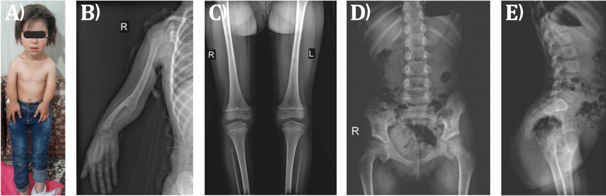 Figure 1
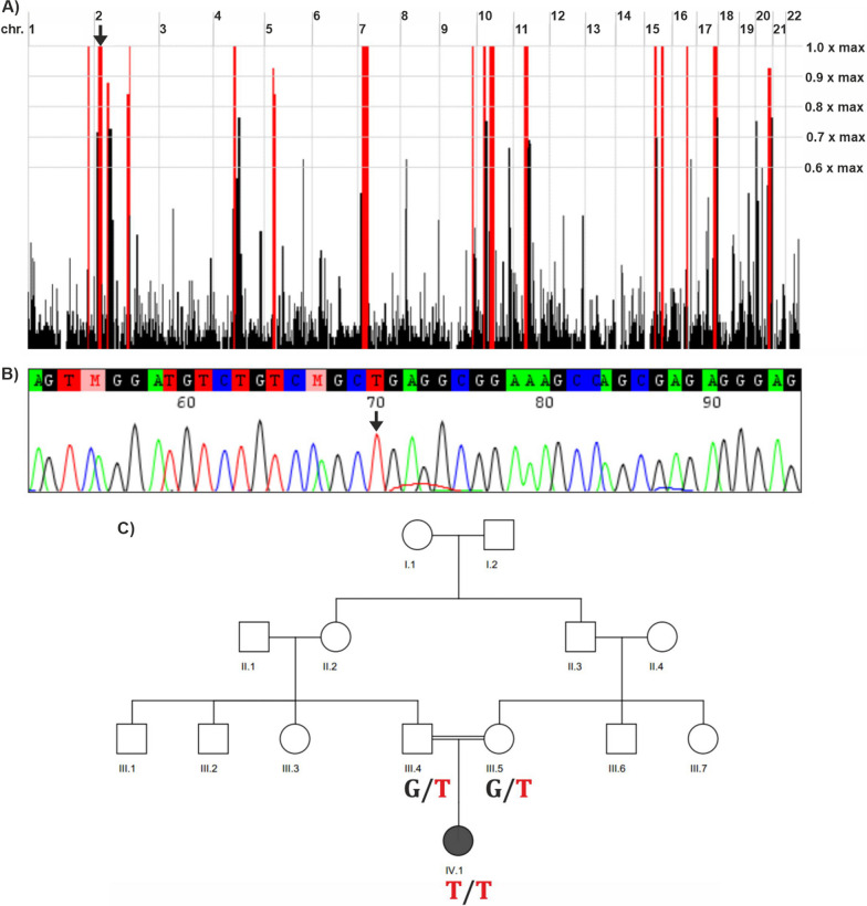 Figure 2
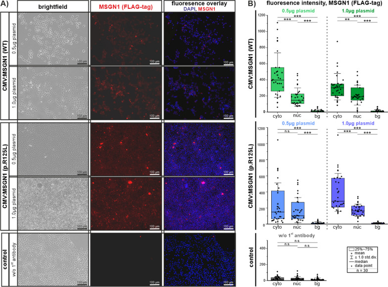 Figure 3
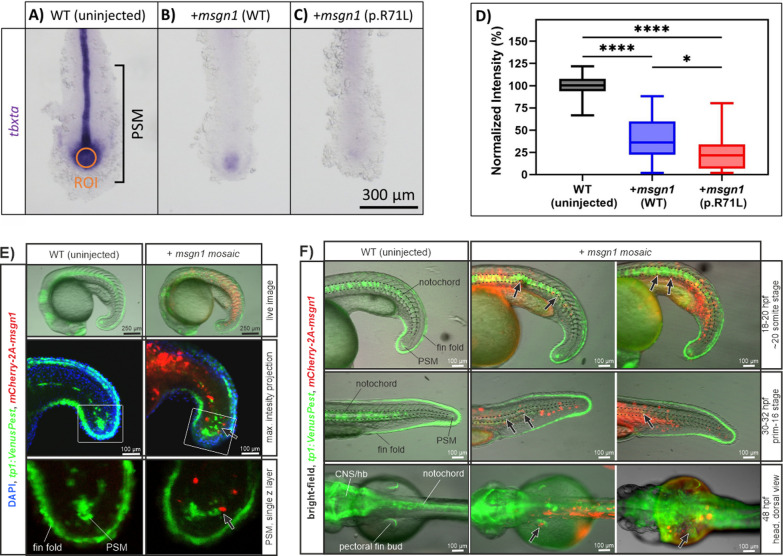 Figure 4
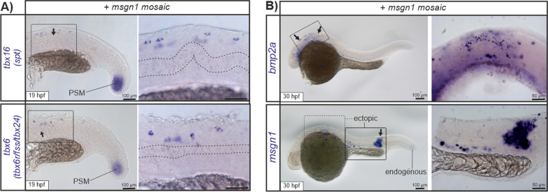 Figure 5
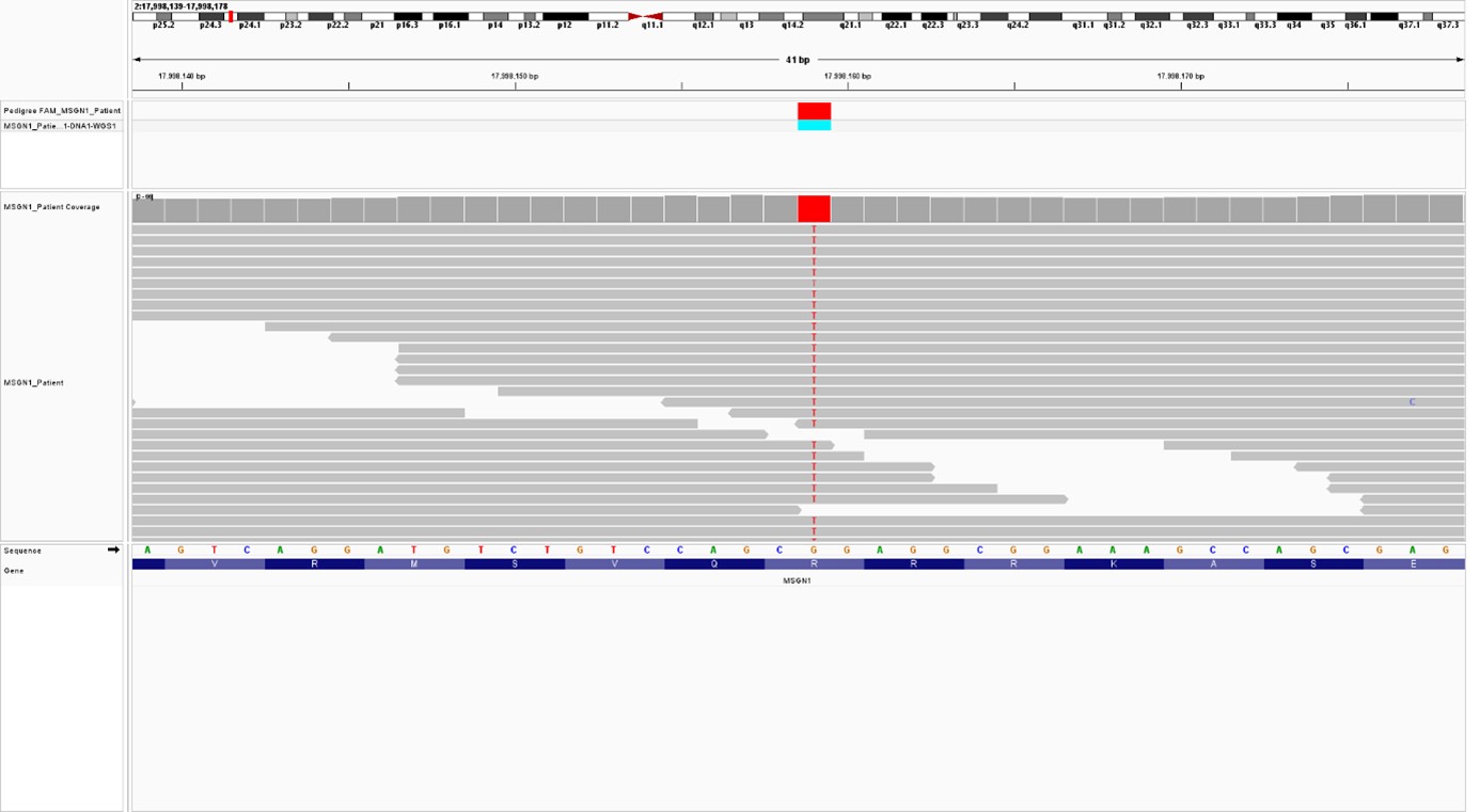 Figure 6
- —http://dx.doi.org/10.13039/501100001659Deutsche Forschungsgemeinschaft
- —Julius-Maximilians-Universität Würzburg (3088)
Peer Reviews
No public reviews on file for this paper yet. If you reviewed it on a platform where reviews are public (OpenReview, ICLR, NeurIPS, ICML), you can paste yours below so the community can read it here.
Videos
No videos yet. Explain this paper in a talk, walkthrough, or lecture? Add one.
Taxonomy
TopicsCongenital heart defects research · Genetics and Neurodevelopmental Disorders · Hippo pathway signaling and YAP/TAZ
Introduction
Skeletal dysplasias are a highly genetically heterogeneous group of skeletal and cartilaginous tissue disorders. The current classification comprises 461 distinct disorders and 437 underlying genes [25, 41]. Because of genetic and phenotypic heterogeneity of skeletal dysplasias, it is often difficult to get an unambiguous clinical diagnosis without molecular analyses. Next generation sequencing (NGS) technologies allow elucidation of the genetic basis of skeletal dysplasias, enabling precise diagnostics and adapted treatments. This method can identify new genetic variants in small patient cohorts or even a single affected individual. However, many of these variants are of uncertain significance/unknown function.
Mesogenin1 (MSGN1, OMIM: *612209), a basic-Helix–Loop–Helix transcription factor, is expressed in the presomitic mesoderm (PSM) only at early stages of vertebrate embryogenesis and plays a crucial role in formation of mesodermal progenitor cells during somitogenesis [47]. Msgn1 null mouse embryos have been shown to be defective in somitogenesis and exhibit somite segmentation defects with absence of all structures arising from the paraxial mesoderm (e.g. musculoskeletal trunk, trunk skeleton, parts of the skin and trunk vasculature). In addition, these mouse embryos displayed prominent tail segmentation defects. Reduced expression of components of the Delta-Notch signaling pathway and the segmentation clock oscillator was observed within the PSM [47]. In addition to the loss-of-function phenotype, ectopic expression of Msgn1 in mice suppresses notochord cell differentiation and promotes a PSM stem-cell fate [2]. The notochord is a primary structural element, a source of patterning signals, and is thereby essential for locomotion by enabling early cartilage and vertebrae development during vertebrate embryogenesis [6, 37]. In accordance with mouse experiments, zebrafish embryos injected with msgn1 mRNA exhibited severe truncation of tail structure, e.g. loss of tailbud and notochord cells [46]. This altered development is induced by disturbed expression of essential PSM regulators like tbxta (alias: TBXT (human), T/brachyury/bra (mouse), no tail/ntla/zft/ta (zebrafish)), which were significantly reduced in the posterior tailbud and in the axial region after gain of Msgn1 function [46]. The correct function of the PSM during early development is the prerequisite to subsequent processes like somite formation, notochord development and the segmentation clock itself. Its function involves a highly sophisticated network of different molecular factors in addition to MSGN1. These comprise WNT, FGF, retinoic acid, Delta-Notch signaling pathways and T-box transcription factors [23, 35] and are evolutionarily conserved among vertebrate species. MSGN1 functions as a transcription factor, which is regulated by these factors, but moreover orchestrates correct transcription of several essential effectors within this network [2].
Here, we have investigated the molecular pathogenesis of a novel form of skeletal dysplasia in the offspring of consanguineous Iranian parents and identified a novel homozygous c.374G > T, p.(Arg125Leu) variant in MSGN1 (NM_001105569) in the affected child. To test the pathogenicity of this variant, we overexpressed either wild-type or mutant msgn1 in zebrafish via mRNA injection and analyzed tbxta expression in the tailbud. Consequences of enhanced Msgn1 activity on early development were further analyzed in vivo in zebrafish embryos. Mosaic transient-transgenic overexpression of msgn1 showed spatial and temporal restricted consequences on tail development by influencing Delta-Notch active cells in the posterior progenitor zone and by inducing ectopic gene expression.
Results
Clinical description
The patient was the first child born to a gravida 2, para 1 mother. The healthy parents were second-degree cousins. On physical examination, the female proband had mild facial dysmorphism including hypertelorism, epicanthus, midface hypoplasia, mild retrognathia, and short stature with noticeable mesomelic shortening of the arms. She also presented dysplastic neck-thorax region with a prominent trapezoid muscle and arched clavicles (Fig. 1A). Radiographic images revealed humero-radial and radio-ulnar synostosis. The radii were severely shortened and hypoplastic, the ulnae were rudimentary with only their proximal part present. The scaphoid bone was only present as a tiny nucleus on the left side, while it was ossified on the right side (Fig. 1B). The diaphysis of the femora was very thin. The fibulae were clearly shortened and too thin (Fig. 1C). The iliac bones were somewhat narrow, the ramus inferior of the pubic bone was missing, and the ischium was only rudimentary (Fig. 1D). In addition, there was a hyperlordosis and mild scoliosis of the spine (Fig. 1E).Fig. 1A Short stature and mesomelic shortening of arms in the patient. B–E Skeletal X-ray surveys of the patient's right upper extremity (B), lower extremities (C), anterior view of the lower spine and pelvis (D), and lateral view of the lower spine and pelvis (E)
Whole exome sequencing revealed a likely disease-causing variant in MSGN1
Whole exome sequencing (WES) analysis detected a rare homozygous missense variant in MSGN1 (NM_001105569.3: c.374G > T, p.(Arg125Leu); NC_000002.11:g.17998159G > T). This variant is listed 11 times in heterozygous but not in homozygous state in the population database gnomAD v.4.0.0. The variant affects a conserved amino acid residue within a highly conserved bHLH domain in MSGN1 (Additional file 2: Fig. S1 and Additional file 3: Fig. S2). Different in-silico prediction tools such as CADD (Combined Annotation Dependent Depletion; [15, 30]), REVEL [11] and AlphaMissense [5] classified this variant as pathogenic (Additional file 8: Table S1). Moreover, the variant was predicted by SpliceAI to have no splicing effect [12]. Furthermore, we visualized the theoretical three-dimensional (3D) representation of both wild-type and mutant forms of the MSGN1 protein, to see any alteration in the structure and conformation of the protein (Additional file 2: Fig. S1A, B). The affected Arginine 125 within the MSGN1 protein is located at the second position of the evolutionary highly conserved basic helix-loop-helix (bHLH) protein domain at the beginning of the larger, first alpha-helix. The exchange from Arginine (negative hydropathy index, amphipathic and polar, positive charged side chain) to Leucine (positive hydropathy index, aliphatic and nonpolar, no charge) is biochemically profound. However, the exchange is predicted to not abrogate the helix structure but to cause misfolding of the encoded protein at the transition between the alpha-helix and the N-terminus (Additional file 2: Fig. S1A, B). According to HomozygosityMapper [34], the detected MSGN1 variant lies within an approximately 13.2 Mb homozygous interval on chromosome 2 (Fig. 2A). Sanger sequencing analysis confirmed WES findings and demonstrated that both parents are heterozygous carriers for the variant (Fig. 2B, C). An additional WGS analysis did not detect other candidate variants with higher pathogenic potential in known skeletal dysplasia genes and surrounding non-coding sequences.Fig. 2. Genetic results of the family. A Homozygous intervals in the patient’s genome, as determined by HomozygosityMapper. MSGN1 resides in a ~ 13.2 Mb homozygous interval on chromosome 2 (black arrow). B Sanger sequencing electropherogram of the patient’s variant in MSGN1. The mutated base is indicated by a black arrow. C Segregation of the MSGN1 variant within the family
In vitro analysis of intracellular MSGN1 localization
At first, we tested whether the newly identified MSGN1 variant and its predicted misfolding results in retention of MSGN1 protein in the cellular cytoplasm and nuclear exclusion, consistent with a loss-of-function of the transcription factor. HEK 293T cells were transfected with CMV-promoter driven expression plasmids at different concentrations incorporating either wild-type or c.374G > T, p.(Arg125Leu) MSGN1-FLAG-tag variants. Subsequent protein visualization was performed after 48 h via immunofluorescence by utilizing the protein tagged (Myc-DDK/FLAG). Transfected HEK 293T cells showed expression of both MSGN1 variants at different plasmid concentrations and did not imply loss of MSGN1 protein or restrained protein localization (Fig. 3A; controls Additional file 4: Fig. S3A).Fig. 3. In vitro transfection of HEK 293T cells with CMV:MSGN1 (WT)-FLAG-tag and CMV:MSGN1 p.(Arg125Leu)-FLAG-tag plasmids at different concentrations. A Immunofluorescence showing expression of tagged MSGN1 proteins (WT) or p.(Arg125Leu) after 48h of transfection. Corresponding experimental controls are shown in Additional file 4: Fig. S3A. B Quantification of MSGN1 localization in transfected cells. Fluorescence signals in a by single z-plane were visualized by confocal laser-scanning microscopy and subsequently signal intensity was measured in single cells (cyto: cytoplasm; nuc: nucleus) and outside of cells (bg: background). Graphs show signal intensity measurements of the RFP channel (MSGN1 Flag-Tag) of 30 cells per experimental group and 30 background positions. Signal intensity measurements of the corresponding DAPI channel for nucleus identification are given in Additional file 4: Fig S3B. Values are given in Additional file 1: Excel file S1
To further quantify intracellular localization of the MSGN1 proteins in the cytoplasm or in the nucleus of transfected cells we subsequently quantified fluorescence signal intensities of MSGN1-FLAG-tag and DAPI by confocal laser-scanning microscopy (LSM; Fig. 3B and Additional file 4: Fig. S3B). Measurement of MSGN1 fluorescence signal intensity did not reveal any difference in localization between MSGN1 (WT)-FLAG-tag or MSGN1 (Arg127Leu)-FLAG-tag variants.
Gain of msgn1 activity results in developmental defects during early stages of zebrafish development
Next we investigated the possible pathogenic effects of the variant on Mesogenin1 activity in zebrafish a in vivo model. To this end, we overexpressed either wild-type or the variant version (p.(Arg71Leu) in zebrafish corresponding to p.(Arg125Leu) in humans) of Msgn1 via mRNA injection at one-cell stage. We examined tbxta expression, which is regulated by Msgn1 [4, 46], in the tailbud at 8-somite stage (Fig. 4A–C). We found that overexpression of both wild-type and msgn1 p.(Arg71Leu) mRNA significantly reduced tbxta expression in the tailbud compared to uninjected wild-type embryos (P < 0.0001). In addition, we also observed a more severe effect by msgn1 p.(Arg71Leu), compared to msgn1 wild-type mRNA (P = 0.0186), suggesting that the variant leads to gain-of-function in Mesogenin1 activity (Fig. 4D).Fig. 4. In situ hybridization images of tbxta (no tail, Brachyury) expression in uninjected wild-type controls (A), wild-type msgn1 RNA (B) and mutant msgn1 RNA p.(Arg71Leu) in zebrafish (≈ p.(Arg125Leu) in humans) injected embryos (C). D Quantification of tbxta ISH signal in posterior PSM cells (orange ROI mark) is shown by normalized intensity comparison between the three groups of three independent experiments (WT (uninjected): 49 embryos; + msgn1 (WT) RNA: 58 embryos; + msgn1 p.(Arg71Leu) RNA: 54 embryos). Mosaic transient-transgenic overexpression of wild-type msgn1 results in partial disruption of Notch signals in PSM cells (E) and influences normal notochord (marked with dashed lines) and pectoral fin bud development (marked with arrow). F In vivo images show representative embryos of three independent injections experiments (overall: 37 imaged embryos; WT: 5 embryos; + msgn1 mosaic: 32 embryos). ISH signal quantification values and injection statistics are given in Additional file 1: Excel file S1
To investigate additional consequences of enhanced Msgn1 activity on embryonic development we utilized a mosaic transient-transgenic overexpression method. In short, microinjection of a DNA plasmid containing a mCherry-2A-msgn1 coding sequence (driven by the zebrafish msgn1 promoter) into single cells of early blastula stage zebrafish embryos drives clonal msgn1 expression during development. Ectopic msgn1 expressing cells are marked by mCherry fluorescence. By application of this method in the tp1:VenusPEST transgenic zebrafish background, we were able to investigate msgn1 overexpressing cells and Notch signaling during development simultaneously by in vivo fluorescence visualization. We observed that mCherry positive/msgn1 expressing cells disturb Notch positive cells within the PSM (18–20 hpf; Fig. 4E). The cell population resembles tbxta expressing posterior PSM cells at the maturation zone, giving rise to notochord and somite tissues [8, 17]. Other Notch positive tissues, e.g. dorsal fin fold cells, were not influenced by msgn1 implying PSM specific effects. PSM disruption is associated with developmental defects whose severity is linked to the amount of msgn1 overexpressing cells in the PSM region (Additional file 5: Fig S4). Investigation of these embryos at later development stages further showed that mosaic embryos displayed altered notochord and pectoral fin structures (e.g. wavy notochord outgrowth, disruption of Notch signals in notochord cells and in pectoral fin buds, marked by arrows in Fig. 4F).
To further decipher potential downstream effects of ectopic Msgn1 activity, we investigated expression of previously described MSGN1 downstream target genes in mosaic zebrafish embryos [2]. Visualization of tbx6 and tbx16 via in situ hybridization showed that both factors are endogenously expressed within the PSM and are additionally expressed within the trunk at regions depicting notochord alterations (19 hpf; Fig. 5A). Moreover, strong ectopic expression of bmp2a and msgn1 was detected within the head and trunk regions of 30 hpf embryos (short staining time with low levels of endogenous ISH transcript signal visible; Fig. 5B). Detection of msgn1 expression via in situ hybridization was in accordance with mCherry-2a-msgn1 fluorescence expression patterns (see Figs. 3F and 5E) and probably detects primarily transgene induced msgn1 expression in these mosaic embryos.Fig. 5A In situ hybridization images of tbx6 (tbx6r/fss/fussed somites/tbx24) and tbx16 (spt/spatetail) expression in injected embryos 19 hpf showing mosaic transient-transgenic overexpression of wild-type msgn1. Expression of tbx6 and tbx16 was detected in PSM cells (endogenous expression) and within the trunk, colocalizing with notochord alterations (shown in higher magnification). B In 30 hpf old msgn1 mosaic embryos, ectopic expression of bmp2a was detected in the head region, consistent with strong ectopic msgn1 expression in trunk and head regions of injected embryos at this stage
General overexpression either of human MSGN1 (WT) or MSGN1 p.(Arg125Leu) variants in zebrafish embryos resulted in similar developmental consequences during early developmental stages as gain of zebrafish msgn1 in mosaic embryos, e.g. notochord bending and disruption of PSM cells (used plasmids: CMV:MSGN1-Flag-tag (WT) and CMV:MSGN1-Flag-tag p.(Arg125Leu), Additional file 6: Fig. S5 and Additional file 1: Excel file S1). This observation implies retained function of human MSGN1 and of the patient derived MSGN1 variant also in zebrafish. Further tissue-restricted mosaic expression of zebrafish msgn1 p.(Arg71Leu) and human MSGN1 p.(Arg125Leu) variants in PSM and trunk cells under msgn1 promoter control in tp1:VenusPEST embryos resulted in similar developmental consequences like msgn1 wild-type injected embryos, but display more severe manifestations and higher rates of severe phenotypes in injected embryos (msgn1:mCherry-2A-msgn1 vector: 15% (n = 197) and 20% (n = 92); msgn1:mCherry-2A-msgn1 p.(Arg71Leu) vector: 32% (n = 50); msgn1:mCherry-2A-MSGN1 p.(Arg125Leu) vector: 27% (n = 104); Additional file 7: Fig. S6 and Additional file 1: Excel file S1).
Discussion
In our study, we identified a homozygous missense variant c.374G > T, p.(Arg125Leu) in MSGN1 via exome sequencing to be potentially associated with prominent skeletal abnormalities in a single patient. MSGN1 missense variants have not yet been associated with a human Mendelian disease. Previously, eleven MSGN1 missense variants have been reported in ClinVar, but all lack detailed zygosity and phenotype information or segregation analyses and are all classified as variants of uncertain significance (Tab. S3). Several structural variants (gains and losses) including the MSGN1 locus are additionally described in ClinVar, but all these alterations affect larger genomic regions incorporating several coding genes and therefore do not allow the conclusion that MSGN1 is disease causing (Tab. S4). The aim of this study was to provide molecular insights into our patient’s phenotype and to link the observed malformations to known MSGN1 functions during development using zebrafish.
In contrast to loss of function phenotypes in MSGN1 animal models (short tail, prenatal lethal, absent notochord), we did not observe prominent vertebral segmentation or axial skeleton defects in our patient. We therefore propose the detected MSGN1 variant has a gain of function effect, which could explain only mildly affected vertebrae of the proband. Initial in vitro investigations clarified, that the corresponding protein of the novel MSGN1 p.(Arg125Leu) variant is still able to localize into the nucleus and therefore may act as a transcription factor. Previously published results [46] in zebrafish showed that elevated msgn1 activity reduces tbxta expression in the PSM. Our own experimental data are in accordance with this observation and further demonstrate that the corresponding msgn1 p.(Arg71Leu) variant results in a stronger gain of function effect hinting to raised Mesogenin1 activity (Fig. 4D and Additional file 7: Fig. S6). TBXT, the prototypical T-box transcription factor, is essential for the formation and differentiation of posterior mesoderm and for axial development in all vertebrates. Homozygous variants in TBXT orthologues cause embryonic lethality, while heterozygous variants are associated with short tail mice [16, 33] and zebrafish (no tail mutant; [27, 38, 42]). In humans, a homozygous TBXT variant (c.512A > G, p.(His171Arg), NM_003181), has been associated with sacral agenesis, abnormal ossification of vertebral bodies and persistent notochord canal [29]. In chicken T is expressed in the lateral plate mesoderm at the onset of limb bud formation [22]. T controls TBX6 expression, which is known to regulate activation of MSGN1 [45]. TBX6 variants cause spondylocostal dysostosis in human [36]. However, ectopic Tbx6 gain-of-function within the segmented paraxial mesoderm and its derivatives in mice are associated with appendicular skeletal defects including hypoplastic scapula, small limbs, shortened and malformed humerus, radius, ulna, femur, tibia and fibula, resembling Tbx15 null mouse embryos and Cousin syndrome in humans (OMIM #260660) [18, 43]. Since gain of Tbx6 function in mice results in short stature and severe limb defects [45], we hypothesize that MSGN1 gain of function is responsible for short stature and limb abnormalities in our patient. The expression of T-box transcription factors like tbxt, tbx6, tbx16, and tbx16l in zebrafish is regulated by Msgn1 [4, 13, 24]. Our transient mosaic experiments in zebrafish and previously published zebrafish experiments further show ectopic tbx6 expression via gain of Msgn1 function (Fig. 4 and [46]). To our knowledge, no skeletal dysplasia has been associated with biallelic pathogenic gain of function variants. However, there are some examples in the literature for biallelic pathogenic gain of function variants, which affect other physiological systems e.g. the immune system (NLRP1, STAT2) [7, 9].
MSGN1 plays an important role in notochord maintenance and development, that expresses genes such as SOX9 which are characteristic of chondrogenesis [37]. Prominent functions of Delta-Notch signals during early posterior tail development have been intensively investigated and are necessary to synchronize PSM cells within the somitogenesis clock of vertebrates [19–21]. We visualized Delta-Notch signalling for in vivo observation of developmental defects in the posterior PSM maturation zone and in the notochord. Our zebrafish results imply that gain of msgn1 function in single cells of the posterior progenitor zone (maturation zone in the posterior PSM) disturbs the structure of Delta-Notch positive cells which give rise to somite, paraxial mesoderm and notochord tissues during later developmental stage [8, 31]. We subsequently observed notochord bending and potential disruption of pectoral fin buds/upper extremity. The later might result from msgn1 ectopic function on mesenchymal stem cells or on chondrogenesis in this region [3, 28, 40]. Interestingly within this context, we found that transcriptional Msgn1 activity results in ectopic bmp2a expression (besides action on tbx6 and tbxta). Bmp2 is described as a direct MSGN1 target [2] and is known to be associated with prominent skeletal anomalies in patients, like brachydactyly type A2 (OMIM #112600), as well as short stature, facial dysmorphism, and skeletal anomalies with or without cardiac anomalies (OMIM #112261). Further studies will have to clarify, if the long-term ectopic expression of downstream effectors of MSGN1 is also the driver of long-term developmental alterations in the current patient, since spatio-temporal MSGN1 expression is usually tightly regulated during development.
Collectively, the studied patient presents a novel MSGN1-associated skeletal syndrome in humans, highlighting the importance of the temporally and spatially appropriate MSGN1 activity in various developmental pathways. However, additional patients/families are needed to unequivocally link MSGN1 gain-of-function to the described syndrome.
Materials and methods
Whole exome sequencing and database research
Exome capture was performed according to the Illumina Nextera Rapid Capture Enrichment library preparation protocol (individuals IV.1, III.4 and III.5) using 50 ng of genomic DNA. Paired-end sequencing of the libraries was performed with a NextSeq500 sequencer and the v2 reagent kit (Illumina, San Diego, California, USA). Sequences were mapped to the human genome reference (NCBI build 37/hg19 version) using the Burrows-Wheeler Aligner. Aligned reads ranged between 85,736,827 and 99,719,268. The mean coverage was ≥ 50 with 91.2%. 99% of the exome were covered at least 10x. A total of 678,344–961,783 variants per sample were called and analyzed using GensearchNGS software (PhenoSystems SA, Braine le Chateau, Belgium). Variants with a coverage of ≤ 10, a Phred-scaled quality of ≤ 15, a frequency of ≤ 15, and a MAF of ≥ 2% were neglected. Six control samples from healthy individuals were used for filtering out platform artefacts. Alamut Visual (Interactive Biosoftware, Rouen, France) software including prediction tools like SIFT, MutationTaster, PolyPhen-2, CADD-, and REVEL-Score was used for variant prioritization. Potential effects of a variant on pre-mRNA splicing were evaluated by SpliceAI, SpliceSiteFinder-like, MaxEntScan, NNSPLICE, GeneSplicer, Human Splicing Finder, ESEfinder, RESCUE-ESE, and EX-SKIP. Population databases like gnomAD v4.0.0, and GME revealed whether a variant has been previously found. Protein expression, structure, and functional aspects were investigated with UniProt and The Human Protein Atlas. Information on mouse and zebrafish models was retrieved from the MGI and ZFIN database, respectively.
Sanger sequencing
MSGN1 exon one was amplified by a touchdown PCR program using primers in the flanking introns (forward: 5´-GGTGGACTACAATATGTTAGCTTTCC-3´ and reverse: 5´-TAGACAGGTGGCAGGTAATTCC-3´). A clean-up step with ExoSAP-IT (Applied Biosystems, Foster City, USA) was followed by the sequencing reaction using the BigDye Terminator Cycle Sequencing Kit v1.1 (Applied Biosystems, Waltham, USA). Sequencing was conducted on a 3130XL capillary sequencer (Applied Biosystems, Waltham, USA) and data analysis was performed with Gensearch (PhenoSystems SA, Braine le Chateau, Belgium).
Whole genome sequencing
Short read genome sequencing was performed to exclude structural variants and non-coding variants in known skeletal dysplasia genes. In brief, genomic DNA was isolated from peripheral blood and sequenced at 30 × coverage using the Illumina TruSeq PCR-free protocol at the West German Genome Center (WGGC). Reads were aligned to the human genome build GRCh37/hg19 using BWA-MEM 0.7.17. The VarFish software was used for filtering and interpretation of variants including SVs (caller: Delly v.0.8.1) according to an in-house SOP [10].
In vitro experiments and intracellular MSGN1 localization measurements
Transfections were performed with 2.5 × 10^5^ HEK 293T cells per well in 12-well plates. HEK 293T cell were initially grown on glass cover slides and subsequently transfected with two different amounts of DNA plasmids containing either the coding sequence of wild-type MSGN1 (WT) or of the MSGN1 p.(Arg125Leu) variant coupled to a FLAG-tag under control of the CMV promoter. 0.5 µg and 1.0 µg plasmid DNA per 100 µl total transfection volume was used with 2 to 3 µl FuGene HD transfection agent per well (Promega, Madison, USA, product-nr. E2311). 48 h after transfection, cells were paraformaldehyde fixed and immunofluorescence staining of Flag-tag was performed by standard protocols for cell HEK 293T cell cultures (primary antibody: monoclonal DYKDDDDK Tag Recombinant, Thermo Fisher Scientific, Waltham, USA, product-nr. 701629, RRID: AB_2532497, 1:250 dilution (2.5 µg/ml); secondary antibody: goat anti-rabbit-Alexa 594, Invitrogen/Thermo Fischer Scientific, product-nr. A-11012, RRID: AB_2534079, 1:1000 dilution (1 µg/ml)) and DAPI (Sigma-Aldrich, St. Louis, USA; product-nr. D9542, 1:5000 dilution (4 µg/ml)). Corresponding controls were performed by transfection of an CMV:GFP-Tag plasmid (positive control), by including non-transfected control cells, by omitting primary or secondary antibody incubations (negative controls). Images were taken with a Keyence BZ-X800 fluorescence microscope (Keyence, Osaka, Japan).
Intracellular localization of MSGN1 proteins was quantified by analysis of single z-level immunofluorescence images acquired by laser scanning confocal microscopy (Nikon A1 + , Nikon NIS-Elements software, Nikon Instruments, Tokyo, Japan) via ImageJ/Fiji software ( https://fiji.sc/) [32]. In contrast to analysis by classical fluorescence microscope techniques, usage of LSM enables high resolution optical sectioning of fluorescence signals in a narrow single z-level at height of the nucleus and removes out-of-focus fluorescence signals outside of the focal plane in conjunction with strict pinhole settings. Images were taken at 600-fold magnification, in two channels (MSGN1-tag: 561 nm laser excitation, 610–620 nm emission; DAPI in nucleus: 405 nm laser excitation, 470–480 nm emission), at a resolution of 1024 × 1024 pixel corresponding to a 205 × 205 µm area. To circumvent light scattering, smallest pinhole settings per laser channel were chosen. For each transfection group, three non-overlapping images were acquired, subsequent 10 cells per image were randomly selected and fluorescence intensity values were measured in ImageJ/Fiji software by straight line histograms (line length: 10 µm (50 pixel), line thickness: 2 µm (10 pixels), mean value per pixel position in length were measured at 50 positions per line and channel). Line positions were adjusted to start in the cytoplasm (low DAPI intensity signal) and end in the nucleus (high DAPI intensity signal), with line midpoint corresponding to nucleus border (position #25). Cytoplasmic values were defined as position #15, 2.5 µm away from nucleolus border. Nuclear values were defined as position #35, 2.5 µm away from nucleolus border. Background values were defined as position #15 and #35 values measured outside of cell bodies at five positions per image.
Zebrafish animal maintenance and lines
Laboratory zebrafish embryos (Danio rerio) of the AB/TU and AB/AB wild-type strain (ZDB-GENO-010924-10; ZDB-GENO-960809-7) and transgenic Tg(EPV.Tp1-Mmu.Hbb:Venus-Mmu.Odc1) (ZDB-TGCONSTRCT-120419-4, tp1:VenusPEST, [26]) were maintained as previously described under standard aquatic conditions at an average of 24 °C water temperature [1, 44]. Embryos were staged by morphological characteristics according to Kimmel et al. [14]. “hpf” and “dpf” indicate embryonic development in hours/days-post fertilization at 28.5 °C incubation temperature, respectively. All procedures involving experimental animals were performed in compliance with local animal welfare laws, guidelines, and policies. All presented experiments have been performed in zebrafish embryos and larvae younger than 5 dpf, before free-swimming and independent feeding, and thus are not regulated as animal experiments in Germany under current legislation.
Mosaic transient-transgenic msgn1 overexpression and in vivo visualization
Mosaic transient-transgenic overexpression was performed by microinjection of a previously published plasmid containing a zebrafish msgn1 promoter fragment driving 2A-coupled expression of mCherry fluorescence protein and wild-type zebrafish msgn1 [46]. Injection of this plasmid results in prominent and severe tail malformations due to gain of Msgn1 function in mesodermal progenitor cells. Injection of msgn1 injection solution (DNA plasmid was diluted in water to a final concentration of 25 ng/µl and admixed with 0.05% Phenol-red (pH 7.0) for visualization of injection solution) into only one cell of early blastula zebrafish embryos (4-cell stage/1.0 hpf up to 16-cell stage/1.5 hpf) resulted in a fraction of msgn1 overexpressing cells within developing embryos. In addition to zebrafish wild-type msgn1 (msgn1:mCherry-2A-msgn1), plasmids containing either a zebrafish msgn1 p.(Arg72Leu) variant (msgn1:mCherry-2A-msgn1 (p.R71L)), or a human MSGN1 p.(Arg125Leu) variant (msgn1:mCherry-2A-MSGN1 (p.R125L)) were injected in a similar way into zebrafish embryos and investigated.
Injected embryos were in vivo analyzed under a Fluorescence Stereomicroscope (Leica S8 APO equipped with Leica GFP and DSR filter sets (filter nr. 10447408 and 10447412), Leica Miscrosystems, Wetzlar, Germany) at different time points during embryonic and early larval development (approximately between 16 and 48 hpf) for cells showing msgn1 overexpression by mCherry fluorescence and for resulting developmental consequences. Injection was performed in embryos of a tp1:VenusPEST transgenic line [26], which enables in vivo visualization of Notch signaling due to expression of a short-half-life version of the fluorescence protein Venus under the tp1 promoter element. The tp1 element contains 12 EBV terminal protein 1 (TP1) gene promoter fragments for endogenous Notch (NICCD) and RBPJ/CB1/Su(H) co-factor binding. Detailed microscopic investigation was performed with Zeiss Imager A1 (in situ hybridizations, Carl Zeiss AG, Jena, Germany) or a Nikon A1 + Laser scanning confocal microscope (in vivo*,* Nikon Corporation, Tokyo, Japan). For detailed microscopic investigations embryos were short time fixed in 4% paraformaldehyde/PBS for 30 min and were mounted in Mowiol. Nuclei were stained by DAPI incubation (1 µg/mL in PBST; 15 min) before mounting. Images were analyzed with ImageJ/Fiji (https://fiji.sc/) and arranged with CorelDraw Graphics Suite (Alludo, Canada) software.
Plasmid vector cloning and mutagenesis
In vitro HEK 293T transfection experiments were performed with CMV promoter driven human tagged MSGN1 (Origene, Rockville, USA, product-nr. RC225212, CDS: NM_001105569). Patient MSGN1 variant was introduced into the plasmid by site-directed mutagenesis (Q5 Site-Directed Mutagenesis Kit, New England Biolabs/NEB, Ipswich, USA, product-nr. E0554S) and was validated via Sanger sequencing.
Zebrafish injection experiments were performed with msgn1 promoter driven zebrafish msgn1 plasmid (sk-tol2-msgn1:mCherry-2A-msgn1) [46]. Human MSGN1 c.374G > T, p.(Arg125Leu) and zebrafish msgn1 c.211AG > CT p.(Arg71Leu) missense variants were introduced by site-directed mutagenesis in subcloned coding sequences without start codons (Q5 Site-Directed Mutagenesis and PCR cloning Kit, New England Biolabs/NEB, product-nr. E0554S and E1202S) and were validated via Sanger sequencing. Subsequently, the newly established variant coding sequences were used to replace zebrafish msgn1 (WT) in sk-tol2-msgn1:mCherry-2A-msgn1 by restriction site cloning. Primer sequences and used plasmids are listed in Additional file 8: Table S2.
Generation of mutant msgn1Arg71Leu mRNA and overexpression in zebrafish
A cDNA library was extracted from embryos and wild-type msgn1 was amplified with PCR using primers listed in Additional file 8: Table S2. Mutant msgn1 p.(Arg71Leu) was created using overlap extension primers. The wild-type msgn1 and mutant msgn1 p.(Arg71Leu) cDNA were cloned into separate pCS2 + vectors between BamHI and EcoRI restriction sites. In vitro transcription was used to create and isolate mRNA (mMESSAGE mMACHINE™ SP6 Transcription Kit, Invitrogen/Thermo Fisher Scientific, Cat# AM1340). Wild-type embryos were either injected with 200 pg wild-type msgn1 mRNA (58 embryos) or msgn1 p.(Arg71Leu) mRNA (54 embryos) at one-cell stage and compared with untreated ones (49 embryos) in three independent experiments. The embryos were fixed at 8-somite stage in 4% paraformaldehyde before in situ hybridization.
Zebrafish embryo RNA in situ hybridization
RNA in situ hybridization was performed according to standard protocols [39]. RNA probes were synthesized from cloned partial mRNA sequences of target genes using the DIG or FLU RNA Labeling Kit (Roche, Basel, Switzerland, product-nr. 11685619910 and 11175025910). All detected expressions patterns with newly established RNA probes were in accordance with previously published and ZFIN database patterns. Used in situ probes were targeted against: tbxta (ZDB-GENE-980526-437; synonyms: T/ta/brachyury/no tail/ntl), msgn1 (ZDB-GENE-030722-1; synonyms: mespo), bmp2a (ZDB-GENE-980526-388), tbx6 (ZDB-GENE-020416-5; synonyms: tbx6r/fss/fused somites/tbx24), tbx16 (ZDB-GENE-990615-5; synonyms: spt/spadetail). In-situ experiments were performed two times independently and included n ≥ 10 embryos per sample and condition. Detailed information and primers used for probe cloning are listed in Additional file 8: Table S2.
Image and statistical analysis of RNA in situ hybridization embryos
The embryos were flat mounted via dissection of the yolk sac and imaged under a Nikon SMZ1500 stereomicroscope (HR Plan Apo 1X WD 54), Nikon DS-Ri1 digital camera with reflected light at 23 °C room temperature. FIJI software (ImageJ 1.54f) [32] was used to assess intensity of tbxta staining in the tailbud using a standardized circular region (120 µm in diameter, ROI seen in Fig. 4A). Images were first inverted, then the mean intensity of anterior tissue background (17 µm in diameter) was subtracted from the mean intensity of tailbud (ROI). Then each intensity was normalized to the average intensity of uninjected embryos in an entire experiment.
We used unpaired two-tailed Kruskal–Wallis nonparametric test without equal standard deviation assumption in Fig. 4D. The statistical tests and distribution calculations (median and quartiles, confidence intervals) were performed in GraphPad Prism 9.5.0 software.
Supplementary Information
Additional file 1. Excel file S1: Raw Data Fig. 3, ISH signal quantification values and injection statisticsAdditional file 2. Figure S1: MSGN1 protein structure computer predictions indicate a conformational change, which can be distinguished between the normal protein structure (A) and the p.(Arg125Leu) variant (B). Amino acid alignment of six vertebrate species indicates evolutionary conservation of Arg125 within the basic helix-loop-helix (bHLH) protein domain (C). Numbers indicate position within the human amino acid sequence. Uniport amino acid sequence IDs are given for different speciesAdditional file 3. Figure S2: Next generation sequencing reads of new MSGN1 missense variant of the affected patientAdditional file 4. Figure S3: Additional data to in vitro transfection of HEK 293T cells with CMV:MSGN1-Tag and CMV:MSGN1-Arg125Leu plasmids at different concentrations presented in Fig. 3. (A) Corresponding immunofluorescence controls after 48h of transfection are shown. (B) Additional data to quantification of MSGN1 localization in transfected cells. Fluorescence signals in a single z-plane were visualized by confocal laser-scanning microscopy and subsequently signal intensity was measured in single cells (cyto: cytoplasm; nuc: nucleus) and outside of cells (bg: background). Graphs show signal intensity measurements of the DAPI channel (nucleus) of 30 cells per experimental group and 30 background positions. Values are given in Excel file S1Additional file 5. Figure S4: Examples of different phenotypes of msgn1 mosaic zebrafish embryos. Injection with the msgn1:mCherry-2A-msgn1 plasmid results in different amounts of mCherry positive/msgn1 expressing cells within the trunk and tailbud region of tp1:VenusPEST transgenic embryos. Phenotype severity in the PSM (marked by arrows) 24 h after injection is correlated with msgn1 overexpressing cell amount and positionAdditional file 6. **Figure S5: **Different phenotypes of CMV:MSGN1 plasmid injected zebrafish embryos. Injection with the CMV:MSGN1-FLAG-tag (WT) or with the CMV:MSGN1-FLAG-tag p.(Arg125Leu) plasmids, which have been used for in vitro cell transfections, results in alteration of tail development also within the trunk and tailbud region of zebrafish embryos at 18 hpf. Injected embryos display axis bending or cell aggregations in the trunk and PSM regions (marked by black arrows). Number of injected embryos are given in Excel file S1.Additional file 7. **Figure S6: **Examples of different phenotypes of msgn1 p.(Arg71Leu) and MSGN1 p.(Arg125Leu) mosaic tp1:VenusPEST zebrafish embryos in vivo. Injection with the sk-tol2-msgn1:mCherry-2A-msgn1 p.(Arg71Leu) or with the sk-tol2-msgn1:mCherry-2A-MSGN1 p.(Arg125Leu) plasmid results in mCherry positive/msgn1 variant expressing cells within the trunk and tailbud region of tp1:VenusPEST transgenic embryos. Phenotype severity in the PSM (marked by arrows) in a time frame between 16 and 35 hpf after injection is shown. The observed phenotypes correlate with msgn1 overexpressing cell amount, position and partly differ in severity between zebrafish and human CDS versions. Number of injected embryos are given in Excel file S1.Additional file 8. Table S1: Pathogenicity prediction of MSGN1 c.374G>T, p.(Arg125Leu) variant. Table S2: PCR Primers and DNA plasmids used in this study. **Table S3: **ClinVar missense MSGN1 VUS variants. **Table S4: **ClinVar structural MSGN1 variants.
The reference list from the paper itself. Each links out to its DOI / PubMed record.
- 1Alestrom PD'Angelo L Midtlyng PJ Schorderet DF Schulte-Merker S Sohm F Warner S Zebrafish: housing and husbandry recommendations Lab Anim 20205421322410.1177/002367721986903731510859 PMC 7301644 · doi ↗ · pubmed ↗
- 2Chalamalasetty RB Garriock RJ Dunty WC Jr Kennedy MW Jailwala P Si H Yamaguchi TP Mesogenin 1 is a master regulator of paraxial presomitic mesoderm differentiation Development 20141414285429710.1242/dev.11090825371364 PMC 4302905 · doi ↗ · pubmed ↗
- 3Chen S Tao J Bae Y Jiang MM Bertin T Chen Y Yang T Lee B Notch gain of function inhibits chondrocyte differentiation via Rbpj-dependent suppression of Sox 9J Bone Miner Res 20132864965910.1002/jbmr.177022991339 PMC 3548081 · doi ↗ · pubmed ↗
- 4Chen W Liu J Yuan D Zuo Y Liu Z Liu S Zhu Q Qiu G Huang S Giampietro PF Progress and perspective of TBX 6 gene in congenital vertebral malformations Oncotarget 20167574305744110.18632/oncotarget.1061927437870 PMC 5302999 · doi ↗ · pubmed ↗
- 5Cheng J Novati G Pan J Bycroft C Zemgulyte A Applebaum T Pritzel A Wong LH Zielinski M Sargeant T Accurate proteome-wide missense variant effect prediction with Alpha Missense Science 2023381 eadg 749210.1126/science.adg 749237733863 · doi ↗ · pubmed ↗
- 6Corallo D Trapani V Bonaldo P The notochord: structure and functions Cell Mol Life Sci 2015722989300810.1007/s 00018-015-1897-z 25833128 PMC 11114051 · doi ↗ · pubmed ↗
- 7Drutman SB Haerynck F Zhong FL Hum D Hernandez NJ Belkaya S Rapaport Fde Jong SJ Creytens D Tavernier SJ Homozygous NLRP 1 gain-of-function mutation in siblings with a syndromic form of recurrent respiratory papillomatosis Proc Natl Acad Sci U S A 2019116190551906310.1073/pnas.190618411631484767 PMC 6754618 · doi ↗ · pubmed ↗
- 8Fior R Maxwell AA Ma TP Vezzaro A Moens CB Amacher SL Lewis J Saude L The differentiation and movement of presomitic mesoderm progenitor cells are controlled by Mesogenin 1Development 20121394656466510.1242/dev.07892323172917 PMC 3509727 · doi ↗ · pubmed ↗