Comparing multi-image and image augmentation strategies for deep learning-based prostate segmentation
Samuel Fransson

TL;DR
This study compares using multiple images versus image augmentation for training deep learning models to segment the prostate in radiotherapy, finding that standard augmentation methods are more effective.
Contribution
The paper evaluates the effectiveness of multi-image strategies versus standard augmentation for prostate segmentation in low-data scenarios.
Findings
Using multiple images showed minimal improvement in DICE and Hausdorff 95% metrics compared to single images.
The rectum showed the maximum difference in performance when training with images from five patients.
Standard augmentation methods outperformed multi-image strategies in most cases.
Abstract
During MR-Linac-based adaptive radiotherapy, multiple images are acquired per patient. These can be applied in training deep learning networks to reduce annotation efforts. This study examined the advantage of using multiple versus single images for prostate treatment segmentation. Findings indicate minimal improvement in DICE and Hausdorff 95% metrics with multiple images. Maximum difference was seen for the rectum in the low data regime, training with images from five patients. Utilizing a 2D U-net resulted in DICE values of 0.80/0.83 when including 1/5 images per patient, respectively. Including more patients in training reduced the difference. Standard augmentation methods remained more effective.
Genes, proteins, chemicals, diseases, species, mutations and cell lines named across the full text — each resolved to its canonical identifier and authoritative record.
Click any figure to enlarge with its caption.
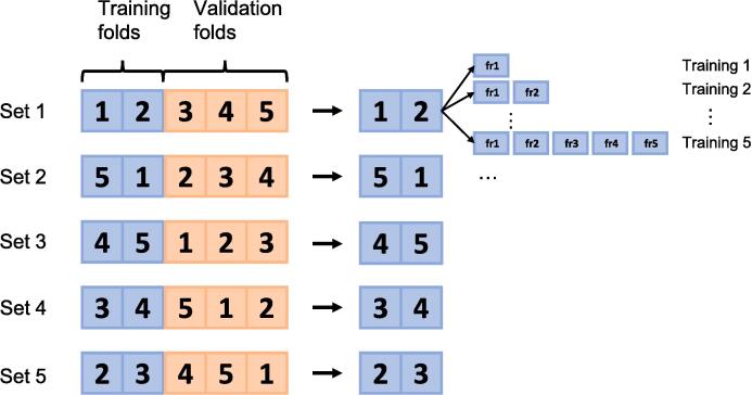 Figure 1
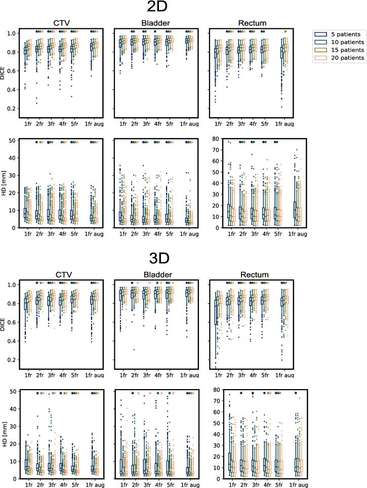 Figure 2
Peer Reviews
No public reviews on file for this paper yet. If you reviewed it on a platform where reviews are public (OpenReview, ICLR, NeurIPS, ICML), you can paste yours below so the community can read it here.
Videos
No videos yet. Explain this paper in a talk, walkthrough, or lecture? Add one.
Taxonomy
TopicsAdvanced Neural Network Applications · Medical Image Segmentation Techniques · Medical Imaging and Analysis