Challenges of automation in quantitative evaluation of liver biopsies: Automatic quantification of liver steatosis
Jessica Darling, Nada Abedin, Paul K. Ziegler, Steffen Gretser, Barbara Walczak, Ana Paula Barreiros, Falko Schulze, Henning Reis, Peter J. Wild, Nadine Flinner

TL;DR
This paper explores how automated methods can improve the accuracy of liver biopsy evaluations for fatty liver disease by reducing variability between pathologists.
Contribution
The study introduces a new area-based algorithm that integrates tissue composition data to improve the accuracy of liver steatosis quantification.
Findings
Area-based approaches showed stronger correlations than cell nucleus-based methods for predicting steatosis grade.
Incorporating tissue composition data reduced prediction errors for both area-based and cell nucleus-based methods.
The final area-based algorithm achieved 80% accuracy and strong correlation with manual evaluations.
Abstract
Die MASLD (metabolische Dysfunktion-assoziierte steatotische Lebererkrankung, oder nichtalkoholische Fettlebererkrankung [NAFLD]) ist eine häufige Erkrankung, deren Diagnose auf der lichtmikroskopischen Auswertung von Leberbiopsien basiert. Diese unterliegt jedoch einer großen Interbetrachtervariabilität (IBV), die durch Hinzunahme von automatisierten Methoden verringert werden kann. Ein Großteil der bestehenden computerbasierenden Methoden reflektiert nicht das, was in der Realität vom Pathologen bewertet wird. Ziel ist es, aufzuzeigen, wie diese Unterschiede die Vorhersage des Verfettungsgrads (VG) beeinflussen. Zusätzlich erschwert die IBV die Validierung von Algorithmen. Insgesamt 40 Gewebeschnitte wurden automatisch mit Bildanalysemethoden zur Fett‑, Zellkern- und Fibroseerkennung ausgewertet. Die Daten wurden verwendet, um den VG zu berechnen. Die IBV bei der Quantifizierung des…
Genes, proteins, chemicals, diseases, species, mutations and cell lines named across the full text — each resolved to its canonical identifier and authoritative record.
Click any figure to enlarge with its caption.
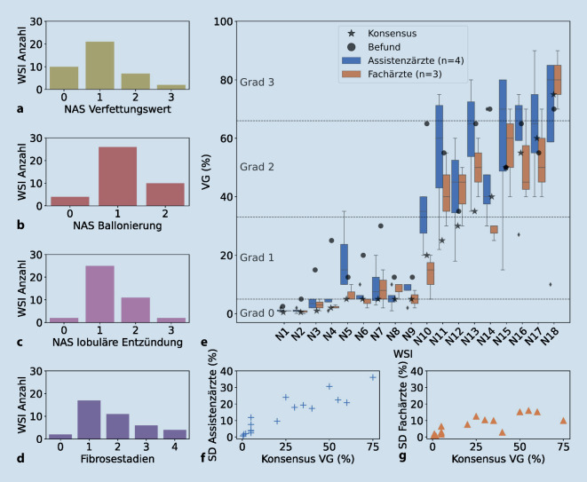 Figure 1
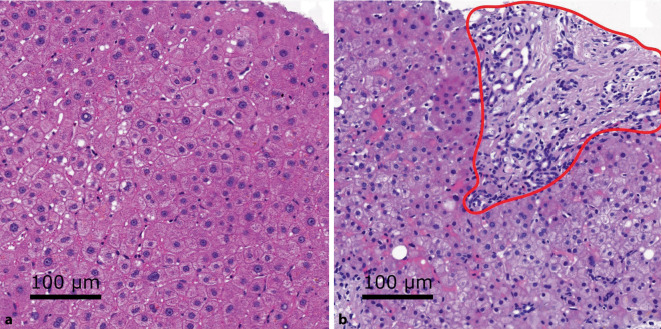 Figure 2
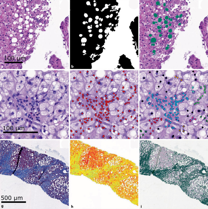 Figure 3
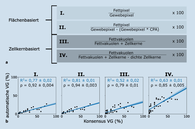 Figure 4
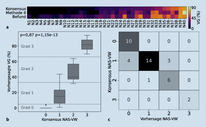 Figure 5
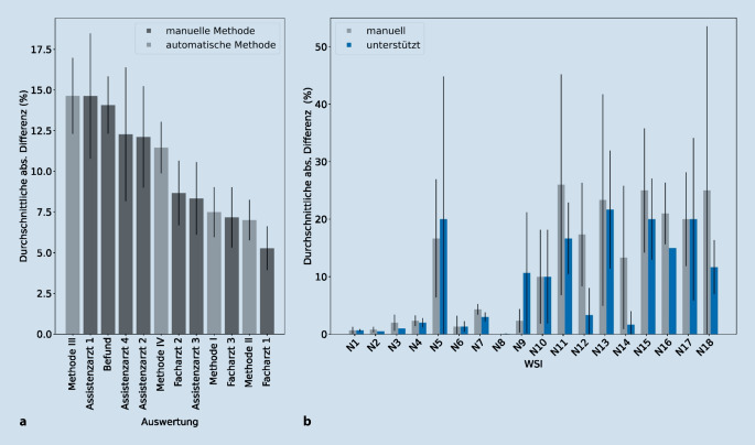 Figure 6
 Figure 7
Peer Reviews
No public reviews on file for this paper yet. If you reviewed it on a platform where reviews are public (OpenReview, ICLR, NeurIPS, ICML), you can paste yours below so the community can read it here.
Videos
No videos yet. Explain this paper in a talk, walkthrough, or lecture? Add one.
Taxonomy
TopicsColorectal Cancer Screening and Detection · Radiomics and Machine Learning in Medical Imaging · COVID-19 diagnosis using AI