Chronic hepatitis B virus infection deteriorates disease outcome of coronavirus disease 2019 in hamster
Lunzhi Yuan, Xuan Liu, Yi Guan, Tong Cheng, Ningshao Xia

Abstract
Genes, proteins, chemicals, diseases, species, mutations and cell lines named across the full text — each resolved to its canonical identifier and authoritative record.
Click any figure to enlarge with its caption.
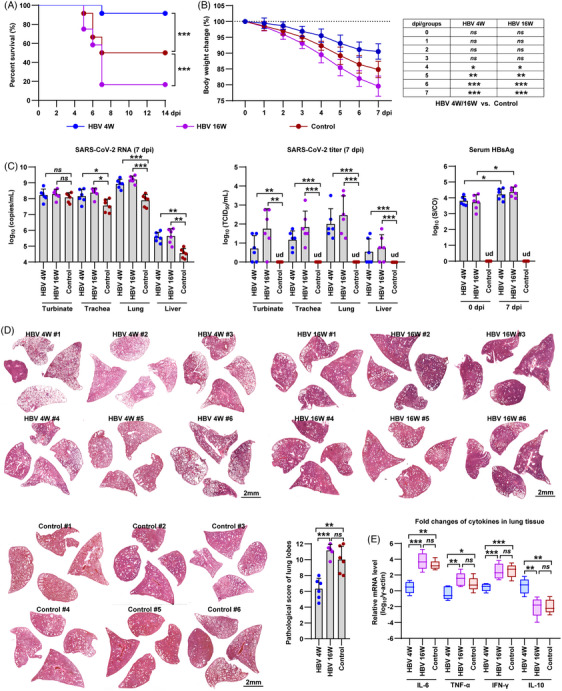 Figure 1
- —National Natural Science Foundation of China 10.13039/501100001809
- —Guangdong Government
- —Shenzhen Science and Technology Program
Peer Reviews
No public reviews on file for this paper yet. If you reviewed it on a platform where reviews are public (OpenReview, ICLR, NeurIPS, ICML), you can paste yours below so the community can read it here.
Videos
No videos yet. Explain this paper in a talk, walkthrough, or lecture? Add one.
Taxonomy
TopicsHepatitis B Virus Studies · Liver Disease Diagnosis and Treatment · Hepatitis C virus research
Dear Editor,
The pandemic of severe acute respiratory syndrome coronavirus 2 (SARS‐CoV‐2) is widely recognized as a global public health crisis. Over the past three years, SARS‐CoV‐2 has been mutating and resulting in varying degrees of coronavirus disease 2019 (COVID‐19), characterized by symptoms such as fever, cough, weakness, severe pneumonia, and even death. After the pandemic waves of the Delta and Omicron sublineages, the XBB variants of SARS‐CoV‐2 are still emerging in local areas. It has been reported that patients with chronic liver diseases, including those with chronic hepatitis B virus (HBV) infection,1 are at a higher risk of experiencing poor outcomes from COVID‐19. However, due to the lack of clinical samples and appropriate animal models, our understanding of various aspects of SARS‐CoV‐2 infection and the progression of COVID‐19 in patients with chronic HBV infection (CHB) remains incomplete. Therefore, in this study, we aimed to replicate the dual infection of HBV and SARS‐CoV‐2 in a sensitive rodent model, namely the hamster. We infected 4‐week‐old male hamsters with AAV‐HBV (1×10^9^ vg per hamster) to establish persistent viremia of HBV surface antigen (HBsAg) (Figure S1).
Subsequently, we mimicked different stages of chronic HBV infection in patients by intranasally infecting the hamsters with 1×10^4^ PFU of the SARS‐CoV‐2 Delta variant at 4 weeks (4 W) and 16 weeks (16 W) after HBV infection. The control group consisted of hamsters infected solely with SARS‐CoV‐2. Interestingly, among the hamsters, six out of 12 control hamsters, one out of 12 HBV 4 W hamsters, and 10 out of 12 HBV 16 W hamsters died within 14 days post‐infection (dpi) with SARS‐CoV‐2 (Figure 1A). Furthermore, at 7 dpi, the control, HBV 4 W, and HBV 16 W hamsters exhibited body weight losses of 15.13 ± 2.59%, 9.45 ± 2.47%, and 20.41 ± 3.18%, respectively (Figure 1B). These findings suggest that HBV 4 W and HBV 16 W hamsters developed contrary disease outcomes following SARS‐CoV‐2 infection.
We proceeded to examine the presence of SARS‐CoV‐2 RNA and its titer in the nasal turbinate, trachea, lung, and liver of the hamsters sacrificed at 7 days post‐infection (dpi). The results obtained through RT‐PCR revealed higher levels of SARS‐CoV‐2 RNA in both the HBV 4 W and HBV 16 W hamsters compared to the control hamsters at 7 dpi (Figure 1C, left). Additionally, titration results indicated the absence of detectable infectious SARS‐CoV‐2 particles in the nasal turbinate, trachea, lung, and liver of the control hamsters at 7 dpi, while a significant viral titer was observed in more than 50% of the HBV 4 W and HBV 16 W hamsters at the same time point (Figure 1C, middle). Notably, there was a substantial variation in both SARS‐CoV‐2 RNA levels and titers among the tissues collected from the HBV 4 W and HBV 16 W hamster groups (Figure 1C). Additionally, SARS‐CoV‐2 infection caused a slight increase in serum HBsAg levels in both the HBV 4 W and HBV 16 W hamsters (Figure 1C, right). Interestingly, both the 4 and 16 W HBV hamsters showed diffuse expression of HBsAg in the liver tissues and delayed clearance of SARS‐CoV‐2 N protein in the lung tissues collected at 7 dpi, respectively (Figure S2).
In addition, we investigated the pathological changes in the lung tissues of hamsters collected at 7 days post‐infection (dpi) as previously described.2 Based on the results of hematoxylin and eosin (H&E) staining of fixed lung lobes, the HBV 16 W hamsters exhibited more severe lung injury compared to the control hamsters, while the HBV 4 W hamsters exhibited milder lung injury than the other two groups (Figure 1D). In addition, we quantified the severity of lung pathology using a comprehensive pathological scoring system that included factors such as alveolar septal thickening and consolidation, hemorrhage, exudation, pulmonary edema and mucus, and inflammatory cell recruitment and infiltration in all lobes of the hamster lungs. The control, HBV 4 W, and HBV 16 W hamsters had mean comprehensive pathologic scores of 10.00 ± 1.69, 6.33 ± 1.32, and 11.17 ± 0.77, respectively (Figure 1D and Table S1). Furthermore, we evaluated the relative mRNA levels of representative cytokines in the lung and liver tissues collected at 7 dpi. Notably, the HBV 4 W hamsters exhibited a significant decrease in proinflammatory cytokines, including interleukin 6 (IL‐6), tumor necrosis factor‐alpha (TNF‐α), and interferon‐gamma (IFN‐γ), and an increase in the anti‐inflammatory cytokine IL‐10 (Figure 1E, Figure S3, and Table S2). In contrast, HBV 16 W hamsters did not show significant changes in the mRNA levels of these cytokines compared to the control group (Figure 1E).
In conclusion, the presence of persistent HBsAg viremia delays viral clearance of SARS‐CoV‐2 in the hamster model. The results of survival rate, body weight changes, pathological changes, and cytokine mRNA levels in lung tissues indicate that long‐term persistence of HBsAg viremia (HBV 16 W) is likely to worsen the disease outcome of COVID‐19 in the hamster model, whereas short‐term persistence of HBsAg viremia (HBV 4 W) may ameliorate the disease severity. Chronic hepatitis B (CHB) typically disrupts innate and adaptive immune responses, and the immunopathophysiological process is complex.3 Based on the immunological characteristics of CHB and COVID‐19 patients and,4, 5 we hypothesized that an exaggerated innate immune response and an inflammatory immune microenvironment in the later stages of CHB contribute to poor disease outcome after SARS‐CoV‐2 co‐infection. Conversely, the early stages of CHB are characterized by immunosuppression, which may result in an inadequate innate immune response and the production of proinflammatory cytokines. Therefore, targeting the abnormal immune response and organ failure are key steps to reserve the complicated pathology progress of SARS‐CoV‐2 co‐infection in CHB patients. For example, intravenous injection of mesenchymal stem cells has been demonstrated available to down‐regulate the severity of liver failure, pneumonia, and cytokine storm. Meanwhile, a combined antiviral treatment might enhance the therapeutic effect of immunotherapy. Overall, Further animal studies and analysis of clinical specimens are needed to validate our hypothesis.
To summarize, this study provides a highly representative animal model of SARS‐CoV‐2 infection in CHB patients and highlights the long‐term persistence of HBsAg viremia as a risk factor. These findings may provide valuable insights into understanding the immunopathophysiological mechanisms underlying HBV/SARS‐CoV‐2 co‐infection and inform clinical treatment strategies.
AUTHOR CONTRIBUTIONS
Y.L.Z. and L.X. contributed equally to this study. Y.L.Z. and L.X. performed the cell and animal experiments. Y.L.Z. and L.X. wrote the manuscript. G.Y., C.T., and N.S.X. supervised the study. All authors have read and approved the final manuscript.
CONFLICT OF INTEREST STATEMENT
The authors declare no conflict of interest.
FUNDING INFORMATION
This work was supported by grants from the National Natural Science Foundation of China (82002139; 32201152), Funding support from the Guangdong Government (HZQB‐KCZYZ‐2021014; 190824215544727), Shenzhen Science and Technology Program (JCYJ20220530143407016).
ETHICS STATEMENT
In this study, the virus and animal studies were approved by the ethics committee of the Guangdong‐Hong Kong Joint Laboratory of Emerging Infectious Diseases/Joint Laboratory for International Collaboration in Virology and Emerging Infectious Diseases (Key Laboratory of Ministry of Education), Joint Institute of Virology (Shantou University/The University of Hong Kong) (Approval number: SUMC2022‐051).
Supporting information
Supporting information
The reference list from the paper itself. Each links out to its DOI / PubMed record.
- 1Marjot T , Moon AM , Cook JA , et al. Outcomes following SARS‐Co V‐2 infection in patients with chronic liver disease: an international registry study. J Hepatol. 2021;74(3):567‐577.33035628 10.1016/j.jhep.2020.09.024PMC 7536538 · doi ↗ · pubmed ↗
- 2Yuan L , Zhu H , Zhou M , et al. Nasal irrigation efficiently attenuates SARS‐Co V‐2 Omicron infection, transmission and lung injury in the Syrian hamster model. i Science. 2022;25(12):105475.36338435 10.1016/j.isci.2022.105475 PMC 9625844 · doi ↗ · pubmed ↗
- 3Iannacone M , Guidotti LG . Immunobiology and pathogenesis of hepatitis B virus infection. Nat Rev Immunol. 2022;22(1):19‐32.34002067 10.1038/s 41577-021-00549-4 · doi ↗ · pubmed ↗
- 4Seto WK , Lo YR , Pawlotsky JM , Yuen MF . Chronic hepatitis B virus infection. Lancet. 2018;392(10161):2313‐2324.30496122 10.1016/S 0140-6736(18)31865-8 · doi ↗ · pubmed ↗
- 5Merad M , Blish CA , Sallusto F , Iwasaki A . The immunology and immunopathology of COVID‐19. Science. 2022;375(6585):1122‐1127.35271343 10.1126/science.abm 8108 PMC 12828912 · doi ↗ · pubmed ↗