Crucial role of TFAP2B in the nervous system for regulating NREM sleep
Ayaka Nakai, Mitsuaki Kashiwagi, Tomoyuki Fujiyama, Kanako Iwasaki, Arisa Hirano, Hiromasa Funato, Masashi Yanagisawa, Takeshi Sakurai, Yu Hayashi

TL;DR
This study shows that the TFAP2B gene in the nervous system is important for regulating NREM sleep and circadian rhythms in mice.
Contribution
The study identifies the specific role of TFAP2B in neurons for sleep regulation and reveals its impact on circadian clock function.
Findings
Nervous system-specific deletion of Tfap2b in mice caused reduced NREM sleep and severe sleep phenotypes.
Postnatal neuronal deletion of Tfap2b also reduced NREM sleep but with less severe effects.
Defective light entrainment of the circadian clock and stereotypic jumping behavior were observed in mice with nervous system-specific Tfap2b deletion.
Abstract
The AP-2 transcription factors are crucial for regulating sleep in both vertebrate and invertebrate animals. In mice, loss of function of the transcription factor AP-2β (TFAP2B) reduces non-rapid eye movement (NREM) sleep. When and where TFAP2B functions, however, is unclear. Here, we used the Cre-loxP system to generate mice in which Tfap2b was specifically deleted in the nervous system during development and mice in which neuronal Tfap2b was specifically deleted postnatally. Both types of mice exhibited reduced NREM sleep, but the nervous system-specific deletion of Tfap2b resulted in more severe sleep phenotypes accompanied by defective light entrainment of the circadian clock and stereotypic jumping behavior. These findings indicate that TFAP2B in postnatal neurons functions at least partly in sleep regulation and imply that TFAP2B also functions either at earlier stages or in…
Genes, proteins, chemicals, diseases, species, mutations and cell lines named across the full text — each resolved to its canonical identifier and authoritative record.
Click any figure to enlarge with its caption.
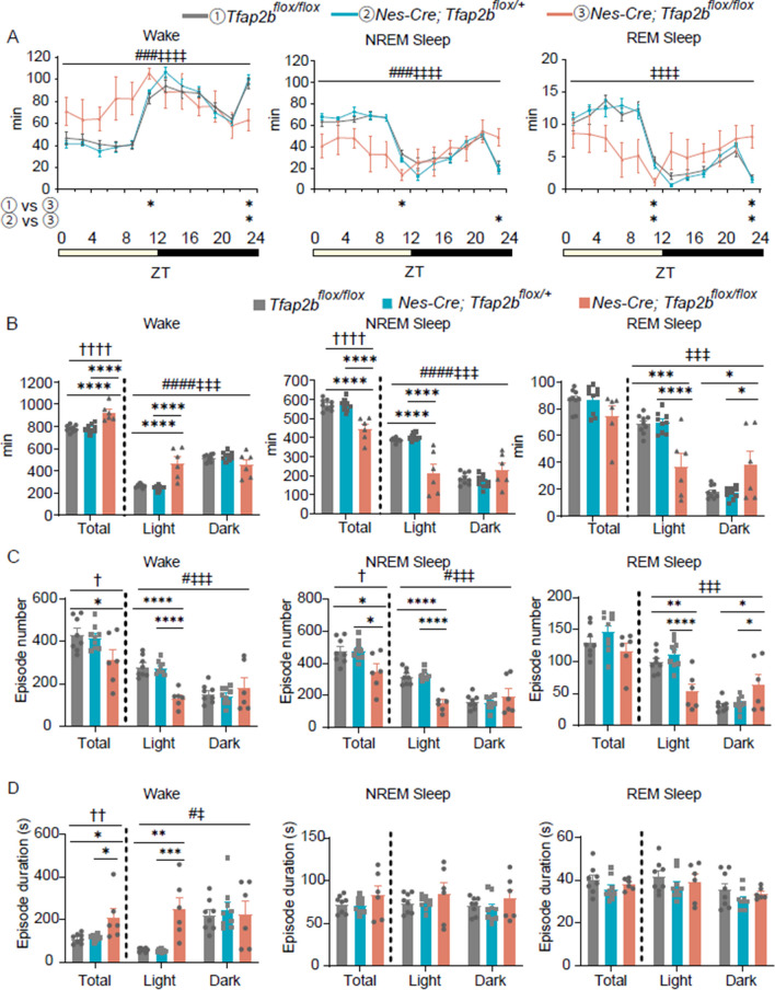 Figure 1
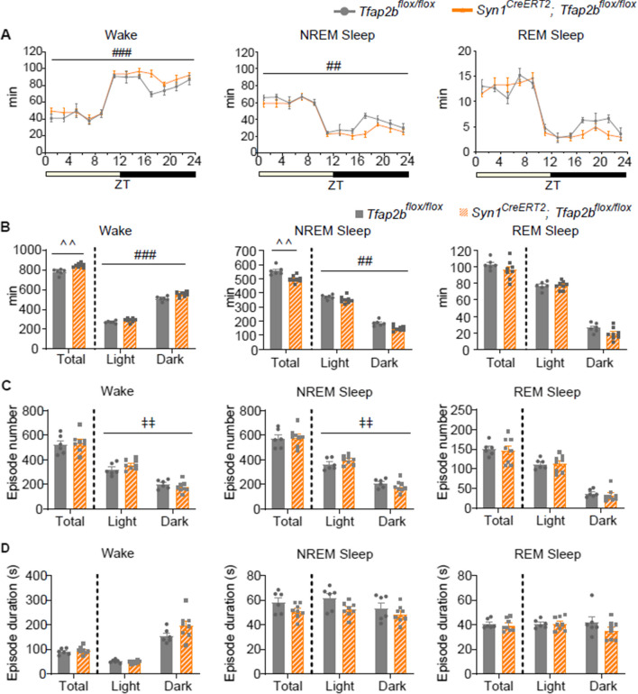 Figure 2
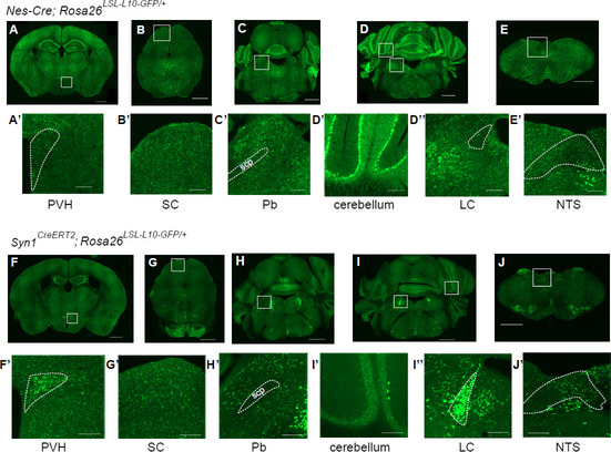 Figure 3
- —http://dx.doi.org/10.13039/501100002241Japan Science and Technology Agency
- —http://dx.doi.org/10.13039/100009619Japan Agency for Medical Research and Development
- —http://dx.doi.org/10.13039/501100001691Japan Society for the Promotion of Science
- —http://dx.doi.org/10.13039/100007684Asahi Glass Foundation
- —Kao Foundation for Research on Health Science
Peer Reviews
No public reviews on file for this paper yet. If you reviewed it on a platform where reviews are public (OpenReview, ICLR, NeurIPS, ICML), you can paste yours below so the community can read it here.
Videos
No videos yet. Explain this paper in a talk, walkthrough, or lecture? Add one.
Taxonomy
TopicsSleep and Wakefulness Research · Circadian rhythm and melatonin · Photoreceptor and optogenetics research
Introduction
Sleep is an evolutionarily conserved state that is crucial for maintaining health in humans [1]. The exact functions and mechanisms of sleep, however, remain elusive. In humans, there are reports of natural short sleepers whose reduced sleep does not lead to daytime fatigue [2]. In cases with an inherited natural short sleep trait, identification and analyses of the causal gene are expected to provide clues to the molecular mechanisms of sleep.
In the present study, we focused on the mouse transcription factor AP-2β (TFAP2B). In humans, partial mutations in TFAP2B cause Char syndrome, which is characterized by morphologic abnormalities of the face, limbs, and heart [3, 4]. Two human kindreds with Char syndrome self-reported abnormalities in sleep, namely parasomnia (sleep-walking) and short sleep, respectively [5]. In nematodes, the orthologous transcription factor APTF-1 is critical for specification of the sleep-promoting RIS neuron, and aptf-1 mutants exhibit severely reduced sleep [6–8]. In fruit flies, knockdown of neuronal TFAP2 results in reduced nighttime sleep [9]. In mammals, homozygous Tfap2b-knock out (KO) mice die shortly after birth [10, 11] whereas heterozygous knockout or partial mutations in mouse Tfap2b lead to reduced or fragmented non-rapid eye movement (NREM) sleep [12, 13]. This phenotype is partly explained by the roles of TFAP2B in GABAergic neurons [14], but considering the rather mild effects on NREM sleep of the conditional knockdown (cKD) of Tfap2b in GABAergic neurons, the entire picture of the mechanisms by which mouse TFAP2B functions to regulate NREM sleep are unclear. During embryogenesis, Tfap2b is expressed in neural crest cells, which generate various cell types, and in the midbrain and hindbrain, where it contributes to the specification of various neurons, including noradrenergic neurons in the locus coeruleus (LC) [15, 16]. Postnatally, Tfpa2b is expressed in the paraventricular hypothalamic nucleus (PVH), superior colliculus (SC), parabrachial nucleus (Pb), cerebellum, LC, and nucleus of the solitary tract (NTS), some of which are regions involved in sleep regulation [12]. It is unknown whether TFAP2B acts during development or postnatally in sleep regulation. Here, using the Cre-loxP system, we tested the effect of homozygous deletion of Tfap2b in either the nervous system during development or in neurons postnatally.
Results
Nervous system-specific Tfap2b deletion induces decreased NREM sleep and increased wakefulness
To elucidate whether TFAP2B functions in the nervous system or other tissues in sleep regulation, we used Nes-Cre mice to delete Tfap2b in a nervous system-specific manner. In Nes-Cre mice, Cre recombinase is expressed in neuronal and glial cell precursors of the central and peripheral nervous systems [17]. Nes-Cre mice were crossed with Tfap2b^flox/flox^ mice, in which the Tfap2b exon 3 is flanked by loxP. We confirmed that Tfap2b exon 3 is deleted in the genomic DNA of the brain in a Nes-Cre-dependent manner (Additional file 1: Fig. S1A). Homozygous Tfap2b KO mice die shortly after birth [10, 11], whereas the nervous system-specific homozygous Tfap2b-conditionally knocked down (cKD) (Nes-Cre; Tfap2b^flox/flox^) mice lived to adulthood. However, when we crossed Nes-Cre; Tfap2b^flox/+^ mice with Tfap2b^flox/flox^ mice to obtain the cKD mice, only 19 out of 176 pups were cKD, which is lower than the expected ratio of 1/4, implying that some of the cKD mice died either prenatally or immediately after birth. The Nes-Cre; Tfap2b^flox/flox^ male mice had a lower body weight than control male mice (Tfap2b^flox/+^, Tfap2b^flox/flox^, or Nes-Cre; Tfap2b^flox/+^ mice; Additional file 1: Fig. S1B). We compared the sleep architecture between Nes-Cre; Tfap2b^flox/flox^ and control male mice (Fig. 1). Nes-Cre; Tfap2b^flox/flox^ mice exhibited increased wakefulness and decreased NREM sleep compared with control mice (Fig. 1B). Decreased NREM sleep could be attributed to a decrease in the episode number (Fig. 1C), whereas the episode duration seemed unaffected (Fig. 1D). In contrast, increased wakefulness could be attributed to a longer episode duration (Fig. 1D), whereas the episode number was rather decreased (Fig. 1C), suggesting that episodes of wakefulness are highly consolidated. When light and dark phases were separately analyzed in Nes-Cre; Tfap2b^flox/flox^ mice, NREM sleep was largely decreased in the light phase with the amount being comparable between the light and dark phases (Fig. 1B). A similar trend was observed for wakefulness, implying that Nes-Cre; Tfap2b^flox/flox^ mice lack typical daily fluctuations in the amount of sleep/wake time (Fig. 1B). This trend was even more obvious in rapid eye movement (REM) sleep; while the total amount of REM sleep was unaffected, the amount of REM sleep in the light phase was decreased, whereas that in the dark phase was increased (Fig. 1B). Analyses of the bi-hourly changes in the sleep–wake cycle (Fig. 1A) also revealed a similar trend; the amount of sleep or wakefulness seemed to lack daily oscillations, and when compared with control mice, the amounts of NREM and REM sleep were decreased at some time points in the light phase and increased at some time points in the dark phase, and vice versa for wakefulness (Fig. 1A). To further address whether Nes-Cre; Tfap2b^flox/flox^ mice exhibit circadian rhythm deficits, we analyzed locomotor activity detected with an infrared sensor. Each individual control mouse exhibited high locomotor activity in the dark phase (Additional file 1: Fig. S2A, B). By contrast, while individual Nes-Cre; Tfap2b^flox/flox^ mice seemed to have a daily locomotor activity rhythm, the start of the active phase varied among individuals and seemed random (Additional file 1: Fig. S2C), suggesting that the circadian clock is not entrained by external light–dark cycles in these mice. When we defined the active phase and inactive phase based on locomotor activity (see Materials and methods for details), Nes-Cre; Tfap2b^flox/flox^ mice appeared to have reduced NREM sleep during both the active and inactive periods, whereas REM sleep seemed unaffected (Additional file 1: Fig. S3), providing further support that the sleep deficits in these mice result from a combination of reduced NREM sleep, increased wakefulness, and abnormal light entrainment of the circadian clock. In addition, when the behavior was monitored with a video camera, Nes-Cre; Tfap2b^flox/flox^ mice frequently displayed stereotypic behaviors such as repeated jumping or climbing (Additional file 2: Video 1). In the electrogram (EEG) power spectrum, Nes-Cre; Tfap2b^flox/flox^ mice exhibited an increase in the EEG power at approximately 5.5–17 Hz during wakefulness, whereas no obvious defects were detected during sleep (Additional file 1: Fig. S4).Fig. 1. Comparison of the sleep architecture in nervous system-specific Tfap2b cKD and control mice. A Bi-hourly amount of wakefulness, NREM sleep, and REM sleep across 24 h in male mice. B Total amount of wakefulness, NREM sleep, and REM sleep during 24 h, light phase, and dark phase in male mice. N = 9 Tfap2b^flox/flox^ mice (grey), N = 9 Nes-Cre; Tfap2b^flox/+^ mice (blue),* N* = 6 Nes-Cre; Tfap2b^flox/flox^ mice (pink). C, D Episode numbers (C) and durations (D) of wakefulness, NREM sleep, and REM sleep during 24 h, light phase, and dark phase.* N* = 8 Tfap2b^flox/flox^ mice (grey), N = 9 Nes-Cre; Tfap2b^flox/+^ mice (blue), N = 6 Nes-Cre; Tfap2b^flox/flox^ mice (pink). ^#^ and ^‡^ indicate a significant main effect of genotype and a significant interaction between genotype and time, respectively, in 2-way repeated-measures ANOVA (^#^, ^‡^p < 0.05, ^###^, ^‡‡‡^p < 0.001, ^####^, ^‡‡‡‡^p < 0.0001). ^†^ indicates significance in the 1-way ANOVA (^†^p < 0.05, ^††^p < 0.01, ^††††^p < 0.0001). * indicates significance in the post-hoc Bonferroni multiple comparison test (*p < 0.05, **p < 0.01, ***p < 0.001, ****p < 0.0001). Data are presented as mean ± SEM
Postnatal neuron-specific Tfap2b deletion induces decreased NREM sleep and increased wakefulness
To test whether TFAP2B in neurons acts postnatally in sleep regulation, we crossed Tfap2b^flox/flox^ mice with Syn1^CreERT2^ mice, which express CreERT2 specifically in neurons [18]. To delete Tfap2b in neurons postnatally, we administered tamoxifen to Syn1^CreERT2^; Tfap2b^flox/flox^ mice at postnatal day (P) 14, 17, and 21. We confirmed that Tfap2b exon 3 is deleted in the genomic DNA of the brain in a Syn1^CreERT2^-dependent manner (Additional file 1: Fig. S5A). These Syn1^CreERT2^; Tfap2b^flox/flox^ mice survived to adulthood and exhibited no significant difference in body weight (Additional file 1: Fig. S5B). Repetitive jumping or climbing behaviors were not observed (Additional file 3: Video 2). We compared the sleep architecture between Syn1^CreERT2^; Tfap2b^flox/flox^ and Tfap2b^flox/flox^ male mice administered with tamoxifen (at P14, 17, and 21) (Fig. 2). Syn1^CreERT2^; Tfap2b^flox/flox^ mice showed increased wakefulness and decreased NREM sleep (Fig. 2A, B), similar to Nes-Cre; Tfap2b^flox/flox^ mice, although the phenotype seemed milder. Perhaps due to the mild phenotype, episode numbers and durations were not significantly affected (Fig. 2C, D). When light and dark phases were analyzed separately, the amount of wakefulness and NREM sleep seemed to be affected throughout both phases (Fig. 2B). Unlike Nes-Cre; Tfap2b^flox/flox^ mice, the circadian rhythm seemed normal, with a high amount of wakefulness during the dark phase and a low amount during the light phase, and vice versa for NREM and REM sleep (Fig. 2A, B). EEG power spectra during wakefulness, NREM sleep, or REM sleep appeared unaffected (Additional file 1: Fig. S6). We also tested whether sleep homeostasis is normal in these mice. Mice were subjected to 5 h of sleep deprivation (SD) at the beginning of the light phase. Both Syn1^CreERT2^; Tfap2b^flox/flox^ mice and Tfap2b^flox/flox^ mice showed an increase in NREM sleep during the following 7 h of the light phase after SD (Additional file 1: Fig. S7A, B). Sleep architecture and EEG power spectra were also similar (Additional file 1: Fig. S7C-E), suggesting that the Syn1^CreERT2^; Tfap2b^flox/flox^ mice exhibit a normal response to SD. Importantly, a previous study showed that the Syn1^CreERT2^ allele does not affect the amount of wakefulness, NREM sleep, and REM sleep [18]. However, we cannot rule out the possibility that it might have had some effect on the sleep architecture or the EEG power spectra, although these factors seemed not to differ between Syn1^CreERT2^; Tfap2b^flox/flox^ and Tfap2b^flox/flox^ mice.Fig. 2. Comparison of the sleep architecture in postnatal neuron-specific Tfap2b cKD and control mice. A Bi-hourly amount of wakefulness, NREM sleep, and REM sleep across 24 h in tamoxifen-injected (at P14, 17, and 21) male mice. B Total amount of wakefulness, NREM sleep, and REM sleep during 24 h, light phase, and dark phase in tamoxifen-injected (at P14, 17, and 21) male mice. C, D Episode numbers (C) and durations (D) of wakefulness, NREM sleep, and REM sleep during 24 h, light phase, and dark phase in tamoxifen-injected (at P14, 17, and 21) male mice. N = 6 Tfap2b^flox/flox^ mice (grey), N = 8 Syn1^CreERT2^; Tfap2b^flox/flox^ mice (orange). ^#^ and ^‡^ indicate a significant main effect of genotype and significant interaction between genotype and time, respectively, in 2-way repeated-measures ANOVA (^##,‡‡^p < 0.01, ^###^p < 0.001). ^ indicates significance in the Welch’s test (^^ p < 0.01). Data are presented as mean ± SEM
Differential patterns of recombination in Nes-Cre and Syn1CreERT2 mice.
To reveal the Cre-mediated recombination patterns in Nes-Cre and Syn1-CreERT2 mice, we crossed these mice with Rosa26^LSL-L10-GFP^ Cre-reporter mice. In the mature brain, TFAP2B is expressed in the PVH, SC, Pb, cerebellum, LC, and NTS [12]. Within these areas, Nes-Cre; Rosa26^LSL-L10-GFP/+^ mice had dense GFP signals in the SC, Pb, cerebellum, and NTS, whereas GFP signals were sparse in the PVH and almost undetectable in the LC (Fig. 3A–E’). Syn1^CreERT2^; Rosa26^LSL-L10-GFP/+^ mice injected with tamoxifen (at P14, 17, and 21) had dense GFP signals in the PVH, SC, Pb, LC, and NTS, whereas GFP signals were sparse in the cerebellum (Fig. 3F–J’). GFP signals in the LC mostly overlapped with tyrosine hydroxylase, suggesting that the recombination occurred in noradrenergic neurons (Additional file 1: Fig. S8).Fig. 3. Comparison of the Cre-mediated recombination patterns in Nes-Cre and Syn1^CreERT2^ mice. (A–J’) Images of areas where GFP signals were detected in Nes-Cre; Rosa26^LSL−L10−GFP/+^ mice (A–E’) and tamoxifen-injected (at P14, 17, and 21) Syn1^CreERT2^; Rosa26^LSL−L10−GFP/+^ mice (F–J’). PVH, paraventricular hypothalamic nucleus (A’, F’); SC, superior colliculus (B’, G’); Pb, parabrachial nucleus (C’, H’); cerebellum (D’, I’), LC, locus coeruleus (D’’, I’’); NTS, solitary tract nucleus (E’, J’), scp, superior cerebellar peduncle. Scale bars in (A–J), 1 mm; scale bars in (A’–J’), 200 µm. Representative images from a replicate of N = 2 Nes-Cre; Rosa26^LSL−L10−GFP/+^ mice, N = 4 Syn1^CreERT2^; Rosa26^LSL−L10−GFP/+^ mice are shown
Discussion
Heterozygous deletion of Tfap2b leads to reduced NREM sleep [12]. The present study showed that homozygous deletion of Tfap2b in the nervous system during development leads to a large reduction in NREM sleep. Moreover, homozygous deletion of Tfap2b in neurons postnatally reduced NREM sleep, although to a milder extent. These findings indicate that TFAP2B in postnatal neurons at least partly functions in sleep regulation and imply that TFAP2B also functions either at earlier stages or in additional cell types.
Consistent with previous studies [19], in Nes-Cre mice, Cre recombination was not detected in various brain regions. Within areas that postnatally express Tfap2b, Cre recombination was detected in the SC in both Nes-Cre and Syn1^CreERT2^ mice. GABAergic neurons in the SC regulate sleep by inhibiting dopaminergic neurons in the ventral tegmental area [20], and loss of function in these neurons might be partly responsible for the reduced NREM sleep in both Syn1^CreERT2^; Tfap2b^flox/flox^ and Nes-Cre; Tfap2b^flox/flox^ mice. This is consistent with a recent study demonstrating that deletion of Tfap2b specifically in GABAergic neurons leads to reduced NREM sleep [14]. The effect of deleting Tfap2b in GABAergic neurons is limited, however, suggesting that TFAP2B in other neuronal types also functions in sleep regulation. Among areas that postnatally express Tfap2b, Cre recombination was also detected in the NTS. Glutamatergic neurons in the NTS promote NREM sleep [21], and loss of function in these neurons might also contribute to reduce NREM sleep. By contrast, among areas that postnatally express Tfap2b, Cre recombination was almost undetectable in the LC in Nes-Cre mice. TFAP2B is required for the synthesis of noradrenaline in the LC [22], which in turn is crucial for maintaining wakefulness [23]. Thus, the milder reduction of NREM sleep in Syn1^CreERT2^; Tfap2b^flox/flox^ mice compared with Nes-Cre; Tfap2b^flox/flox^ mice might be partly due to the additional functional loss of LC noradrenergic neurons in the Syn1^CreERT2^; Tfap2b^flox/flox^ mice, which might have counteracted the NREM sleep-reducing effect of deleting Tfap2b. Another possibility might be that TFAP2B also functions in glial cells or neural progenitor cells to regulate sleep and that deletion of Tfap2b in these cells are responsible for the more severe sleep phenotypes of Nes-Cre; Tfap2b^flox/flox^ mice compared with Syn1^CreERT2^; Tfap2b^flox/flox^ mice. Indeed, homozygous Tfap2b KO affects expression of genes involved in glial cell differentiation [14]. These possibilities should be tested in the future using mice that express Cre specifically in either neurons or glial cells.
Nes-Cre; Tfap2b^flox/flox^ mice had prolonged wake episodes, during which they exhibited stereotypic behaviors, including repeated jumping and climbing. Similar stereotypic behaviors are observed in mice with knockout of dopamine transporter 1 (Dat1) and those with knockout of SH3 and multiple ankyrin repeat domains 2 (Shank2) [24, 25] and are thought to share properties with human stereotypic behaviors observed in various psychiatric or developmental disorders [26]. This phenotype is likely due to loss of Tfap2b either during the fetal period or in glial cells as such abnormal behavior was not observed in Syn1^CreERT2^; Tfap2b^flox/flox^ mice.
In addition, Nes-Cre; Tfap2b^flox/flox^ mice exhibited a deficit in light entrainment of the circadian rhythm. Intrinsically photosensitive retinal ganglion cells (ipRGCs) function as photosensors for this light entrainment [27]. Within the retina, TFAP2B is crucial for the development of horizontal and amacrine cells [28, 29], and considering the deficits in light entrainment, TFAP2B might also have a role in the development of ipRGCs.
Whereas Syn1^CreERT2^; Tfap2b^flox/flox^ mice showed reduced basal NREM sleep, they responded normally to SD with increased amounts of NREM sleep in the subsequent hours. Thus, TFAP2B might not be required in postnatal neurons for the homeostatic regulation of sleep, but TFAP2B in other cell types may still play a role. We could not conduct a similar SD experiment with Nes-Cre; Tfap2b^flox/flox^ mice because the beginning of the inactive phase varied among individual mice.
This study showed that postnatal neurons require TFAP2B to regulate sleep. To further narrow down the brain regions and neuronal subtypes in which Tfap2b is involved in regulating sleep, additional studies using region-specific Cre-driver lines or local microinjection of adeno-associated viral vectors carrying Cre are required. Once the responsible populations of neurons are identified, further studies using single-cell RNA sequencing may identify the target genes of TFAP2B that are required for regulating NREM sleep and contribute to a deeper understanding of the molecular mechanisms of sleep.
Materials and methods
Mice
All animal experiments were approved by the institutional animal care and use committee of the University of Tsukuba. All animals were maintained according to the institutional guidelines of the animal facilities of the Laboratory of Animal Resource Center, University of Tsukuba. Mice were maintained and bred within the International Institute for Integrative Sleep Medicine under a 12-h light/dark cycle and with free access to food and water. Male Nes-Cre; Tfap2b^flox/flox^, Syn1^CreERT2^; Tfap2b^flox/flox^, and corresponding control mice aged 10–13 weeks were used for sleep analyses. To obtain Tfap2b cKD mice, Tfap2b^tm1a(EUCOMM)Wtsi/+^ mice were crossed with mice carrying the CAG-flp allele [30] to remove a genomic DNA fragment containing IRES:lacZ and a neomycin-resistant cassette flanked by FRT. Homozygous floxed Tfap2b exon 3 (Tfap2b^flox/flox^) mice were mated to mice carrying the Nes-Cre transgene [17] or the Syn1^CreERT2^ allele [18] to induce Cre-mediated recombination in a nervous system-specific manner or a neuron-specific manner, respectively.
To confirm the recombination sites in the Nes-Cre and Syn1^CreERT2^ mice, Nes-Cre and Syn1^CreERT2^ mice were mated with the GFP reporter strain B6.129S4-Gt (ROSA)26Sor^tm1(CAG-EGFP/Rpl10a-birA) Wtp/J^ (Rosa26^LSL-L10-GFP^, Jackson Laboratory ; #022367) [31].
To induce CreERT2-mediated recombination, 500 μg of tamoxifen (Toronto Research Chemicals Inc.; T006000) dissolved in corn oil was administered intraperitoneally at P14, P17, and P21.
Genotyping
Genotyping of mice carrying Tfap2b^flox^, Nes-Cre, Syn1^CreERT2^, or Rosa26^LSL-L10-GFP^ was performed using the following primers. Tfap2b^flox^: 5′-AAGGCGCATAACGATACCAC-3′, 5′-CCGCCTACTGCGACTATAGAGA-3′, 5′-GACATCCTACAATGCACAGCT-3′, and 5′- TTGCTGTGAGCTAAGAGCTTC-3′ (Tfap2b^flox^: 218 bp, Tfap2b^+^: 381 bp), Nes-Cre: 5′-GACGATGCAACGAGTGATGA-3′ and 5′-AGCATTGCTGTCACTTGGTC-3′ (Nes-Cre: 300 bp), Syn1^CreERT2^: 5′-TGCCTCCACCTTGTCTCTCT-3′, 5′-AACAAAGGCATGGAGCATCT-3′, and 5′-GATCTGGAGGTGACCAGGAA-3′ (Syn1^CreERT2^: 443 bp, Syn1^+^: 387 bp), Rosa26^LSL-L10-GFP^: 5′-AAGGGAGCTGCAGTGGAGTA-3′, 5′-CCGAAAATCTGTGGGAAGTC-3′, 5′-AAGATCCGCCACAACATCG-3′, and 5′-TTCTCGTTGGGGTCTTTGCT-3′ (Rosa26^LSL-L10-GFP^: 146 bp, Rosa26^+^: 297 bp).
PCR using genomic DNA extracted from brain
Each brain was harvested and divided in half at the midline and frozen with liquid nitrogen. Genomic DNA was extracted using ISOSPIN Tissue DNA Kit (NIPPON GENE; 316-08891). PCR was performed using the following primers. Tfap2b^−^: 5′-AAGGCGCATAACGATACCAC-3′ and 5′-ACTGATGGCGAGCTCAGACC-3′ (Tfap2b^−^: 174 bp, Tfap2b^flox^: 1024 bp), Chn1: 5′-AGGGCTTTCCTTGCTGTGTC-3′ and 5′-TAGGTCCCTTCTCATGAACC-3′ (Chn1^+^: 120 bp).
EEG/ electromyogram (EMG) and locomotor recording
EEG and EMG signals were recorded from freely moving mice at the age of 10–13 weeks according to the method described in a previous study [32], with some modifications. First, 8–10 weeks-old mice were implanted with EEG/EMG electrodes. Stainless steel EEG electrodes were implanted epidurally over the cerebellum and parietal cortex, and EMG electrodes were embedded into the trapezius muscles bilaterally. Mice were allowed to recover from the surgery in their home cage for at least 4 days. Then, mice were placed in a sleep recording chamber and habituated for at least 5 days. Subsequently, EEG/EMG signals were recorded from the onset of the light phase. EEG/EMG signals were filtered (bandpass 0.5–250 Hz), collected, and digitized at a 512 Hz sampling rate using VitalRecorder (Kissei Comtec). Video and infrared signals were also recorded from the start of the light phase. Locomotor activity detected by infrared signals was used to determine the onset of the active phase, which was automatically done using a template-matching algorithm in the ClockLab software. Briefly, the onset of the active phase was defined as the start point of 5 consecutive hours of high locomotor activity (above the 20% percentile of average locomotor activity) that followed 5 consecutive hours of low locomotor activity. 12 consecutive hours from the onset of the active phase were categorized as the active phase, and the remaining 12 consecutive hours were categorized as the inactive phase.
EEG/EMG analysis
EEG/EMG data were divided into 4-s epochs (time window), and EEG data were further subjected to fast Fourier transform analyses using SleepSign (Kissei Comtec). The vigilance state in each epoch was manually classified as REM sleep, NREM sleep, or wake based on absolute delta (0.5–4 Hz) power, theta (6–10 Hz) power to delta power ratio, and the integral of EMG signals. If a single epoch contained multiple states, the state with the highest occupancy was assigned. The EEG power spectrum of each state was calculated and normalized by EEG power at 16–30 Hz averaged across 24 h. Epochs that contained multiple stages or presumable large movement-derived artifacts in the EEG data were included in the stage analysis but excluded from the EEG power spectrum analysis.
Sleep deprivation (SD)
SD was performed for 5 h during ZT0-ZT5. We monitored EEG/EMG to detect sleep. The cage was changed at the start of SD. Whenever sleep was detected, Kimwipes or marbles were placed in or removed from the cage, or the cage was gently tapped.
Histology
Deeply anesthetized mice were killed by injecting a lethal dose of anesthetic and transcardially perfused with 0.1 M phosphate-buffered saline (PBS) followed by 4% paraformaldehyde (w/v) in 0.1 M PBS or 10% formalin neutral buffer solution. The brains were postfixed in the same fixative for 1 day and subsequently equilibrated in 30% sucrose (w/v) in PBS. The brains were sectioned at 40 μm using a sliding microtome (Yamato Kohki) or a cryostat (Leica).
Immunohistochemistry
Sections were washed 3 times in tris-buffered saline with Tween20 (TBST; 50 mM Tris-HCl, pH 7.6, 150 mM NaCl, 0.05% Tween20), once in 0.3% H_2_O_2_/TBST, and 3 times again in TBST. The sections were then blocked for 30 min in blocking buffer [tris-buffered saline with 0.5% Blocking Reagent (Perkin Elmer; FP1012)]. The sections were incubated with a primary antibody [1/2000 mouse anti-tyrosine hydroxylase (Sigma; T2928)]. After washing 3 times in TBST, the sections were incubated with a secondary antibody [1/500 horseradish peroxidase-conjugated donkey anti-mouse IgG (Abcam; ab7061)] for 2 h. After washing 3 times in TBST, the sections were incubated with Tyramide Signal Amplification (TSA) plus Cyanine 5 reagent (Perkin Elmer; NEL745001KT) for 30 min. Finally, after washing 4 times in TBST, the sections were mounted on a slide glass with mounting medium (ThermoFisher; TA-006-FM). Images were captured with a digital slide scanner (Carl Zeiss; AxioScan.Z1) or a confocal microscope (Carl Zeiss; LMS800).
Quantification and statistical analysis
The experimenter was blinded to the genotype during sleep scoring. All statistical analyses were performed using Prism9 (GraphPad), and statistical significance was set at p < 0.05. Where applicable, all statistical tests were 2-tailed.
Supplementary Information
Additional file 1. Fig. S1. Confirmation of Tfap2b deletion and comparison of bodyweight in nervous system-specific Tfap2b cKD and control mice. Fig. S2. Locomotor activity of nervous system-specific Tfap2b cKD mice. Fig. S3. Comparison of the amount of sleep/wake in nervous system-specific Tfap2b cKD and control mice during the active and inactive phase determined by locomotor activity. Fig. S4. Comparison of EEG power spectra in nervous system-specific Tfap2b cKD and control mice. Fig. S5. Confirmation of Tfap2b deletion and comparison of bodyweight in postnatal neuron-specific Tfap2b cKD and control mice. Fig. S6. Comparison of EEG power spectra in postnatal neuron-specific Tfap2b cKD and control mice. Fig. S7. Comparison of the sleep architecture after sleep deprivation (SD) in postnatal neuron-specific Tfap2b cKD and control mice. Fig. S8. Analyses of the Cre-mediated recombination pattern in Syn1^CreERT2^ mice at the LC. **Additional file 2. Video 1. **Representative video of nervous system-specific Tfap2b cKD and control mice. (A-D) Video recordings of an individual Nes-Cre; Tfap2b^flox/+^ (A), Tfap2b^flox/flox^ (B), Nes-Cre; Tfap2b^flox/+^ (C), or Nes-Cre; Tfap2b^flox/flox^ (D) male mouse during the light phase.Additional file 3: Video 2. Representative video of postnatal neuron-specific Tfap2b cKD and control mice. (A, B) Video recordings of an individual Syn1^CreERT2^; Tfap2b^flox/flox^ (A) or Tfap2b^flox/flox^ (B) male mouse injected with tamoxifen (at P14, 17, and 21) during the light phase.
The reference list from the paper itself. Each links out to its DOI / PubMed record.
- 1Miyazaki S Liu C-Y Hayashi Y Sleep in vertebrate and invertebrate animals, and insights into the function and evolution of sleep Neurosci Res 201711831210.1016/j.neures.2017.04.01728501499 · doi ↗ · pubmed ↗
- 2Shi G, Wu D, Ptáček LJ, Fu YH. Human genetics and sleep behavior. Curr Opin Neurobiol. 2017; 43–9.10.1016/j.conb.2017.02.015PMC 551108328325617 · doi ↗ · pubmed ↗
- 3Davidson HRA large family with patent ductus arteriosus and unusual face J Med Genet 19933050350510.1136/jmg.30.6.5038326495 PMC 1016426 · doi ↗ · pubmed ↗
- 4Satoda M Pierpont MEM Diaz GA Bornemeier RA Gelb BD Char syndrome, an inherited disorder with patent ductus arteriosus, maps to chromosome 6p 12-p 21Circulation 1999993036304210.1161/01.CIR.99.23.303610368122 · doi ↗ · pubmed ↗
- 5Mani A Radhakrishnan J Farhi A Carew KS Warnes CA Nelson-Williams C Syndromic patent ductus arteriosus: evidence for haploinsufficient TFAP 2B mutations and identification of a linked sleep disorder Proc Natl Acad Sci 20051022975297910.1073/pnas.040985210215684060 PMC 549488 · doi ↗ · pubmed ↗
- 6Konietzka J Fritz M Spiri S Mc Whirter R Leha A Palumbos S Epidermal growth factor signaling promotes sleep through a combined series and parallel neural circuit Curr Biol 202030116.e 1310.1016/j.cub.2019.10.04831839447 · doi ↗ · pubmed ↗
- 7Turek M Lewandrowski I Bringmann H An AP 2 transcription factor is required for a sleep-active neuron to induce sleep-like quiescence in C. elegans Curr Biol 2013232215222310.1016/j.cub.2013.09.02824184105 · doi ↗ · pubmed ↗
- 8Turek M Besseling J Spies J-PKönig S Bringmann H Sleep-active neuron specification and sleep induction require FLP-11 neuropeptides to systemically induce sleep Elife 2016511810.7554/e Life.12499 PMC 480553826949257 · doi ↗ · pubmed ↗