The expression of VEGF and cyclin D1/EGFR in common primary liver carcinomas in Egypt: an immunohistochemical study
Dina Sweed, Shaymaa Sabry El Gammal, Shimaa Kilany, Shimaa Abdelsattar, Sara Mohamed Abd Elhamed

TL;DR
This study examines the expression of VEGF, cyclin D1, and EGFR in liver cancers in Egypt, finding their overexpression may influence cancer development and prognosis.
Contribution
The study identifies distinct roles of VEGF, cyclin D1, and EGFR in hepatocellular and cholangiocarcinoma liver tumors in Egyptian patients.
Findings
Cyclin D1, EGFR, and VEGF are significantly overexpressed in HCC and CCA compared to controls.
EGFR and VEGF overexpression is higher in HCC from non-cirrhotic livers.
Cyclin D1 correlates with better prognosis in HCC, while EGFR correlates with worse prognosis in CCA.
Abstract
The most common types of primary malignant liver tumours are hepatocellular carcinoma (HCC) and cholangiocarcinoma (CCA). Treatment options for patients who are inoperable/advanced, or recurring are challenging. Cyclin D1, epidermal growth factor (EGFR) and vascular endothelial growth factor (VEGR) are common carcinogenic proteins that have potential therapeutic targets in various cancers. They have been implicated in the development of HCC and CCA. In this study, we aimed to evaluate the oncogenic function expression of cyclin D1, EGFR and VEGF in HCC and CCA of Egyptian patients. This could help to validate their therapeutic potential. Tumour cases were selected from 82 cases of primary liver carcinomas, with 58 cases being from HCC and 24 cases from CCA compared to 51 non-tumour adjacent liver cases and 18 from normal liver tissue. The immunohistochemical study of cyclin D1, EGFR…
Genes, proteins, chemicals, diseases, species, mutations and cell lines named across the full text — each resolved to its canonical identifier and authoritative record.
Click any figure to enlarge with its caption.
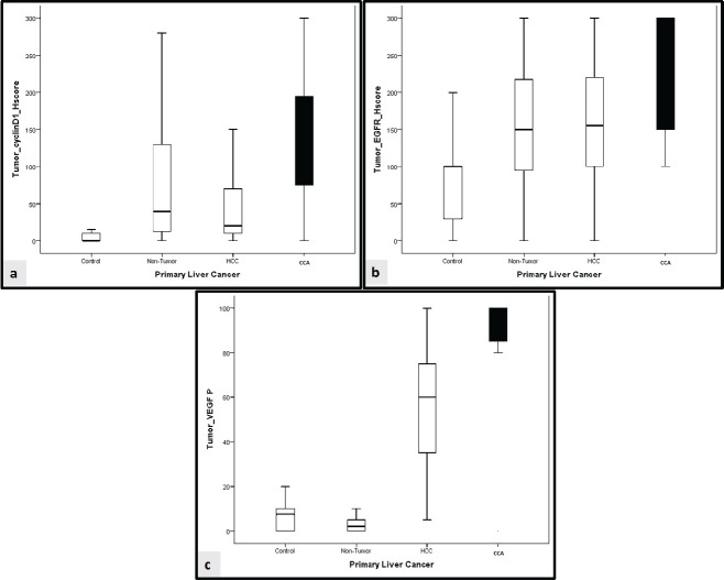 Figure 1
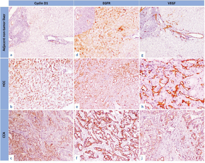 Figure 2
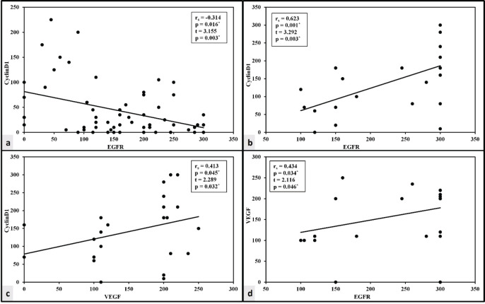 Figure 3
Peer Reviews
No public reviews on file for this paper yet. If you reviewed it on a platform where reviews are public (OpenReview, ICLR, NeurIPS, ICML), you can paste yours below so the community can read it here.
Videos
No videos yet. Explain this paper in a talk, walkthrough, or lecture? Add one.
Taxonomy
TopicsHistorical Studies in Science · Geography and Education Methods
Background
Primary liver carcinomas are common malignancies originating from the liver with high mortality and morbidity. Primary liver carcinomas are divided mainly into hepatocellular carcinoma (HCC) represents about 85%, and cholangiocarcinoma (CCA) represents about 10% [1]. Primary liver carcinomas are the sixth most common cancer worldwide, and the fourth leading cause of cancer-related death globally [2]. The highest incidence rates in the world are found in Asia and Africa [3]. In Egypt, HCC is the leading cause of cancer-related mortality among both genders [4]. The major risk factors for HCC vary according to geographic distribution with chronic hepatitis B (HBV) and hepatitis C virus (HCV) infection accounting for 56% and 20% of HCC-related deaths worldwide, respectively. On the other hand, non-viral risk factors including steatotic liver disease a leading cause of HCC in Western countries [5]. CCA risk factors included sclerosing cholangitis, biliary cysts, ulcerative colitis, hepatic lithiasis, hepatic infection and toxins [6]. Despite improved HCV eradication, the incidence of HCC remains elevated due to liver cirrhosis and established genetic abnormalities [7]. CCA cases are increasing annually indicating a need for improved surveillance and management [8].
HCC is potentially curable in its early stages when treated with hepatic resection, transplantation and ablation [5]. The adoption of treatment stage migration (TSM) was proposed in the 2022 update of The Barcelona Clinic Liver Cancer system treatment algorithm of HCC. TSM is used when a certain patient profile or therapy failure/infeasibility may cause the advice to shift to the alternative that would be considered for a more advanced stage [9]. Targeted agents are frequently associated with significant resistance and adverse effects. Furthermore, for advanced patients, chemotherapy lacks its survival advantage [10]. Sorafenib is well-established as a targeted therapy for late-stage HCC; however, it is complemented by a high risk of tumour resistance [11].
Radical surgery with a negative resection margin is the best curative management for CCA with liver transplantation not considered a standard treatment. Local regional therapy such as ablation and transarterial chemoembolisation could be optional therapies in advanced liver-limited disease. However, 40%–85% of patients experience recurrence of the disease following radical excision [12]. In addition, CCA has a very poor prognosis with no current effective pharmacological treatment available [13].
HCC and CCA share some commonly known risk factors and possible carcinogenic pathways [14]. Cyclin D1 is a cyclin-dependent kinase (CDK) 4/6 regulatory protein. Cyclin D1 increases cell proliferation, and its activation is assumed to be the first step in the progression of HCC and CCA through enhanced cell-cycle progression [13, 15]. Receptor tyrosine kinases (RTKs) are being recognised as important participants in tumour progression and cancer dissemination [16]. Some RTKs, such as epidermal growth factor (EGFR), can contribute to tumour spreading by establishing a molecular complex with integrin, inducing genetic aberrations and inducing treatment resistance [17]. Targeting EGFR may participate in the treatment of primary liver cancer [18].
Angiogenesis is an essential step in the development of HCC. Vascular endothelial growth factor (VEGF) is regarded as a driving factor in both healthy and pathological angiogenesis [19]). Sorafenib, a VEGF inhibitor, has been found to enhance survival in late-stage HCC. Resistance to anti-VEGF therapy, on the other hand, prompts research into combinational or alternative medications [20]. Although biliary tract inflammation is the first step in carcinogenesis, the microenvironment also plays an important role in pathogenesis, encouraging tumour angiogenesis and spread. VEGF plays a role in angiogenesis and has been studied as a prognostic marker in CCA [21].
As a result, we designed this study to study the protein expression of cyclin D1, EGFR and VEGF in Egyptian patients with HCC and CCA with the prognostic factors. Furthermore, determining the expression level of these proteins could help to elucidate their therapeutic potential.
Material and methods
This is retrospective, case-control research conducted at the Pathology Department, National Liver Institute, Egypt. The cases were obtained as part of the patients' medical management between December 2020 and December 2022. Tumour samples were collected from 82 primary liver cancer patients who were candidates for curative surgical resection. The cases were divided into 58 HCC cases and 24 CCA cases. As a control, 51 cases of adjacent non-tumour liver tissue and 18 cases of healthy liver tissue were included. The control, healthy group included potential liver transplant donors with normal liver function tests and ultrasound findings. Serological results for autoimmune and viral liver disorders were negative, and there was no history of diabetes or metabolic syndrome. Upon institution approval (NLIIRB protocol Number 00485/2023), clinical and survival information was obtained.
After surgery, there will be a 1-month follow-up with ultrasound, triphasic computed tomography and serum tumour markers, followed by a 2-month follow-up and then 3-month follow-ups for 1 year. Then every 6 months for 3 years. The overall survival (OS) was calculated from the diagnosis date through the last follow-up visit or death.
Inclusion criteria
Primary liver carcinomas approved to be classic HCC or CCA based on clinical, laboratory, pathological and immunohistochemical confirmation.
Exclusion criteria
Primary liver carcinomas in children are not considered. All patients who had undergone local ablation or had systemic therapy such as neo-adjuvant chemotherapy or sorafenib before surgery were excluded.
Pathological studies
Tumour size and multiplicity, pathological grade, lymph vascular invasion (LVI), perineural invasion and pathological stage were all included in the pathological data for tumour cases. Using the tumour-node-metastasis classification method, the pathologic stage was evaluated. The following was the pathological stage for HCC: T1a, solitary ≤ 2 cm on greatest dimension with or without LVI, T1b, solitary > 2 cm on greatest dimension without LVI, T2, solitary > 2 cm on greatest dimension with LVI or multiple tumours, none > 5 cm on greatest dimension, T3, multiple tumours any > 5 cm on greatest dimension, T4, tumour(s) involving a major branch of the portal or hepatic vein or with direct invasion of adjacent organs or with perforation of visceral peritoneum. The following was the pathological stage for CCA: T1a, solitary ≤ 5 cm on greatest dimension without LVI, T1b, solitary > 5 cm on greatest dimension without LVI, T2, solitary tumour with LVI or multiple tumours with or without LVI, T3, tumour perforating the visceral peritoneum, T4, tumour involving local extrahepatic structures by direct hepatic invasion [22, 23].
Immunohistochemical technique
The tissue microarray technique was done for both tumour and non-tumour tissue samples [24]. For the normal liver tissue, sections from needle liver biopsy samples were used.
The cyclin D1, EGFR and VEGF primary antibodies were all acquired from (Bioss, Massachusetts, USA) and diluted at 1:200. Sections were treated for 20 minutes in high PH Dako antigen retrieval (Ref K8000) before cooling for another 20 minutes. The slides were covered by primary antibodies overnight at 4°C. The immunostaining was visualised using 3-diaminobenzidine chromogen (DAKO A/S). For each run, positive and negative controls were used. Cyclin D1 revealed nuclear expression while EGFR and VEGF revealed cytoplasmic expression. The histoscore (H score) approach has been used by two blinded histopathologists to independently evaluate the expression of markers. H score is calculated via multiplication of a percentage by the intensity (0–3): H-score = [(0 × % negative cells) + (1 × % mildly positive cells) + (2 × % moderately positive cells) + (3 × % strongly positive cells)] with a total score of 0 to 300 [25].
Statistics
The Statistical Package for the Social Sciences software version 20.0 was used to analyse the data. The chi-square test, along with Fisher exact and Monte Carlo correction tests, was used to compare the two groups. The normality of continuous data was investigated. For abnormally distributed quantitative variables, the Mann-Whitney and Kruskal Wallis tests were used to compare two groups and more than two studied groups, respectively. For paired comparisons, use post hoc (Dunn's multiple comparisons test). The Pearson correlation coefficient was applied to examine the relationship between the two variables. The multivariate Cox proportional hazard regression model includes factors that were significantly associated with OS. The significance of the results was assessed at the 5% level.
Results
Table 1 shows the detailed clinicopathological data for both tumour groups.
The expression of cyclin D1, EGFR and VEGF in the tumour and control groups
In the normal control group, cyclin D1 was observed in 40% of cases, while EGFR and VEGF were detected in 88.9% and 61.1%, respectively. Cyclin D1 was observed in 40% of cases in adjacent non-tumour liver, while EGFR and VEGF were detected in 88.9% and 61.1%, respectively.
In HCC cases, cyclin D1 was observed in 84.3% of cases, while EGFR and VEGF were detected in 96.1% and 66.7%, respectively.
In CCA cases, cyclin D1 was observed in 95.8% of cases, while EGFR and VEGF were detected in 100% and 91.7%, respectively. Detailed markers expression is illustrated in Table 2.
In comparison to the control group, cyclin D1 H score was overexpressed in non-tumorous liver tissue and tumour groups (p < 0.001, for both). Similarly, overexpression of EGFR H score was significantly observed in non-tumorous liver tissue and tumour groups (p = 0.007 and p < 0.001, respectively). Furthermore, the VEGF H score was significantly overexpressed in the tumour group (p < 0.001, for both) (Figures 1 and 2).
Correlation between the cyclin D1, EGFR and VEGF in the primary liver carcinoma groups
There was a lack of correlation between VEGF and either cyclin D1 or EGFR in the HCC group. Cyclin D1 and the EGFR H score of expression, however, have a negative correlation. The expression of three markers exhibited a significant positive correlation in the CCA group (Figure 3).
This is confirmed using the linear regression analysis (Table 3).
Relation between the studied markers and the different clinicopathological parameters in tumour groups
The female gender and old age were both significantly linked with the cyclin D1 H score in the HCC group (p = 0.032 and p = 0.011, respectively). A high cyclin D1 H score was also strongly related to a well-differentiated grade and an early tumour stage (p = 0.016 and p = 0.042, respectively VEGF H score was significantly higher in well-differentiated grade (p = 0.040) (Table 4).
The EGFR H score had a significant association with large tumour size in the CCA group (p = 0.047), as shown in Table 5.
Multivariate COX regression analysis for the parameters affecting mortality in primary liver carcinomas
Furthermore, multivariate COX regression analysis for factors influencing cancer mortality did not reveal any effect of clinicopathological or marker expression on patient survival in cases of primary liver carcinoma, as shown in Table 6.
Regarding the etiological background of the adjacent non-tumour liver
There was significant overexpression of EGFR and VEGF in HCC raised in the non-cirrhotic (virology negative) liver compared to those developed in post-hepatitic liver cirrhosis (p = 0.003 and p = 0.014) (Table 3).
Discussion
In the current study, HCC and CCA shared overexpression of cyclin D1, EGFR and VEGF with the dominant expression in the CCA group. Furthermore, there is a lack of significant correlation between cyclin D1/EGFR and VEGF expression in HCC cases, in contrast to their positive correlation with VEGF expression in CCA.
Through the activation of cell cycle progression, the expression of cyclin D1 in the HCC and CCA groups in the current study may enhance cell proliferation. Cyclin D1 is created in the G1 phase, where it then interacts with CDK4/6 to control the G1/S phase transition. In HCC, cyclin D1 overexpression could be induced through gene amplification [26, 27]. Other studies emphasise the regulatory role of the autophagy pathway on cyclin D1 activation [26, 28]. Similarly, CCA overexpressed cyclin D1 to evade the inhibitory effect of transformed growth factor-β [29]. Another in-vitro study found that cyclin D1-enriched CCA tissue significantly reduced the count of cells that were exposed to CDK4/6 inhibitors [13].
The EGFR has also been linked to the development of HCC and CCA tumours in previous studies [30, 31]. EGFR overexpression is common in HCC, and its activation could be an alarming sign of primary resistance to sorafenib [32]. EGFR family members were expressed in CCA contributing to tumour development and aggressiveness [33]. In addition, angiogenesis is considered a cornerstone in the progression and metastasis of human HCC [34]. Angiogenesis occurs when tumour microenvironmental cells, hepatic stellate cells and inflammatory cells begin to release VEG [35]. VEGF has been reported to be overexpressed in different classes of CCA (intrahepatic and extrahepatic) [36]. We postulate that the treatment of HCC and CCA malignancies may be affected by the overexpression of cyclin D1 and EGFR.
In the CCA group, the present study showed a direct association between cyclin D1/EGFR and VEGF. The production of cyclin D1 induces the activation of VEGF, which then modulates blood vessels. Cyclin D1 modulates STAT 3 activity and so promotes VEGF production [37]. Cyclin D1 and VEGF are the downstream target proteins of the NF-κB pathway in CCA cells [38]. Additionally, the VEGF and EGFR pathways both share downstream carcinogenic signalling [39]. VEGF signalling is upregulated by EGFR expression [40].
In HCC and CCA cases, there was a controversial relationship between cyclin D1 and EGFR. This conflict was observed in previous studies. Hernandez-Garcia et al [41] found EGFR tyrosine-phosphorylation and activation in the cyclin D1 expressed HCC cell lines and HCC samples. The possible mechanism could be through activating the EGFR/Akt/NF-κB/cyclin D1 [42, 43]. A negative association between EGFR and cyclin D1 in HCC, on the contrary, could be the result of alternative pathway activity. EGFR activation leads to β-catenin phosphorylation mediated by PI3K leading to decreased expression of cyclin D1 [44].
HCC raised in the non-cirrhotic liver significantly overexpressed EGFR and VEGF in comparison to those developed in post-hepatitic liver cirrhosis. There is debate regarding the functional relevance of VEGF in HCC amongst patient groups with and without cirrhosis. According to Fodor et al [45], VEGF plays a detrimental function in the development of immature vasculature in individuals with cirrhosis. However, other studies found high serum VEGF expression in HCC on top of the non-cirrhotic liver and assumed that the downregulation of VEGF levels in patients with portal hypertension impacts hepatocyte regeneration [46]. There is little information available regarding the relative expression of EGFR and cyclin D1 on HCC that develops in livers with and without cirrhosis. The involvement of EGFR in HCC could be altered by EGFR overexpression or functional polymorphism with no accompanying copy number gain [47].
The potential therapeutic role of VEGF, cyclin D1 and EGFR has been reported in association with sorafenib therapy. Sorafenib prevents tumour cell growth and angiogenesis, which significantly slows the course of HCC and increases patient survival time [48]. Sorafenib's antiangiogenic effect could be mediated through VEGFR activation [49, 50]. In addition, sorafenib reduces the production of cyclin D1 and arrests the cell cycle to prevent the proliferation of tumour cells [11, 51]. On the contrary, EGFR activation could induce sorafenib resistance in primary HCC cells [32]. As a result, decreasing EGFR expression or limiting its kinase activity could increase sorafenib sensitivity [52].
Regarding the prognostic role of the studied markers, cyclin D1 and VEGF shared a similar good prognostic impact on the HCC group which agrees with the previous studies. It may be possible to determine an early role for cyclin D1 in hepatocarcinogenesis and tumour differentiation from the significant association between cyclin D1 expression and a well-differentiated HCC histology [53]. Well-differentiated HCCs had the highest levels of VEGF expression, which was followed by moderately and poorly differentiated HCCs [54, 55]. On the other hand, other studies found an association between cyclin D1 and high-grade and advanced stages of HCC [15, 56]. In CCA, EGFR was associated with large tumour size which could be explained by the role of EGFR in induction of the proliferative activity and tumour growth [57, 58]. Cyclin D1 and VEGF did not significantly correlate with the prognostic factors in CCA, which may be due to the small sample size of the studied cases. In CCA, VEGF expression is linked to poor prognostic indicators such as nodal metastases and advanced stage [59].
Conclusion
Overexpression of cyclin D1, EGFR and VEGF in HCC and CC contributes to their pathogenesis. Cyclin D1, EGFR and VEGF shared significant overexpression in HCC and CCA. EGFR and VEGF may play an oncogenic function in the development of HCC in non-cirrhotic liver. Furthermore, Cyclin D1 and VEGF may play a good prognostic function in HCC, but EGFR may play a bad prognostic role in CCA.
Limitations and future recommendations
Future genetic studies are recommended to determine the wild/mutant form of EGFR in relation to VEGF. Clinical investigations are recommended to determine the value of sorafenib in combination with cyclin D1 and/or EGFR inhibitors in the management of sorafenib-resistant HCC and CCA.
Conflicts of interest
The authors declare no conflicts of interest.
Funding
This study received no specific funding from government, commercial, or non-profit organisations.
Ethics approval
The study was approved by the ethics committee/Institutional Review Board of (National Liver Institute, Menoufia University, (NLIIRB protocol Number 00485/2023)).
Author contributions
DS, wrote the manuscript, contributed to the study design and supervised the implementation of the research and corresponding author; SSE, implemented the research and contributed to writing the manuscript; SK, contributed to the concept and design of the work, performed the clinical part and collecting the data and agreed on the final version; SA, contributed to the concept and design of the work, and revising the paper; SMA, contributed to study design, supervised implementing the research, and revising writing process of the manuscript.
The reference list from the paper itself. Each links out to its DOI / PubMed record.
- 1Rumgay H Ferlay J Martel C Global, regional and national burden of primary liver cancer by subtype Eur J Cancer 2022161108118[Internet]10.1016/j.ejca.2021.11.02334942552 · doi ↗ · pubmed ↗
- 2Llovet JM Kelley RK Villanueva A Hepatocellular carcinoma Nat Rev Dis Prim 2021716[Internet]10.1038/s 41572-020-00240-333479224 · doi ↗ · pubmed ↗
- 3Petrick JL Florio AA Znaor A International trends in hepatocellular carcinoma incidence, 1978–2012 Int J Cancer 20201472317330[Internet]10.1002/ijc.3272331597196 PMC 7470451 · doi ↗ · pubmed ↗
- 4Sung H Ferlay J Siegel RL Global cancer statistics 2020: GLOBOCAN estimates of incidence and mortality worldwide for 36 cancers in 185 countries CA Cancer J Clin 202171320924910.3322/caac.2166033538338 · doi ↗ · pubmed ↗
- 5Kulik L El-Serag HB Epidemiology and management of hepatocellular carcinoma Gastroenterology 20191562477.e 1491.e 1[Internet]10.1053/j.gastro.2018.08.06530367835 PMC 6340716 · doi ↗ · pubmed ↗
- 6Tyson GL El-Serag HB Risk factors for cholangiocarcinoma Hepatology 2011541173184[Internet]10.1002/hep.2435121488076 PMC 3125451 · doi ↗ · pubmed ↗
- 7Ezzat R Eltabbakh M El Kassas M Unique situation of hepatocellular carcinoma in Egypt: a review of epidemiology and control measures World J Gastrointest Oncol 2021131219191938[Internet]10.4251/wjgo.v 13.i 12.191935070033 PMC 8713321 · doi ↗ · pubmed ↗
- 8Javle M Lee S Azad NS Temporal changes in cholangiocarcinoma incidence and mortality in the United States from 2001 to 2017 Oncologist 2022271087488310.1093/oncolo/oyac 15035972334 PMC 9526482 · doi ↗ · pubmed ↗