Review of Anaka Dworakowska & Viraktamath, 1975 (Hemiptera, Cicadellidae, Typhlocybinae) with the descriptions of five new species from China

Abstract
Genes, proteins, chemicals, diseases, species, mutations and cell lines named across the full text — each resolved to its canonical identifier and authoritative record.
Click any figure to enlarge with its caption.
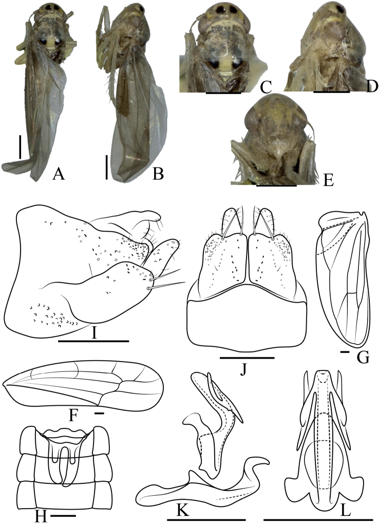 Figure 1
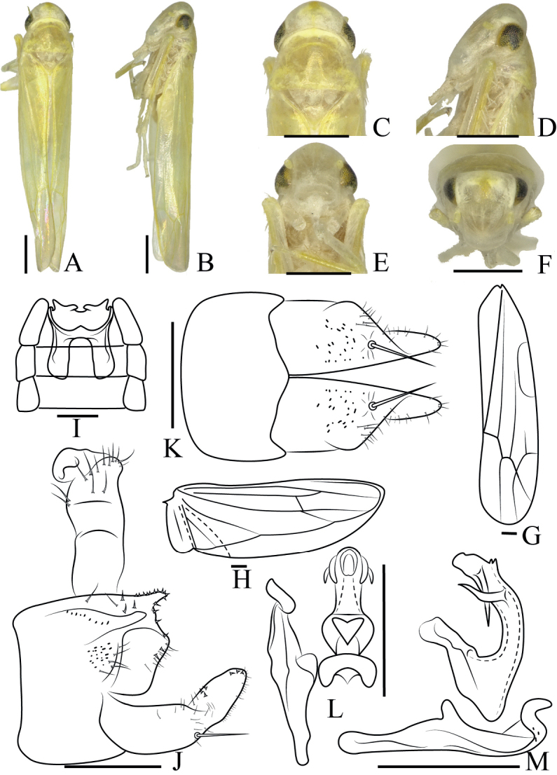 Figure 2
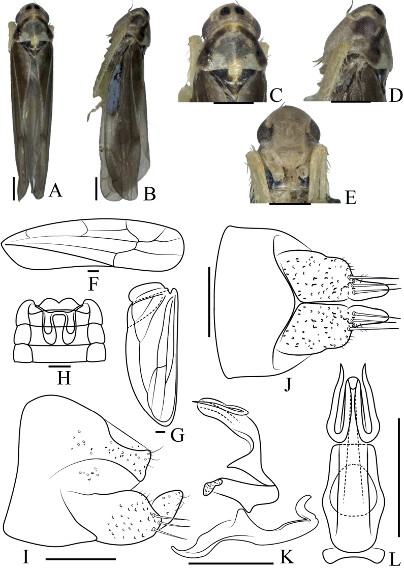 Figure 3
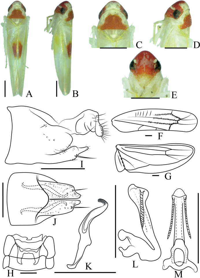 Figure 4
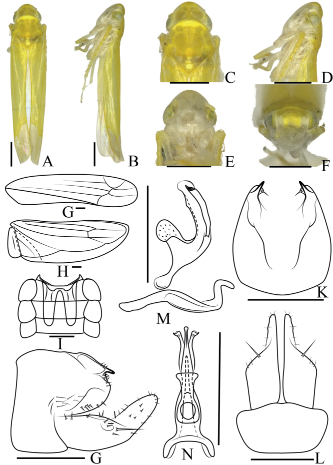 Figure 5
| 1 | Aedeagus with processes basally |
|
| – | Aedeagus with processes apically |
|
| 2 | Aedeagal processes extended beyond apex of shaft |
|
| – | Aedeagal processes shorter than or equal to shaft |
|
| 3 | Aedeagal processes sculptured |
|
| – | Aedeagal processes smooth |
|
| 4 | Aedeagal processes with areolate sculpture distally and parallel grooves basally |
|
| – | Aedeagal processes with distal areolate sculpture only |
|
| 5 | Aedeagal shaft with minute corrugation on ventral side |
|
| – | Aedeagal shaft without minute corrugation on ventral side |
|
| 6 | Apices of aedeagal processes twisted |
|
| – | Apices of aedeagal processes straight |
|
| 7 | Aedeagal stem straight, close to basal appendages |
|
| – | Aedeagal stem curved, well separated from to basal appendages |
|
| 8 | Aedeagus with one pair of apical processes |
|
| – | Aedeagus with two pairs of apical processes |
|
| 9 | Apex of aedeagal stem not curved |
|
| – | Apex of aedeagal stem curved |
|
| 10 | Apices of aedeagal processes long and sculptured |
|
| – | Apices of aedeagal processes short and not sculptured |
|
| 11 | Aedeagal apical processes unbranched |
|
| – | Aedeagal apical processes branched |
|
| 12 | Aedeagal apical processes broadly curved |
|
| – | Aedeagal apical processes narrowly curved |
|
Peer Reviews
No public reviews on file for this paper yet. If you reviewed it on a platform where reviews are public (OpenReview, ICLR, NeurIPS, ICML), you can paste yours below so the community can read it here.
Videos
No videos yet. Explain this paper in a talk, walkthrough, or lecture? Add one.
Taxonomy
TopicsPhytoplasmas and Hemiptera pathogens · Plant Pathogenic Bacteria Studies · Plant Virus Research Studies
Introduction
The typhlocybine leafhopper genus Anaka Dworakowska & Viraktamath, 1975 belonging to the tribe Dikraneurini was erected by Dworakowska and Viraktamath (1975) with Anakacolorata from India as the type species. Thapa and Sohi (1986) added A.nepalica and A.spinosa from Nepal. Dworakowska (1993) added A.blada (India), A.sumatrana (Indonesia), A.shashidhari (India), A.burmensis (Upper Burma), and A.roryi (China). Thus, eight species of the genus have been reported to date. Here, we review the previously known species and describe five new species, Anakaauricula sp. nov., Anakacruciata sp. nov., Anakacurvata sp. nov., Anakarosacea sp. nov. and Anakaspiralis sp. nov. from China. We also provide a key to males of all recognized species of the genus.
Material and methods
All specimens identified in this study were collected by net trapping in southern China and are housed at the Institute of Entomology, Guizhou University, Guiyang, Guizhou, P. R. China (GUGC:10657).
Dry specimens were used for preparing descriptions and illustrations. External morphology was observed under a stereoscopic microscope. Body length was measured with an ocular micrometer, in millimeters, from the apex of the head to the apex of the forewing in repose. The genital segments of the specimens examined were macerated in 10% NaOH, washed in water and transferred to glycerin. Male specimens were dissected under a MOTIC B1 SMS-168 SERIES microscope. Figures were made using an OLYMPUS CX41 compound microscope. Photographs were taken with Keyence VHX-1000 and an Olympus E-520 digital camera. The digital images were then imported into Adobe Photoshop CS6 for labeling and figure composition.
Taxonomic account
Anaka
Taxon classificationAnimaliaHemipteraCicadellidae
Genus
Dworakowska & Viraktamath, 1975
7C8111A7-4B1F-56F8-BD6E-9D4D8BF91C54
Anaka Dworakowska & Viraktamath, 1975: 521.
Type species.
Anakacolorata Dworakowska & Viraktamath, 1975 (type locality: India).
Description.
Vertex rounded, coronal suture distinct. Face rounded gradually to vertex, flat, lorum broad. Ocelli vestigial. Pronotum ~ 3× longer than head in dorsal view, as broad as head with eyes. Scutum with scutellum distinctly shorter than pronotum. Forewing broad, 3^rd^ apical cell stalked. Hind wing veins RP and MA confluent in male. Hindwing narrow, membrane smoked, veins dark, area bordered.
Abdominal apodemes well developed, reaching caudal margin of 4^th^ abdominal sternite. Pygofer side dark, well sclerotized, dorsal lobe the darkest, hind and ventral margins not pigmented, row of microsetae caudad. Subgenital plate has group of few big macrosetae at approximately mid-length, numerous thin short microsetae present at apical 1/3, several small rigid setae near macrosetae, and a row of thin long setae on basal 1/2 of ventral margin. Paramere hooked at apex, strongly attached to subgenital plate. Connective fused with aedeagus (Dworakowska and Viraktamath 1975). Aedeagus tubular, with basal or apical processes. Dorsoatrium well developed. Gonopore apical.
Distribution.
China (Guizhou, Guangdong, Guangxi, Yunnan, Sichuan, Chongqing, Hunan, Zhejiang, Fujian, Taiwan), India, Nepal, Sumatra, Indonesia, Burma.
Checklist of Anaka Dworakowska & Viraktamath, 1975
1. Anakaauricula sp. nov.
Distribution. China (Guizhou).
2. Anakablada Dworakowska, 1993
Anakablada Dworakowska, 1993a: 161.
Distribution. India.
3. Anakaburmensis Dworakowska, 1993
Anakaburmensis Dworakowska, 1993a: 163.
Distribution. China (Guizhou, Sichuan, Yunnan, Chongqing, Guangdong, Fujian), India.
4. Anakacolorata Dworakowska & Viraktamath, 1975
Anakacolorata Dworakowska & Viraktamath, 1975a: 523.
Distribution. India.
5. Anakacruciata sp. nov.
Distribution. China (Yunnan).
6. Anakacurvata sp. nov.
Distribution. China (Guangdong, Guangxi).
7. Anakanepalica Thapa & Sohi, 1986
Anakanepalica Thapa & Sohi, 1986a: 54.
Distribution. Nepal.
8. Anakaroryi Dworakowska, 1993
Anakaroryi Dworakowska, 1993c: 116.
Distribution. China (Taiwan).
9. Anakarosacea sp. nov.
Distribution. China (Guizhou).
10. Anakashashidhari Dworakowska, 1993
Anakashashidhari Dworakowska, 1993a: 162.
Distribution. India.
11. Anakaspinosa Thapa & Sohi, 1986
Anakaspinosa Thapa & Sohi, 1986a: 56.
Distribution. India, Nepal.
12. Anakaspiralis sp. nov.
Distribution. China (Yunnan).
13. Anakasumatrana Dworakowska, 1993
Anakasumatrana Dworakowska, 1993a: 162.
Distribution. Sumatra.
Key to males of the genus Anaka Dworakowska & Viraktamath, 1975
**: **
Anaka
auricula
sp. nov.
Taxon classificationAnimaliaHemipteraCicadellidae
EF6486A8-F31C-5A16-8F27-746DCFE39CE9
https://zoobank.org/FFD18D4F-C13E-4AA9-9327-72711E53EE19
Type material.
Holotype, 1♂, China: Guizhou Province, Daozhen. 28.1892°N, 107.4294°E, H, 1700 m, 14.V.2006, collected by Yang Zaihua.
Description.
Length: male 4.2 mm. Body (Fig. 1A, B) sandy beige. Crown (Fig. 1C) with two black patches. Face (Fig. 1D, E) yellowish, frontoclypeal area protuberant, anteclypeus broad. Pronotum yellowish brown, wider than crown. Scutellum yellowish with two blackish patches at lateral corner. Forewing (Fig. 1F) infuscate 3^rd^ apical cell stalked, hind wing (Fig. 1G) transparent.
Anakaauricula sp. nov. A male body, dorsal view B male body, lateral view C head and thorax, dorsal view D head and thorax, lateral view E face F forewing G hindwing H abdominal apodeme I male pygofer, lateral view J subgenital plate, ventral view K aedeagus, connective, and paramere, lateral view L aedeagus and connective, ventral view. Scale bars: 0.5 mm (A–E); 0.1 mm (F–L).
Male abdomen (Fig. 1H) well developed and reaching 4^th^ abdominal sternite. Pygofer side (Fig. 1I) broad, single row of thin setae on central and apical parts. Basal 1/2 of subgenital plate (Fig. 1J) broad, distal 1/2 slender in lateral view, two large macrosetae at approximately mid-length, several small setae near macrosetae, and scattered setae on distal 1/2. Paramere (Fig. 1K) hooked at apex. Aedeagus (Fig. 1K, L) tubular in lateral view, stem curved at middle, apical processes auricle-shaped with small spine at middle. Gonopore apical.
Etymology.
The specific epithet is derived from the Latin word auricula (an ear) referring to the shape of the aedeagal processes.
Remarks.
This species has an aedeagus very similar in form to that of A.blada, but it differs from that species in having elongated apical processes.
Anaka
cruciata
sp. nov.
Taxon classificationAnimaliaHemipteraCicadellidae
8DBC217B-13A4-5560-BEB0-9E15C9322768
https://zoobank.org/71769A36-C830-407C-8082-E4575284A965
Type material.
Holotype, 1♂, China: Yunnan Province, Pingbian. 22.9101°N, 103.7008°E, H, 2084 m, 22.V.2015, collected by Yan Bin.
Description.
Length: male 4.2 mm. Body (Fig. 2A, B) yellowish. Crown (Fig. 2C) obtuse. Coronal suture distinct. Face (Fig. 2D–F) white, frontoclypeal area protuberant, anteclypeus broad. Pronotum yellowish, wider than crown. Scutellum small. Wings (Fig. 2G, H) without patches.
Anakacruciata sp. nov. A male body, dorsal view B male body, lateral view C head and thorax, dorsal view D head and thorax, lateral view E face F head, frontal view G forewing H hindwing I abdominal apodeme J male pygofer, lateral view K subgenital plate, ventral view L aedeagus, connective, and paramere, dorsal view M aedeagus, connective, and paramere, lateral view. Scale bars: 0.5 mm (A–F); 0.1 mm (G–M).
Male abdomen (Fig. 2I) reaching 4^th^ abdominal sternite. Pygofer side (Fig. 2J) broad, with a small extension and thin setae on central and apical parts. Basal 1/2 of subgenital plate (Fig. 2K) broad, distal 1/2 slender in lateral view, one large macroseta at approximately midlength, several small setae near macrosetae, and scattered setae on distal 1/2. Paramere (Fig. 2L, M) hooked at apex. Connective fused with aedeagus. Aedeagus (Fig. 2L, M) tubular, curved, with two pairs of apical processes, of which each pair are crossed. Gonopore apical.
Etymology.
The specific epithet is derived from the Latin word cruciatus (marked by a cross) referring to the shape formed by the two pairs of aedeagal processes.
Remarks.
This species with two pairs of aedeagal processes differs from all other species of Anaka, and two pairs of processes originate from subapical of stem, but in different positions.
Anaka
curvata
sp. nov.
Taxon classificationAnimaliaHemipteraCicadellidae
E0369A01-A6FB-5532-96C1-88383906C096
https://zoobank.org/2B819388-B5DB-4879-948A-162E2242B86B
Type material.
Holotype, 1♂, China: Guangdong Province, Nanling National Natural Reserve, 24.8796°N, 113.0137°E, H, 1340 m. 4.VIII.2006, collected by Zhou Zhonghui. Paratypes, 4♂♂, China: Guangxi Province, Damingshan National Natural Reserve, 23.5049°N, 108.4153°E, H, 1290 m. 15.IV.2012, collected by Long Jiankun; 6♂♂, China: Guangxi Province, Damingshan National Natural Reserve, 23.4898°N, 108.4411°E, H, 1250 m. 14.V.2012, collected by Huang Rong and Yu Xiaofei.
Description.
Length: male 4.4–4.5 mm. Body (Fig. 3A, B) brown. Crown (Fig. 3C) with two black patches. Coronal suture distinct. Face (Fig. 3D, E) yellowish brown, frontoclypeal area protuberant, anteclypeus broad. Pronotum brown, wider than crown. Scutellum with a vertical yellow stripe in the median. Forewing (Fig. 3F) infuscate, 3^rd^ apical cell stalked, hind wing transparent (Fig. 3G).
Anakacurvata sp. nov. A male body, dorsal view B male body, lateral view C head and thorax, dorsal view D head and thorax, lateral view E face F forewing G hindwing H abdominal apodeme I male pygofer, lateral view J subgenital plate, ventral view K aedeagus, connective, and paramere, lateral view L aedeagus, connective, ventral view. Scale bars 0.5 mm (A–E); 0.1 mm (F–L).
Male abdomen (Fig. 3H) reaching 4^th^ abdominal sternite. Pygofer side (Fig. 3I) broad, thin setae on central and apical parts. Basal 1/2 of subgenital plate (Fig. 3G) broad, distal 1/2 slender in lateral view, three large macrosetae at approximately mid-length, several small setae near macrosetae, and scattered setae on distal 1/2. Paramere (Fig. 3K) hooked at apex. Connective fused with aedeagus. Aedeagus (Fig. 3K, L) tubular, curved, with a pair of apical processes, which are curved like a paper clip. Gonopore apical.
Etymology.
The specific epithet is derived from the Latin word curvatus (curved) referring to the shape of the aedeagal processes.
Remarks.
This species is similar to A.blada, but it differs in having the aedeagus processes more strongly curved and less divergent from the stem.
Anaka
rosacea
sp. nov.
Taxon classificationAnimaliaHemipteraCicadellidae
A73E0969-4B51-56F8-9931-A2155EF00F55
https://zoobank.org/43F14E58-F199-4013-86FB-2ED54327CB64
Type material.
Holotype, 1♂, China: Guizhou Province, Jinsha, 27.4553°N, 106.2667°E, H, 1300 m, 5.VIII.2015, collected by Zhang Yaowen. Paratypes, 3♂9♀, same data as holotype.
Description.
Length: male 4.4–4.5 mm. Body (Fig. 4A, B) white with red patches. Crown (Fig. 4C) obtuse, yellowish white. Coronal suture distinct. Face (Fig. 4D, E) red, frontoclypeal area protuberant, anteclypeus broad, yellowish. Pronotum yellowish, with red patches in the central part, wider than crown. Scutellum yellowish. Forewing (Fig. 4F) white with red patches along inside margin, hind wing transparent (Fig. 4G).
Anakarosacea sp. nov. A male body, dorsal view B male body, lateral view C head and thorax, dorsal view D head and thorax, lateral view E face F forewing G hindwing H abdominal apodeme I male pygofer, lateral view J subgenital plate, ventral view K paramere, lateral view L aedeagus and connective, lateral view M aedeagus and connective, dorsal view. Scale bars 0.5 mm (A–E); 0.1 mm (F–M).
Male abdomen (Fig. 4H) weakly developed and reaching 4^th^ abdominal sternite. Pygofer side (Fig. 4I) broad, apical part elliptical. Basal 1/2 of subgenital plate (Fig. 4J) broad, distal 1/2 slender in lateral view, one large macroseta at approximately midlength. Paramere (Fig. 4K) hooked at apex. Connective fused with aedeagus. Aedeagus (Fig. 4L, M) tubular, stem inflated at apex, with one pair of apical processes, apical processes straight and sculptured, oriented basad. Gonopore apical.
Etymology.
The specific epithet is derived from the Latin word rosaceus (rose-colored) referring to the color of the head.
Remarks.
This species marked with rose-red spots. The aedeagal processes are similar to A.blada and A.spinosa but differs in having the aedeagus with two long apical processes and the processes straight with spiral pattern.
Anaka
spiralis
sp. nov.
Taxon classificationAnimaliaHemipteraCicadellidae
E46BA7D8-4F4D-5C3F-A799-F15C9A8B26C7
https://zoobank.org/FAB85DAA-EBFE-4621-ADED-BBB32FFD5514
Type material.
Holotype, 1♂, China: Yunnan Province, Baoshan, 25.1581°N, 99.0814°E, H, 1500 m, 14.V.2016, collected by Li Bin and Ren Guoru. Paratypes, 3♂4♀, same data as holotype.
Description.
Length: male 4.4–4.5 mm. Body (Fig. 5A, B) yellow. Crown (Fig. 5C) obtuse. Coronal suture distinct. Face (Fig. 5D–F) white, frontoclypeal area protuberant, anteclypeus broad. Pronotum yellow, wider than crown. Forewing (Fig. 5G) yellow with apical part white, hind wing (Fig. 5H) transparent.
Anakaspiralis sp. nov. A male body, dorsal view B male body, lateral view C head and thorax, dorsal view D head and thorax, lateral view E face F head, frontal view G forewing H hindwing I abdominal apodeme J male pygofer, lateral view K male pygofer lobe, dorsal view L subgenital plate, ventral view M aedeagus, connective and paramere, lateral view N aedeagus and connective, dorsal view. Scale bars 0.5 mm (A–F); 0.1 mm (G–N).
Male abdomen (Fig. 5I) well developed and reaching 5^th^ abdominal sternite. Pygofer side (Fig. 5J, K) broad, with small extension on superior margin, setae along periphery. Basal 1/2 of subgenital plate (Fig. 5L) broad, distal 1/2 slender in lateral view, one large macroseta at approximately mid-length. Paramere (Fig. 5M) hooked at apex. Connective fused with aedeagus. Aedeagus (Fig. 5M, N) tubular, with one pair of basal processes, apical part of processes spiral and not exceeding the stem. Gonopore apical.
Etymology.
The specific epithet is derived from the Latin word spiralis (spiraling) referring to the shape of the aedeagal processes.
Remarks.
In this species the aedeagus has a pair of basal processes like A.burmensis and A.shashidhari, but these basal processes have spiral-shaped top, and do not exceed the stem. These features are also not as long as in A.nepalica.
Supplementary Material
XML Treatment for Anaka
XML Treatment for Anaka auricula
XML Treatment for Anaka cruciata
XML Treatment for Anaka curvata
XML Treatment for Anaka rosacea
XML Treatment for Anaka spiralis
The reference list from the paper itself. Each links out to its DOI / PubMed record.
- 1Dworakowska I (1993) Some Dikraneurini (Auchenorrhyncha: Cicadellidea: Typhlocybinae) from south-east Asia.Oriental Insects 27(1): 151–173. 10.1080/00305316.1993.10432268 · doi ↗
- 2Dworakowska I Viraktamath CA (1975) On some Typhlocybinae from India (Auchenorrhyncha, Cicadellidae).Bulletin of the Academie Polonaise Des Sciences 223(8): 521–530.
- 3Thapa VK Sohi AS (1986) A review of Dikraneurini (Homoptera, Cicadellidae) of Nepal.Colemania 3: 53–60.