Optical coherence tomography for the diagnosis and differentiation of cutaneous cysts: a case series
Sarah Hobelsberger, Frank Friedrich Gellrich, Julian Steininger, Stefan Beissert, Jörg Laske

TL;DR
This case series explores how optical coherence tomography can help diagnose and differentiate types of skin cysts.
Contribution
The study demonstrates the use of optical coherence tomography to distinguish between various cutaneous cyst types based on structural features.
Findings
Optical coherence tomography visualized cysts as hyporeflective round lesions with clear boundaries.
Epidermal, trichilemmal cysts, and hidrocystomas showed linear borders representing epithelium.
Mucoid pseudocysts lacked linear borders, aiding in differentiation from other cyst types.
Abstract
Kutane zystische Läsionen (n = 35) wurden mit optischer Kohärenztomographie untersucht. Zysten waren sichtbar als hyporeflektive rundliche Raumforderung mit klarer Abgrenzung unter teils verdünnter Epidermis. Epidermalzysten, trichilemmale Zysten und Hidrozystome hatten einen linearen Rand, der das Zystenepithel darstellt, während mukoide Pseudozysten keinen linearen Rand aufwiesen. Trichilemmal- und Epidermoidzysten wiesen zudem einen hyperreflektiven Inhalt auf, welcher Keratin entspricht. Durch die Visualisierung des Randsaums und des Inhalts der Zyste war es möglich, zwischen verschiedenen Entitäten von Zysten zu differenzieren.
Genes, proteins, chemicals, diseases, species, mutations and cell lines named across the full text — each resolved to its canonical identifier and authoritative record.
Click any figure to enlarge with its caption.
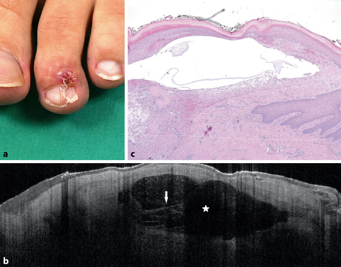 Figure 1
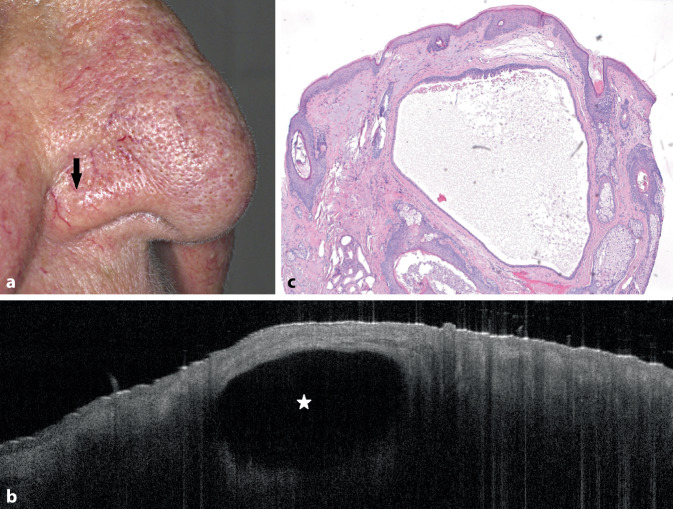 Figure 2
 Figure 3
Peer Reviews
No public reviews on file for this paper yet. If you reviewed it on a platform where reviews are public (OpenReview, ICLR, NeurIPS, ICML), you can paste yours below so the community can read it here.
Videos
No videos yet. Explain this paper in a talk, walkthrough, or lecture? Add one.
Taxonomy
TopicsOptical Coherence Tomography Applications · Cancer and Skin Lesions · Advanced Fluorescence Microscopy Techniques