Kinetic Modeling of Brain [18-F]FDG Positron Emission Tomography Time Activity Curves with Input Function Recovery (IR) Method
Marco Bucci, Eleni Rebelos, Vesa Oikonen, Juha Rinne, Lauri Nummenmaa, Patricia Iozzo, Pirjo Nuutila

TL;DR
This paper introduces a method to improve PET data quality by recovering poor plasma input curves using a model trained on optimal data, enhancing brain imaging analysis.
Contribution
The novel contribution is an input recovery (IR) method that rescues sub-optimal PET input functions using tail curve information and a reference model.
Findings
Recovered plasma curves showed comparable AUC and maxSUV to original curves in the reference set.
The IR method produced biologically plausible results for CM parameters and FUR in brain PET studies.
The method successfully rescued poor-quality plasma inputs, enabling kinetic modeling for previously excluded cases.
Abstract
Accurate positron emission tomography (PET) data quantification relies on high-quality input plasma curves, but venous blood sampling may yield poor-quality data, jeopardizing modeling outcomes. In this study, we aimed to recover sub-optimal input functions by using information from the tail (5th–100th min) of curves obtained through the frequent sampling protocol and an input recovery (IR) model trained with reference curves of optimal shape. Initially, we included 170 plasma input curves from eight published studies with clamp [18F]-fluorodeoxyglucose PET exams. Model validation involved 78 brain PET studies for which compartmental model (CM) analysis was feasible (reference (ref) + training sets). Recovered curves were compared with original curves using area under curve (AUC), max peak standardized uptake value (maxSUV). CM parameters (ref + training sets) and fractional uptake rate…
Genes, proteins, chemicals, diseases, species, mutations and cell lines named across the full text — each resolved to its canonical identifier and authoritative record.
Click any figure to enlarge with its caption.
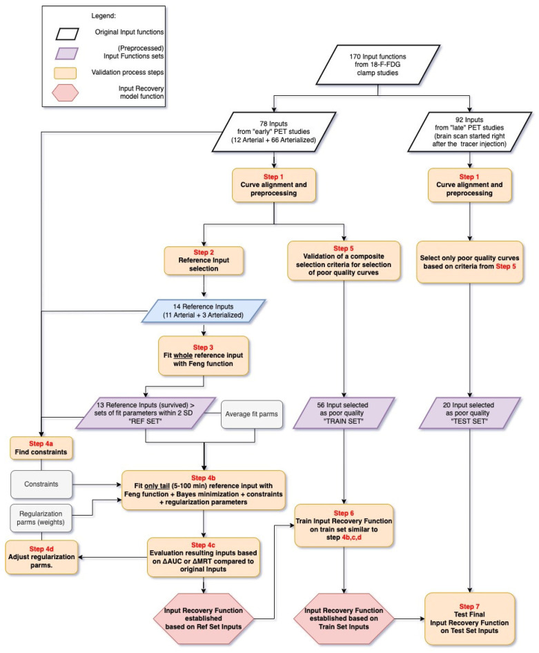 Figure 1
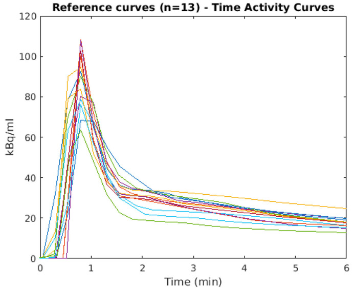 Figure 2
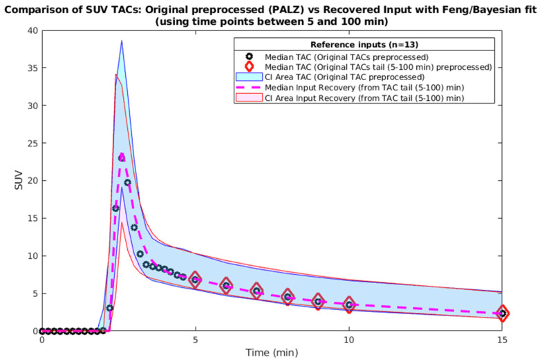 Figure 3
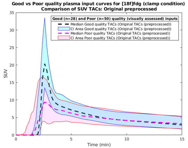 Figure 4
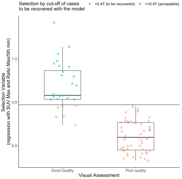 Figure 5
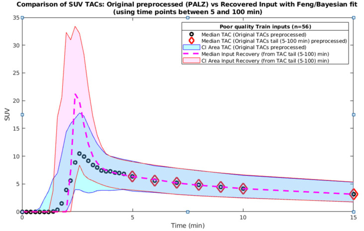 Figure 6
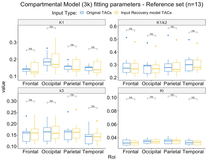 Figure 7
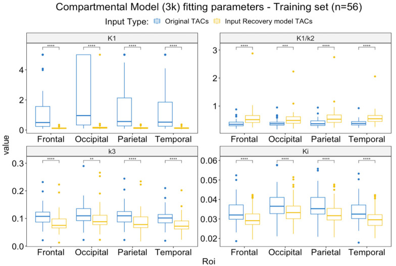 Figure 8
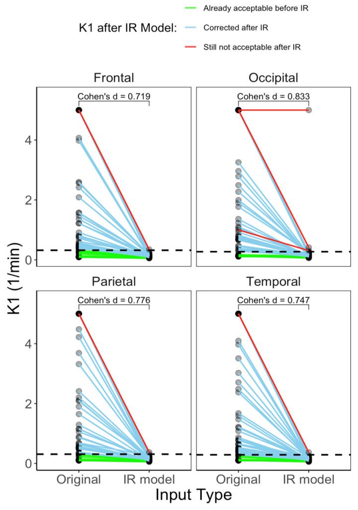 Figure 9
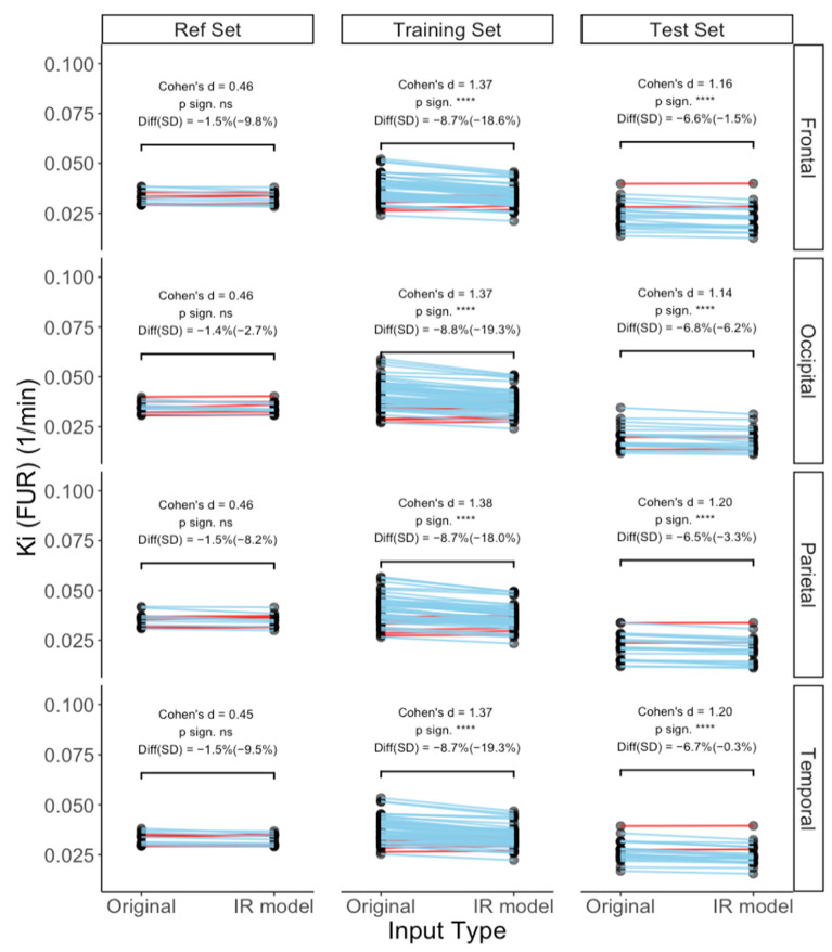 Figure 10
Peer Reviews
No public reviews on file for this paper yet. If you reviewed it on a platform where reviews are public (OpenReview, ICLR, NeurIPS, ICML), you can paste yours below so the community can read it here.
Videos
No videos yet. Explain this paper in a talk, walkthrough, or lecture? Add one.
Taxonomy
TopicsMedical Imaging Techniques and Applications · Advanced MRI Techniques and Applications · MRI in cancer diagnosis