Comparative Analysis of Vascular Structures in OLIF51 and the Lateral Corridor Approach under Supine MRI and Intraoperative Enhanced CT in the Lateral Decubitus Position
Yoshihisa Kotani, Hiroyuki Tachi, Atsushi Ikeura, Takahiro Tanaka, Takanori Saito

TL;DR
This study compares vascular structures at the L5/S1 level using MRI and CT scans to improve safety in spinal surgeries like OLIF51 and LCA.
Contribution
Provides precise vascular measurements and movement data between supine and lateral decubitus positions for safer spinal surgery planning.
Findings
OLIF51 had larger vascular windows (22.8 mm and 34.1 mm) compared to LCA (14.2 mm and 12.6 mm) at L5/S1 levels.
The left common iliac vein shifted significantly (3.8 mm and 6.9 mm) to the right when moving from supine to lateral decubitus position.
Iliolumbar vein was detected in 52% of MRI scans and located 31 mm from the midline.
Abstract
Background and Objectives: As the oblique lateral interbody fusion at L5/S1 (OLIF51) and the lateral corridor approach (LCA) have gained popularity, an understanding of the precise vascular structure at the L5/S1 level is indispensable. The objectives of this study were to investigate the vascular anatomy at the L5/S1 level, and to compare the movement of vascular tissue between the supine and lateral decubitus positions using intraoperative enhanced CT and MRI. Materials and Methods: A total of 43 patients who underwent either OLIF51 or LCA were investigated with an average age at surgery of 60.4 (37–80) years old. The preoperative MRI was taken to observe the axial and sagittal anatomy of the vascular position under the supine position. The intraoperative vein-enhanced CT was taken just before incision in the right decubitus position, and compared to supine MRI anatomy. Iliolumbar…
Genes, proteins, chemicals, diseases, species, mutations and cell lines named across the full text — each resolved to its canonical identifier and authoritative record.
Click any figure to enlarge with its caption.
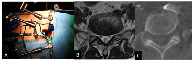 Figure 1
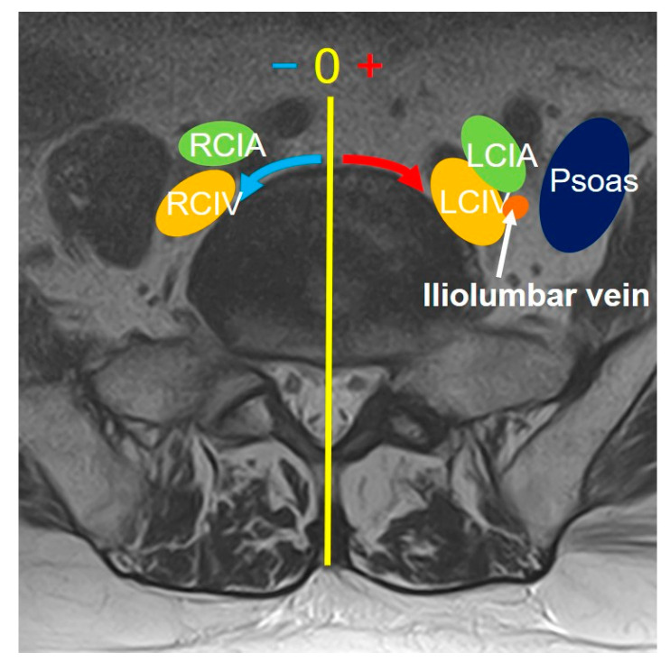 Figure 2
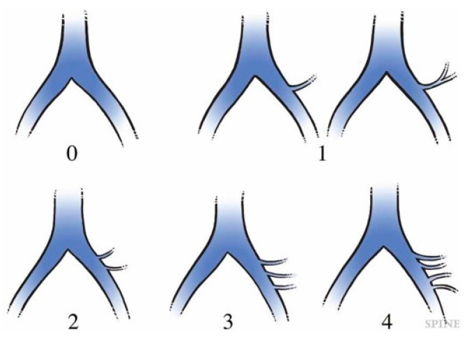 Figure 3
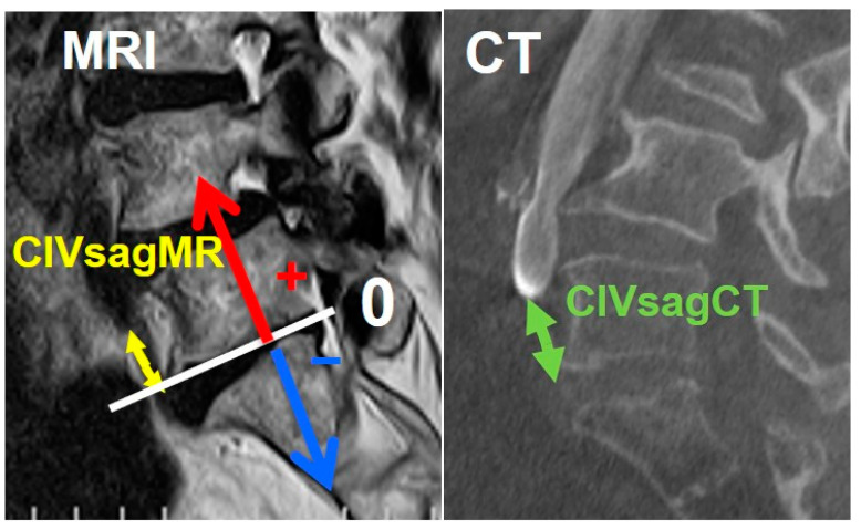 Figure 4
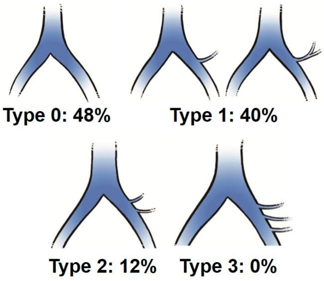 Figure 5
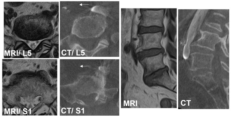 Figure 6
 Figure 7
Peer Reviews
No public reviews on file for this paper yet. If you reviewed it on a platform where reviews are public (OpenReview, ICLR, NeurIPS, ICML), you can paste yours below so the community can read it here.
Videos
No videos yet. Explain this paper in a talk, walkthrough, or lecture? Add one.
Taxonomy
TopicsSpine and Intervertebral Disc Pathology · Spinal Fractures and Fixation Techniques · Pelvic and Acetabular Injuries