Reflectance confocal microscopy features of chronic radiodermatitis: A useful tool for presurgical mapping
Luca Rapparini, Federico Venturi, Chiara Gelati, Federico Giorgini, Marco Pignatti, Michelangelo La Placa, Biagio Scotti, Giulia Veronesi, Emi Dika

Abstract
Genes, proteins, chemicals, diseases, species, mutations and cell lines named across the full text — each resolved to its canonical identifier and authoritative record.
Click any figure to enlarge with its caption.
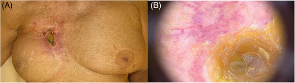 FIGURE 1
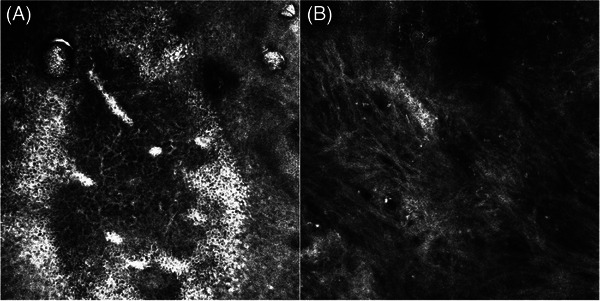 FIGURE 2
Peer Reviews
No public reviews on file for this paper yet. If you reviewed it on a platform where reviews are public (OpenReview, ICLR, NeurIPS, ICML), you can paste yours below so the community can read it here.
Videos
No videos yet. Explain this paper in a talk, walkthrough, or lecture? Add one.
Taxonomy
TopicsInfrared Thermography in Medicine · Cutaneous Melanoma Detection and Management · Effects of Radiation Exposure
Dear Editor,
Reflectance confocal microscopy (RCM) is a useful tool for the diagnosis of skin cancers and inflammatory disorders.1, 2, 3, 4, 5 We present the case of a 75‐year‐old woman with a history of infiltrating lobular right breast cancer (pT2N0M0, UICC TNM 5th edition), treated with radical mastectomy, cobalt radiotherapy and breast reconstruction with an abdominal flap in 1997. Recent mammography and CT scan were negative for tumour recurrence. In October 2023, the patient was referred to our unit for the evaluation of an erythematous plaque located in the right mammary area. At clinical examination, an erythematous macule surmounted by a large crusted, fibrinous plaque was observed (Figure 1A). Dermoscopic examination of the lesion revealed an atypical vascular pattern at the periphery of the central keratin mass (Figure 1B). The whole skin appeared intensely photodamaged. Two incisional biopsies of the ulcerated plaque were performed. Histopathological evaluation led to the diagnosis of a post‐radiotherapy atypical vascular lesion (sec. WHO 2020). We further performed RCM for presurgical mapping of the lesion, which revealed, at the level of the stratum corneum, a severely disrupted corneal layer with presence of parakeratoses and detached corneocytes, whereas at the level of the stratum granulosum areas of necrosis corresponding to dark spaces with irregular demarcation and pronounced disruption of the epidermal architecture. Surrounding keratinocytes displayed the presence of spongiosis, seen as increased brightness of intercellular spaces, accentuating the honeycombed pattern of the granular layer. RCM changes observed were compatible with chronic radiodermitis (Figure 2A). Due to the presence of severe photodamage, RCM was performed contralaterally on the left mammary skin, and displayed an irregular epidermal honeycomb pattern with curled collagen in the dermis (Figure 2B). Subsequently, the patient underwent complete surgical excision of the atypical vascular lesion confirming the histopathologic analysis of the first incisional biopsies. Post‐radiation vascular lesions (including angiosarcoma and atypical vascular lesions) are a rare complication of irradiated skin in breast cancer patients. The latter present clinically as solitary or multiple reddish‐brown papules and enter the differential diagnosis with breast cancer recurrence, post‐traumatic haemorrhage, and benign dermatological conditions. They rarely progress to angiosarcoma, so the treatment of choice is surgical excision and follow‐up. In the literature, RCM has been used for the evaluation of acute radiation dermatitis toxicity, but there are no described cases of the application in chronic radiodermatitis. In acute radiation dermatitis histopathologic changes were detectable by RCM after a mean time of 15 days, compared with 30 days for clinical manifestations, and include spongiosis, exocytosis and inflammatory cells, dendritic‐shaped cells, streaming‐like figures, broken geographic papillae, epidermal architectural disarray, effacement of rete ridges, melanophages and hyperpigmentation of the basal layer5. Conversely, from what can be deduced from RCM examination in our case, the characteristics of chronic radiodermatitis with concomitant post‐radiation atypical vascular lesion are severe parakeratoses and detached corneocytes, together with areas of necrosis and spongiosis. To date, this is the first report of RCM features of chronic radiodermatitis. Further studies with larger samples are needed to clearly define RCM presentation of such entity.
Clinical and dermoscopic presentation of radiodermatitis: clinical examination showed an erythematous macule surmounted by a large crusted, fibrinous plaque (A). Dermoscopy revealed an atypical vascular pattern characterized by large vessels at the periphery of the central keratin mass (B).
RCM features of radiodermitis revealed, at the level of the stratum corneum, a severely disrupted corneal layer with presence of parakeratoses and detached corneocytes, whereas at the level of the stratum granulosum we observed areas of necrosis corresponding to dark spaces with irregular demarcation and pronounced disruption of the epidermal architecture. Surrounding keratinocytes displayed the presence of spongiosis, seen as increased brightness of intercellular spaces, accentuating the honeycombed pattern of the granular layer (A). Due to the presence of severe photodamage, RCM was performed also contralaterally on the left mammary skin, which displayed an irregular epidermal honeycomb pattern with curled collagen in the dermis (B).
CONFLICTS OF INTEREST STATEMENT
The authors have no relevant financial or non‐financial interests to disclose.
The reference list from the paper itself. Each links out to its DOI / PubMed record.
- 1Venturi F , Pellacani G , Farnetani F , Maibach H , Tassone D , Dika E . Noninvasive diagnostic techniques in the preoperative setting of Mohs micrographic surgery: A review of the literature. Dermatol Ther. 2022;35(11):e 15832.36106464 10.1111/dth.15832 · doi ↗ · pubmed ↗
- 2Pellacani G , Scope A , Gonzalez S , et al. Reflectance confocal microscopy made easy: the 4 must‐know key features for the diagnosis of melanoma and nonmelanoma skin cancers. J Am Acad Dermatol. 2019;81(2):520–526.30954581 10.1016/j.jaad.2019.03.085 · doi ↗ · pubmed ↗
- 3Zhai H , Dika E , Goldovsky M , Maibach HI . Tape‐stripping method in man: comparison of evaporimetric methods. Skin Res Technol. 2007;13(2):207–210.17374064 10.1111/j.1600-0846.2007.00218.x · doi ↗ · pubmed ↗
- 4De Giorgi V , Scarfì F , Silvestri F , et al. Genital piercing: A warning for the risk of vulvar lichen sclerosus. Dermatologic Therapy. Portico. 2020;34(1). doi:10.1111/dth.14703 33368949 · doi ↗ · pubmed ↗
- 5Vano‐Galvan S , Fernandez‐Lizarbe E , Truchuelo M , et al. Dynamic skin changes of acute radiation dermatitis revealed by in vivo reflectance confocal microscopy: Confocal features of radiation dermatitis. J Eur Acad Dermatol Venereol. 2013;27(9):1143–1150.22882643 10.1111/j.1468-3083.2012.04680.x · doi ↗ · pubmed ↗