Hepatic fascioliasis: a rare case of “fake” biliary gallstones
Michele Amata, Giovanni Boncori, Daniela Scimeca, Filippo Mocciaro, Ambra Bonaccorso, Claudia Colomba, Roberto Di Mitri

Abstract
Genes, proteins, chemicals, diseases, species, mutations and cell lines named across the full text — each resolved to its canonical identifier and authoritative record.
Click any figure to enlarge with its caption.
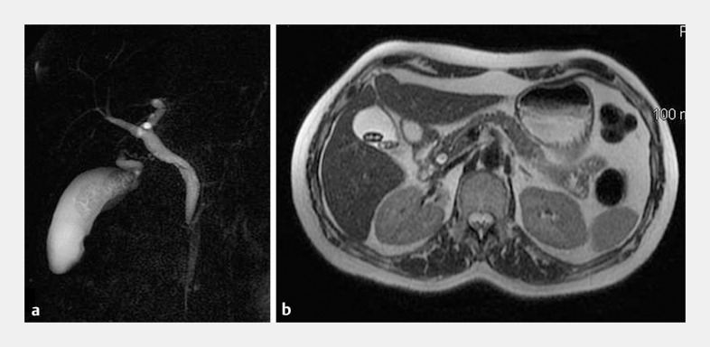 Fig. 1
 Fig. 2
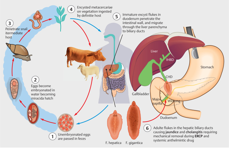 Fig. 3
Peer Reviews
No public reviews on file for this paper yet. If you reviewed it on a platform where reviews are public (OpenReview, ICLR, NeurIPS, ICML), you can paste yours below so the community can read it here.
Videos
No videos yet. Explain this paper in a talk, walkthrough, or lecture? Add one.
Taxonomy
TopicsHelminth infection and control · Gallbladder and Bile Duct Disorders · Biliary and Gastrointestinal Fistulas
A 52-year-old woman who had previously traveled to a developing country was admitted to our emergency department with fever, colicky abdominal pain, and pruritus. Laboratory tests showed hypereosinophilia (1432 eosinophils/µL, limit value <500) and direct hyperbilirubinemia (2.5 mg/dL, reference range 0–1.3 mg/dL). Magnetic resonance cholangiopancreatography revealed endoluminal filling defects into a dilated common bile duct (CBD) and in the gallbladder ( Fig. 1 ). Endoscopic retrograde cholangiopancreatography (ERCP) was then planned to treat choledocholithiasis.
Imaging studies showing a small defect inside the common bile duct, with two suspected stones inside the gallbladder with flat shape and atypical morphology. a Magnetic resonance cholangiopancreatography. b Axial fast imaging employing steady-state acquisition magnetic resonance imaging.
Using the double-wire technique, deep biliary cannulation was performed and fluoroscopic cholangiogram confirmed an irregular filling defect in the distal CBD. Surprisingly, after biliary sphincterotomy, a live and mobile flat-shaped worm was extracted from the CBD by a balloon catheter and retrieved using a biopsy forceps ( Fig. 2 , Video 1 ). Macroscopically, the parasitic trematode presented with a whitish, flat, and elongated morphology, 25 × 10 mm in size, compatible with Fasciola hepatica ; it was also confirmed at the microscopic analysis by the evidence of a pathognomonic spiny tegument.
Fasciola hepatica extracted from the biliary tree. At the macroscopic evaluation, the trematode presented as a flat leaf-shaped hermaphrodite fluke, gray in color. The adult worm may live in the biliary tract of the definitive host for many years (5 years in sheep and 10 years in humans).
Endoscopic extraction of Fasciola hepatica from the biliary tree. Video 1
Following a course of antiparasitic drug (triclabendazole 10 mg/kg oral solution, twice a day for 2 days), laparoscopic cholecystectomy was performed and confirmed only the presence of two gallstones. The patient was asymptomatic at the 4-month follow-up.
F. hepatica is a leaf-shaped trematode that usually attacks cattle and sheep ( Fig. 3 ), and is frequently found in endemic and developing countries. Humans may become accidental hosts through drinking water or ingesting raw green vegetables contaminated with encysted metacercariae. The parasite larva penetrates the intestinal wall and Glisson’s capsule, colonizing the biliary tree 1 . Living or dead F. hepatica may occlude the bile ducts, causing obstruction and sometimes cholangitis. ERCP is fundamental for diagnosis and the mechanical removal of F. hepatica from the CBD 2 , and triclabendazole allows complete eradication as it is active against both immature and adult parasites.
Life cycle of Fasciola hepatica (graphical illustration by Michele Amata, MD). CHD, common hepatic duct; ERCP, endoscopic retrograde cholangiopancreatography; IHBD, intrahepatic bile duct.
Endoscopy_UCTN_Code_CCL_1AC_2AG
The reference list from the paper itself. Each links out to its DOI / PubMed record.
- 1Rajan E Taha K Kalandar K Endoscopic management of biliary fascioliasis: a case report J Med Case Rep 201048320205932 10.1186/1752-1947-4-83PMC 2841078 · doi ↗ · pubmed ↗
- 2Hauser SC Bynum TE Abnormalities on ERCP in a case of human fascioliasis Gastrointest Endosc 198430808210.1016/s 0016-5107(84)72323-66714606 · doi ↗ · pubmed ↗