Dual ectopy: Unique appearance of ectopic thyroid
Liam du Preez, Francis Flaherty, Ragaa Elkabbani

TL;DR
A 67-year-old woman had two ectopic thyroid tissues found at the base of her tongue and hyoid bone, confirmed through imaging and biopsy.
Contribution
This case highlights the unique occurrence of dual ectopy in thyroid tissue and emphasizes the importance of embryologic development in diagnosing heterotopic tissue.
Findings
Two ectopic thyroid lesions were identified at the base of the tongue and hyoid bone.
The lesions were confirmed as ectopic thyroid tissue through imaging and biopsy.
The case underscores the need to consider thyroid developmental anomalies in midline neck lesions.
Abstract
A 67-year-old female underwent a computed tomography angiogram (CTA) of the head in the setting of acute, short-term memory loss. Two lobulated hyperattenuating lesions were incidentally discovered at the base of the tongue and the hyoid bone. Upon further investigation in the outpatient setting including further imaging and ultrasound-guided biopsy, the lesions were confirmed to be ectopic thyroid tissue with dual ectopy. Heterotopic tissue, especially when arising at separate sites, can be concerning for a broad differential diagnosis including malignancy, and further evaluation is certainly recommended. When evaluating possible heterotopic tissue, one must always keep in mind the expected embryologic development of the organ in question. Further, in cases where biopsy is less favorable, consideration of the heterotopic tissue's expected physiology is equally important. With these 2…
Genes, proteins, chemicals, diseases, species, mutations and cell lines named across the full text — each resolved to its canonical identifier and authoritative record.
Click any figure to enlarge with its caption.
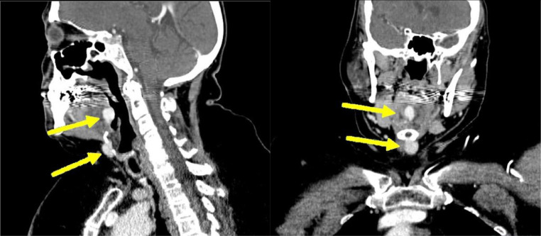 Figure 1
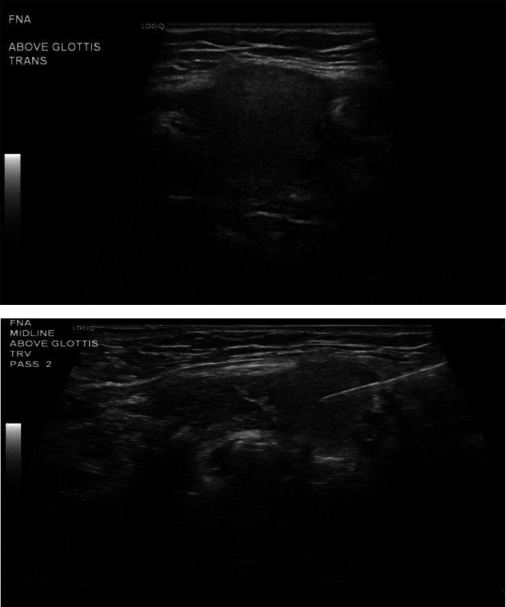 Figure 2
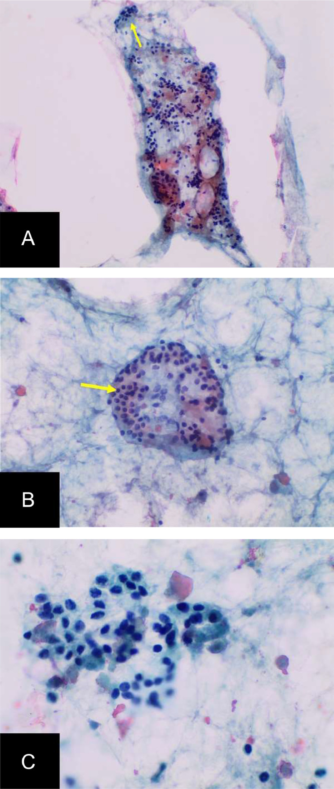 Figure 3
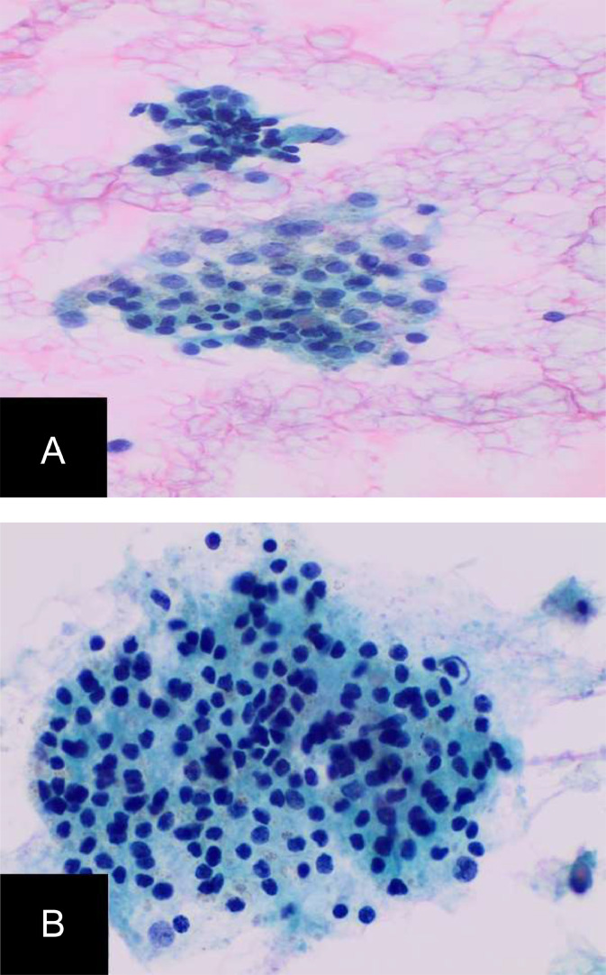 Figure 4
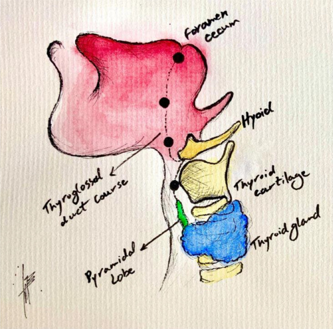 Figure 5
Peer Reviews
No public reviews on file for this paper yet. If you reviewed it on a platform where reviews are public (OpenReview, ICLR, NeurIPS, ICML), you can paste yours below so the community can read it here.
Videos
No videos yet. Explain this paper in a talk, walkthrough, or lecture? Add one.
Taxonomy
TopicsHead and Neck Anomalies · Teratomas and Epidermoid Cysts · Tumors and Oncological Cases
Introduction
Heterotopic locations of organs have myriad manifestations, pathophysiology, and clinical significance. With modern imaging techniques, heterotopias can be seen as early as the prenatal period or may go undetected until autopsy. Differential diagnoses for incidentally noted heterotopic tissue can include malignancy (both primary and metastatic), infection, sequelae of prior trauma, or developmental events. When incidentally identified masses occur in specific patterns, synthesis of laboratory results, imaging, pathology, and embryological development can narrow the differential significantly. This case report features a rare presentation of dual ectopic thyroid in a 67-year-old adult with hypothyroidism and exemplifies this process.
Ectopic thyroid tissue is the most common form of thyroid dysgenesis, occurring once in every 100,000-300,000 people [1]. Greater than 75% of cases occur in females, and presentation can be variable. Hypothyroidism is a common association, but many euthyroid patients are incidentally found on imaging [2]. Other reported cases describe dysphagia, dysphonia, and rarely symptoms of airway obstruction [3]. Not only are symptoms and associations broad, but the location of the ectopia is highly variable. Ectopic thyroid tissue is usually found along the pathway of descent of the thyroglossal duct, extending from the foramen cecum to the level of the thyroid cartilage. In extremely rare cases, primitive thyroidal tissue can seed multiple locations, such as in the case of dual thyroid ectopy.
Case report
A 67-year-old female with a significant past medical history of hypothyroidism underwent a computed tomography angiogram (CTA) of the head and neck at an outside hospital for evaluation of cerebrovascular accident in the setting of acute short-term memory loss. While there was no finding of large vessel occlusion, 2 incidental neck masses were identified. Outpatient follow-up with ENT was recommended, and upon further assessment, the patient underwent a contrast-enhanced computed tomography (CT) of the neck. Two similar lesions were identified: a 1.3 × 1.0 × 1.7 cm hyperdense mass of the mid-tongue base and a 1.5 × 1.2 cm hyperdense lesion just inferior to the hyoid bone. The thyroid gland was noted to be absent (Fig. 1).Fig. 1. Contrast-enhanced CT of Neck Masses: Sagittal and Coronal images of a contrast-enhanced CT of the neck showing the 2 sites of incidentally noted hyperdense tissue, the more superior of the 2 being at the base of the tongue, the inferior being anterior and adjacent to the hyoid bone.Fig 1
While the presumptive diagnosis was ectopic thyroid tissue, the differential diagnosis remained broad and included possible malignancy. Thus, ultrasound-guided percutaneous fine needle aspiration (FNA) of both lesions (Fig. 2) was performed by Interventional Radiology. Samples were placed in Cytolyt and Thyroseq suspensions and sent to Pathology.Fig. 2. Ultrasound-guided biopsy images: Two intraprocedural images show the hyoid lesion in transverse orientation under ultrasound, with the second image showing percutaneous fine needle aspiration of the same lesion. The tongue-based lesion was sampled as well (not shown).Fig 2
Samples from both sites showed clusters of benign follicular epithelial cells and colloids, consistent with ectopic thyroid tissue (Fig. 3.1, Fig. 3.2). The patient did not experience any side effects from the procedure and continued with the management of her hypothyroidism without issue.Fig. 3.1Pathology Images from fine needle aspiration of hyoid bone lesion: Cytologic pictures of direct alcohol-fixed papanicolaou-stained smears show colloid (arrow, a) and clusters of benign follicular epithelial cells (arrow, b) at 20x magnification (A and B), and 40x (C), consistent with benign heterotopic thyroid tissue.Fig 31. Fig. 3.2Pathology Images from fine-needle aspiration from base of the tongue lesion: This shows clusters of bland follicular cells and (B) Hürthle cells in a background of thin watery colloid; Direct, alcohol fixed pap-stained smear, magnification 20x magnification (A), ThinPrep CytoLyt Solution, 40x magnification (b).Fig 32
Discussion
Ectopic thyroid tissue is secondary to incomplete migration of primitive foregut tissue and may appear anywhere along the migration pathway from the foramen cecum to the paratracheal region, as well as rarely in the mediastinum, and subdiaphragmatic regions (Fig. 4). The most common ectopic location is lingual, which appears in approximately 90% of cases [4]. Symptoms manifest broadly and include dysphonia, dysphagia, globus sensation, and airway obstruction. There is also an association with hypothyroidism [5]. Thyroid ectopy has a female predominance and can manifest at any age, but most commonly within the first 3 decades of life.Fig. 4. Foramen cecum and thyroglossal duct illustration: illustrated diagram of the thyroid gland's developmental descent via the foramen cecum. Image courtesy of Dr. Behnaz Khazai, MD.Fig 4
Ectopic thyroid, as well as various other thyroid abnormalities, has been found to be associated with congenital hypothyroidism [2]. Specifically in cases such as ours with lingual ectopy, hypothyroidism occurs in as much as 70% of patients [3]. The increased attenuation within the lesions on noncontrast CT is secondary to physiologic iodine content. Our case demonstrates a rare occurrence of dual thyroid ectopy, with ectopic thyroid tissue in the tongue base and infra-hyoid neck. Important considerations in the differential diagnosis include lymphoma, metastatic thyroid cancer, neurogenic tumor, and mesenchymal/thymic tumors.
This case was deemed valuable for both imaging and clinical insights into those with heterotopic tissue. For example, while tissue biopsy is certainly the most definitive means of confirming benign thyroid tissue, multiple nuclear medicine scans could also have helped confirm the diagnosis. Thallium-201 especially is useful in not only the detection of thyroid tissue but also in separating malignant from benign based on radiotracer washout characteristics [6]. However, nuclear medicine scans such as this weren't readily available at our institution. Further, in this case, both lesions were amenable to biopsy. In many patients, one or both lesions may not be possible to be biopsied, like cases that involve mediastinum. Again, a nuclear medicine scan would be a less invasive option for securing the diagnosis.
Finally, while this manifestation remains exceedingly rare, dual ectopic thyroid is an important differential diagnosis to consider with multiple homogeneously enhancing neck masses specifically in the pattern of midline distribution along the expected descent of the thyroglossal duct, especially alongside absence of the typical thyroid gland.
Patient consent
Consent was obtained by the corresponding author and a consent form was completed and scanned into the patient's electronic health record.
The reference list from the paper itself. Each links out to its DOI / PubMed record.
- 1Santangelo G Pellino G De Falco N Colella GD'Amato S Maglione MG Prevalence, diagnosis and management of ectopic thyroid glands Int J Surg 282016 S 1S 610.1016/j.ijsu.2015.12.04326708843 · doi ↗ · pubmed ↗
- 2Léger Juliane Marinovic Daniella Garel Catherine Bonaïti-PelliéCatherine Polak Michel Czernichow Paul Thyroid developmental anomalies in first degree relatives of children with congenital hypothyroidism J Clin Endocrinol Metab 872200257558010.1210/jcem.87.2.826811836288 · doi ↗ · pubmed ↗
- 3Kumar Choudhury B Kaimal Saikia U Sarma D Saikia M Dutta Choudhury S Barua S Dual ectopic thyroid with normally located thyroid: a case report J Thyroid Res 2011201115970310.4061/2011/159703 PMC 313418021765986 · doi ↗ · pubmed ↗
- 4Ibrahim NA Fadeyibi IO.Ectopic thyroid: etiology, pathology and management Hormones (Athens)10201126126910.14310/horm.2002.131722281882 · doi ↗ · pubmed ↗
- 5Yoon Ji Sung Won Kyu Chang Cho Ihn Ho Lee Jae Tae Lee Hyoung Woo Clinical characteristics of ectopic thyroid in Korea Thyroid 171120071117112110.1089/thy.2007.000417887928 · doi ↗ · pubmed ↗
- 6Hsu Chien-Chin Chen Yu-Wen Huang Ying-Fong Chuang Ya-Wen Routine 201Tl scintigraphy in the follow-up of patients with differentiated thyroid carcinoma: diagnostic accuracy and clinical impact”Nucl Med Commun 289200768168710.1097/MNM.0b 013e 3282742090 https://journals.lww.com/nuclearmedicinecomm/abstract/2007/09000/routine_201tl_scintigraphy_in_the_follow_up_of.2.aspx 17667746 · doi ↗ · pubmed ↗