The sad tale of a youthful right ventricle struggling with endomyocardial fibrosis
Samah El-Mhadi, Zineb Agoumy, Belghait El Hajjaj, Nesma Bendagha, Aida Soufiani, Said Moughil

TL;DR
A 15-year-old girl from Guinea was diagnosed with rare heart disease called endomyocardial fibrosis, which led to severe heart failure and ultimately cardiac arrest.
Contribution
This case highlights the challenges in diagnosing and managing endomyocardial fibrosis in young patients.
Findings
Right ventricular endomyocardial fibrosis was diagnosed in a 15-year-old girl.
The patient declined surgery and suffered cardiac arrest two months later.
Early diagnosis and intervention are critical for better outcomes in EMF.
Abstract
Endomyocardial fibrosis (EMF) is a rare and often underdiagnosed form of restrictive cardiomyopathy. Prognosis is generally unfavorable. Early diagnosis, along with surgical and medical intervention, is crucial for improved outcomes. We report the case of a 15-year-old girl from Guinea who presented with suspected Ebstein’s anomaly and severe right heart failure. Multimodal cardiac imaging revealed right ventricular EMF. Despite counseling on prognosis, the family declined surgery. The patient experienced cardiac arrest two months later.
Genes, proteins, chemicals, diseases, species, mutations and cell lines named across the full text — each resolved to its canonical identifier and authoritative record.
Click any figure to enlarge with its caption.
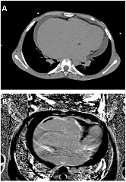 Figure 1
Peer Reviews
No public reviews on file for this paper yet. If you reviewed it on a platform where reviews are public (OpenReview, ICLR, NeurIPS, ICML), you can paste yours below so the community can read it here.
Videos
No videos yet. Explain this paper in a talk, walkthrough, or lecture? Add one.
Taxonomy
TopicsEosinophilic Disorders and Syndromes · Eosinophilic Esophagitis · Cardiac tumors and thrombi
CASE DESCRIPTION
A 15-year-old Guinean girl was referred to our cardiology department due to suspected Ebstein’s anomaly presenting with severe right heart failure.
The physical examination revealed a murmur indicative of tricuspid regurgitation and signs consistent with right-sided heart failure.
Her electrocardiogram indicated right atrial hypertrophy.
Transthoracic echocardiography revealed an ectatic right atrium that hindered the analysis of other cardiac chambers, accompanied by signs of diastolic dysfunction on Doppler imaging.
Peripheral blood count didn’t show hypereosinophilia.
Cardiac CT scan demonstrated severe dilation of the right atrium, associated with thrombosis in right atrium and ventricle (Fig. 1A).
(A) Cardiac CT-scan revealing severe dilation of the right atrium associated with thrombosis in right atrium and ventricle. (B) CMR: 4-chambers view LGE sequence showing right ventricular endomyocardial fibrosis, with enhancement alongside thrombosis consistent with typical ‘three-layered’ pattern.
Cardiac magnetic resonance (CMR) allowed for the diagnosis of right ventricular EMF with late gadolinium enhancement alongside thrombosis, consistent with typical ‘three-layered’ pattern (Fig. 1B).
The family was informed of the prognosis but declined surgical intervention. The patient was discharged home with oral anticoagulation.
Unfortunately, she experienced a cardiac arrest two months later.
EMF is a rare and often underdiagnosed cause of restrictive cardiomyopathy. The etiology of this condition remains undefined, potentially arising from a convergence of clinical factors interacting with genetic predispositions in individuals susceptible to an inflammatory process leading to fibrotic formation [1].
Suspecting EMF requires attention to signs and symptoms of restrictive heart failure, particularly in endemic regions. Key ECG evidence includes manifestations of right atrial overload. Echocardiographic indicators encompass increased atrial volume, normal ventricular volume, atrioventricular valve dysfunction due to subvalvular fibrosis, and apical obliteration of one or both ventricles [1].
The gold standard for confirming the diagnosis of EMF is CMR, playing a pivotal role in its accurate assessment.
The disease prognosis is generally unfavorable, marked by a high incidence of sudden cardiac death, thromboembolic complications, and end-stage heart failure [2].
Early diagnosis and appropriate management are crucial for enhancing outcomes, involving a combination of medical therapy and surgical intervention [3].
The reference list from the paper itself. Each links out to its DOI / PubMed record.
- 1Espinoza Romero C, Lima ICV, Hotta VT, Bocchi EA, Salemi VMC. Endomyocardial fibrosis of the right ventricle in a patient with schistosomiasis: a case report. Eur Heart J Case Rep 2022;6:ytac 312.35949701 10.1093/ehjcr/ytac 312PMC 9356724 · doi ↗ · pubmed ↗
- 2García-Granja PE, Pombo-Otero J, Barriales-Villa R. Isolated right ventricle endomyocardial fibrosis. An increasingly frequent disease in Spain. Med Clin (Barc) 2019;153:219–20.30146360 10.1016/j.medcli.2018.06.014 · doi ↗ · pubmed ↗
- 3Sutter JS, Suboc TM, Rao AK. Tropical endomyocardial fibrosis. JACC Case Rep 2020;2:819–22.34317354 10.1016/j.jaccas.2020.02.020PMC 8301721 · doi ↗ · pubmed ↗