A Rare Presentation of Polypoid Endometriosis of the Douglas Pouch: Case Report
Emilie Demondion, Yves Borghesi, Nathalie Trouillet

TL;DR
A 46-year-old woman with abnormal bleeding had a rare case of polypoid endometriosis in the Douglas pouch, initially mistaken for a tumor on MRI.
Contribution
Highlights the rare presentation of polypoid endometriosis and its potential misdiagnosis as a malignancy.
Findings
MRI showed a mass near the uterus, initially suspected to be an ovarian tumor.
Histology confirmed the lesion was polypoid endometriosis from the Douglas pouch.
The case emphasizes the importance of MRI features in distinguishing benign from malignant lesions.
Abstract
A case is reported of a 46-year-old woman referred to a magnetic resonance imaging (MRI) for menometrorrhagia. MRI revealed a mass lesion lateral to the uterus fundus, suspicious of an ovarian granulosa cell tumor. Extensive surgery was performed. Histological examination revealed a polypoid endometriosis lesion arising from the Douglas pouch. Teaching point: Polypoid endometriosis is a rare benign entity with a challenging differential diagnosis from malignancy. Specific MRI features can contribute to the diagnosis and thus avoid excessive surgical resection.
Genes, proteins, chemicals, diseases, species, mutations and cell lines named across the full text — each resolved to its canonical identifier and authoritative record.
Click any figure to enlarge with its caption.
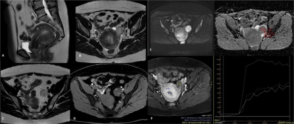 Figure 1
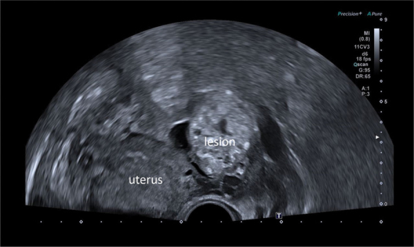 Figure 2
 Figure 3
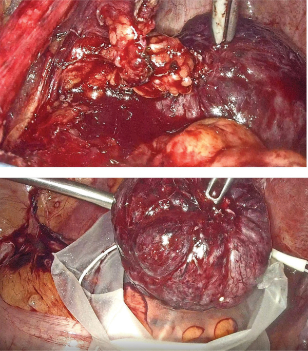 Figure 4
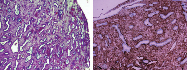 Figure 5
Peer Reviews
No public reviews on file for this paper yet. If you reviewed it on a platform where reviews are public (OpenReview, ICLR, NeurIPS, ICML), you can paste yours below so the community can read it here.
Videos
No videos yet. Explain this paper in a talk, walkthrough, or lecture? Add one.
Taxonomy
TopicsEndometriosis Research and Treatment · Uterine Myomas and Treatments · Endometrial and Cervical Cancer Treatments