Identification and characterization of stromal-like cells with CD207+/low CD1a+/low phenotype derived from histiocytic lesions – a perspective in vitro model for drug testing
Agnieszka Śmieszek, Klaudia Marcinkowska, Zofia Małas, Mateusz Sikora, Martyna Kępska, Beata A. Nowakowska, Marta Deperas, Marta Smyk, Carlos Rodriguez-Galindo, Anna Raciborska

TL;DR
This study identifies and characterizes stromal-like cells from histiocytic lesions, offering a new in vitro model for understanding and testing treatments for histiocytoses.
Contribution
The paper introduces novel stromal-like cell models derived from histiocytic lesions for drug testing and disease research.
Findings
The derived cells express CD1a/CD207 markers and show features of both dendritic and mesenchymal cells.
The cells display distinct mitochondrial morphology and transcriptomic profiles, including non-coding RNA biomarkers.
The models show variable sensitivity to vemurafenib and trametinib, suggesting potential for drug testing.
Abstract
Histiocytoses are rare disorders manifested by increased proliferation of pathogenic myeloid cells sharing histological features with macrophages or dendritic cells and accumulating in various organs, i.a., bone and skin. Pre-clinical in vitro models that could be used to determine molecular pathways of the disease are limited, hence research on histiocytoses is challenging. The current study compares cytophysiological features of progenitor, stromal-like cells derived from histiocytic lesions (sl-pHCs) of three pediatric patients with different histiocytoses types and outcomes. The characterized cells may find potential applications in drug testing. Molecular phenotype of the cells, i.e. expression of CD1a and CD207 (langerin), was determined using flow cytometry. Cytogenetic analysis included GTG-banded metaphases and microarray (aCGH) evaluation. Furthermore, the morphology and…
Genes, proteins, chemicals, diseases, species, mutations and cell lines named across the full text — each resolved to its canonical identifier and authoritative record.
Click any figure to enlarge with its caption.
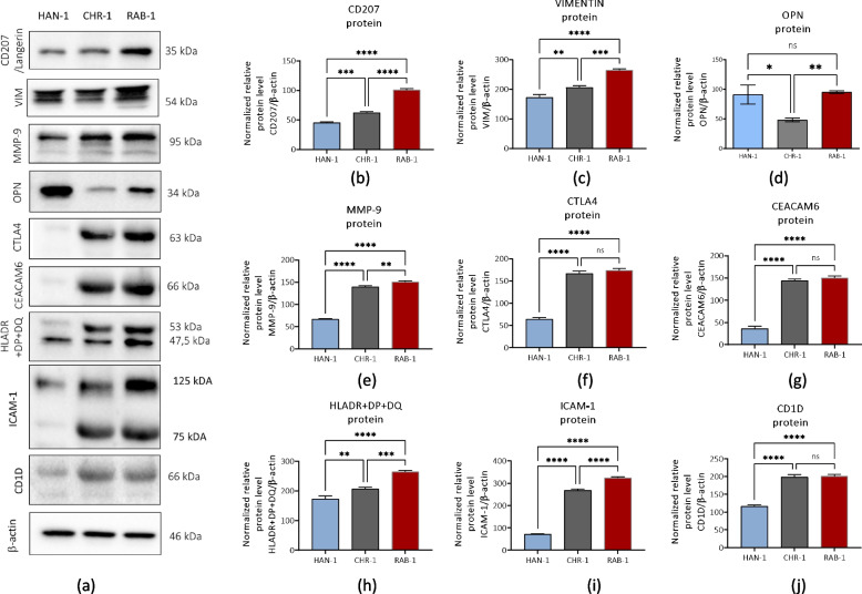 Figure 10
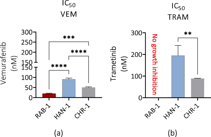 Figure 11
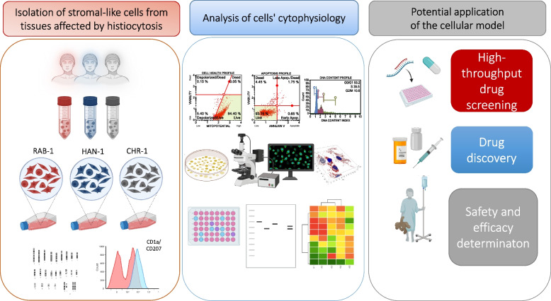 Figure 1
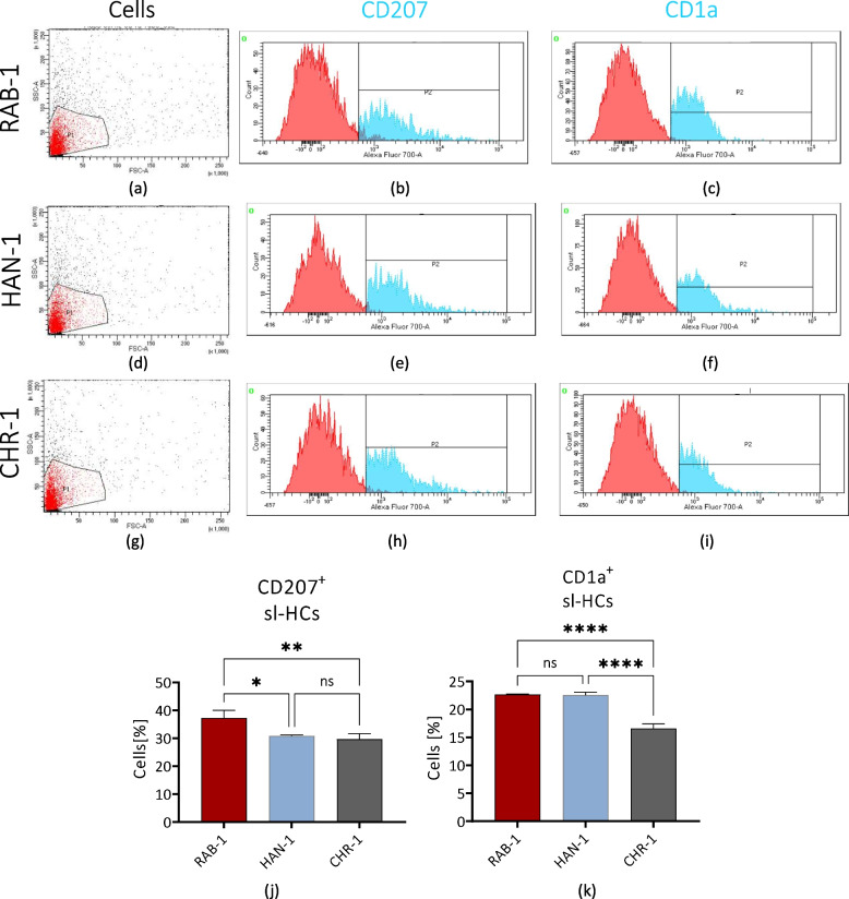 Figure 2
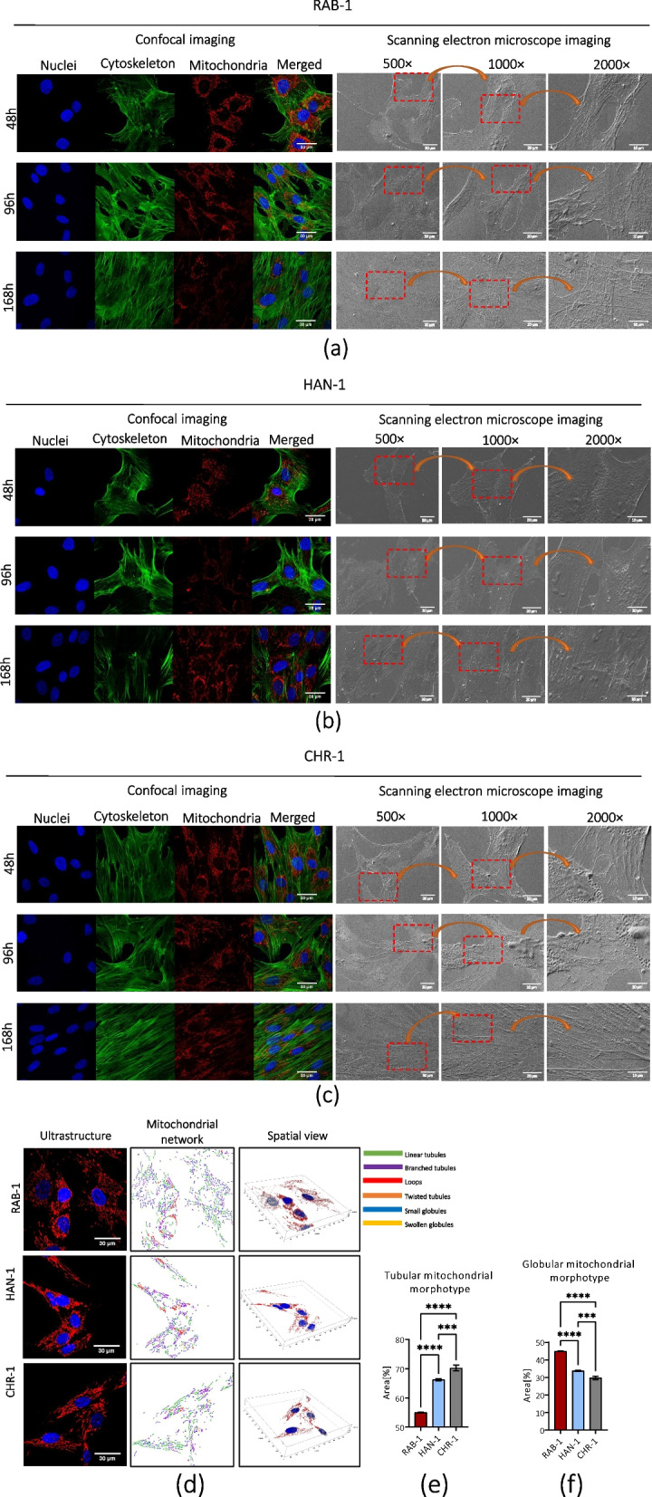 Figure 3
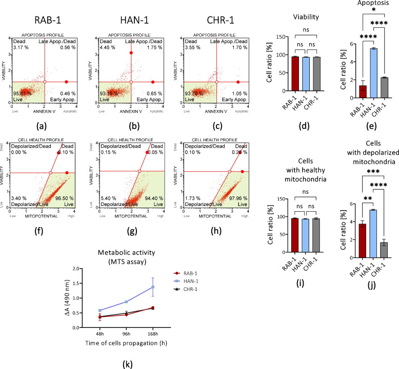 Figure 4
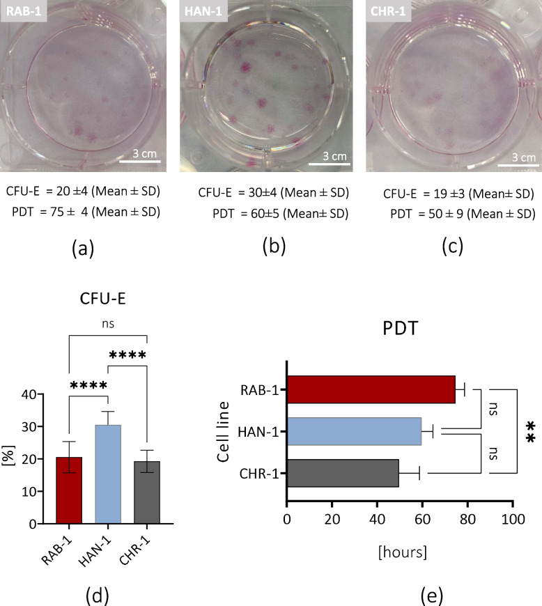 Figure 5
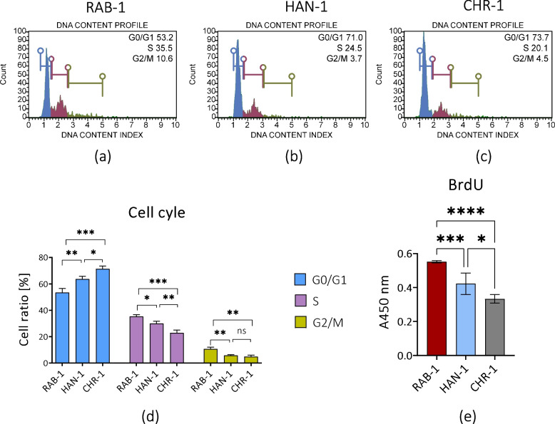 Figure 6
 Figure 7
 Figure 8
 Figure 9
Peer Reviews
No public reviews on file for this paper yet. If you reviewed it on a platform where reviews are public (OpenReview, ICLR, NeurIPS, ICML), you can paste yours below so the community can read it here.
Videos
No videos yet. Explain this paper in a talk, walkthrough, or lecture? Add one.
Taxonomy
TopicsHistiocytic Disorders and Treatments · Extracellular vesicles in disease · Tumors and Oncological Cases