Quantitative assessments of retinal macular structure among rural-dwelling older adults in China: a population-based, cross-sectional, optical coherence tomography study
Qinghua Zhang, Cong Zhang, Yongxiang Wang, Lin Cong, Keke Liu, Zhe Xu, Chunyan Jiang, Weiyan Zhou, Chunxiao Zhang, Yi Dong, Jianli Feng, Chengxuan Qiu, YiFeng Du

TL;DR
This study compares retinal macular structures in older rural Chinese adults using two OCT scanners and finds associations with age, sex, education, and cardiovascular disease.
Contribution
The study provides new insights into macular structure differences using two OCT models and identifies demographic and health factors influencing retinal thickness in rural older adults.
Findings
Spectralis OCT showed higher macular thickness but lower volume compared to Primus 200 OCT.
Older age and female sex were associated with lower macular thickness and volume.
Education level and cardiovascular disease were linked to specific macular thickness changes.
Abstract
To quantitatively assess and compare retinal macular structures of rural-dwelling older adults in China using two different optical coherence tomography (OCT) scanners and to examine their associations with demographic, lifestyle, clinical and ocular factors. This population-based, cross-sectional study included 971 participants (age ≥60 years) derived from the Multimodal Interventions to Delay Dementia and Disability in Rural China study. We collected data on demographics, lifestyle factors, clinical conditions (eg, cardiovascular disease (CVD)) and ocular factors (eg, visual acuity and spherical equivalent). We used two models of spectral-domain OCT to measure macular parameters in nine Early Treatment Diabetic Retinopathy Study subfields. Data were analysed using the multiple general linear models. Spectralis OCT demonstrated higher macular thickness but a lower macular volume than…
Genes, proteins, chemicals, diseases, species, mutations and cell lines named across the full text — each resolved to its canonical identifier and authoritative record.
Click any figure to enlarge with its caption.
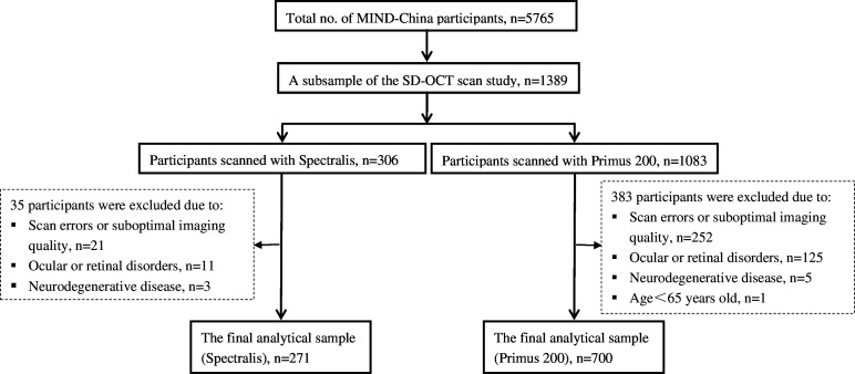 Figure 1
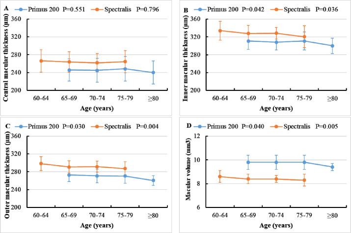 Figure 2
Peer Reviews
No public reviews on file for this paper yet. If you reviewed it on a platform where reviews are public (OpenReview, ICLR, NeurIPS, ICML), you can paste yours below so the community can read it here.
Videos
No videos yet. Explain this paper in a talk, walkthrough, or lecture? Add one.
Taxonomy
TopicsRetinal Imaging and Analysis · Retinal and Optic Conditions · Retinal Diseases and Treatments