Bullous Pemphigoid Causing Successive Emergency Department Visits
Edmund Hsu, Andrew T. Kinoshita, C. Eric McCoy

TL;DR
An elderly man with bullous pemphigoid had multiple emergency visits before receiving proper dermatology care.
Contribution
This case highlights the diagnostic challenges and clinical progression of bullous pemphigoid in older adults.
Findings
Bullous pemphigoid can cause multiple emergency department visits due to blistering and pruritus.
Diagnosis and treatment require dermatology care rather than initial emergency interventions.
BP incidence is rising, particularly in older adults with atypical presentations.
Abstract
In this case presentation, an 84-year-old male with Fitzpatrick type IV skin tone experienced blistering due to bullous pemphigoid (BP), first on the distal upper left extremity and then on the distal lower extremities, chest, and back. These symptoms resulted in three visits to the emergency department within a month, as well as an episode of hospitalization. Despite treatment, the blistering did not resolve until future outpatient care with dermatology. Bullous pemphigoid is a rare autoimmune disease where autoantibodies target hemidesmosomal proteins causing basement membrane destruction and tense subepithelial bullae with pruritus. While uncommon, the incidence of BP is increasing. Bullous pemphigoid tends to affect older adults, appearing as a rash prior to bullae formation on the abdomen, extremities, groin, axillae, or mucosa. Bullous pemphigoid may also be drug-related with…
Genes, proteins, chemicals, diseases, species, mutations and cell lines named across the full text — each resolved to its canonical identifier and authoritative record.
Click any figure to enlarge with its caption.
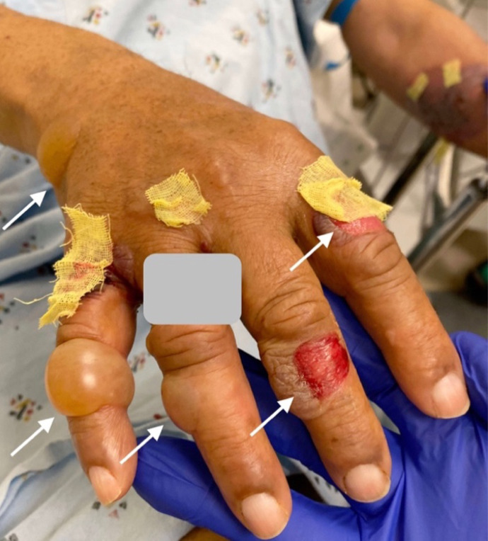 Image 1
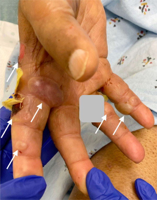 Image 2
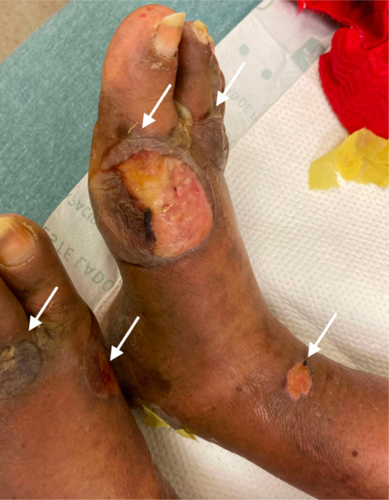 Image 3
Peer Reviews
No public reviews on file for this paper yet. If you reviewed it on a platform where reviews are public (OpenReview, ICLR, NeurIPS, ICML), you can paste yours below so the community can read it here.
Videos
No videos yet. Explain this paper in a talk, walkthrough, or lecture? Add one.
Taxonomy
TopicsAutoimmune Bullous Skin Diseases · Coagulation, Bradykinin, Polyphosphates, and Angioedema · Urticaria and Related Conditions
CASE PRESENTATION
An 84-year-old male with a history of diabetes mellitus, Alzheimer’s disease, Parkinson’s disease, and coronary artery disease with previous coronary artery bypass graft presented to the emergency department (ED) with hyperglycemia and blistering on his distal upper and lower extremities at multiple stages of healing (Images 1–3). Four weeks prior, the patient went to the ED for painless pruritic blistering on the left upper extremity that had spread toward the axilla and groin over a period of two months. He was treated for presumptive bullous pemphigoid (BP) and discharged on doxycycline and clobetasol. Symptoms improved until two weeks following ED discharge, and the patient returned to the ED for now painful blistering of his distal lower extremities, as well as blistering of the chest and back. He was admitted to the hospital for three days and seen by dermatology, who entertained the diagnosis of bullous lymphedema versus bullous diabeticorum. A biopsy was not performed due to concern for poor wound healing. Leg compression and elevation was recommended upon discharge.
Dorsal aspect of hand with bullous pemphigoid blisters, some ruptured; arrows indicate blisters. (Gray box covers patient identifier.)
Ventral aspect of hand with bullous pemphigoid, some ruptured; arrows indicate blisters. (Gray box covers patient identifier.)
Foot and ankle with bullous pemphigoid, some ruptured. Arrows indicate blisters.
The patient returned to the ED one week after discharge due to ongoing blisters and extremity swelling. At this visit, vital signs were stable with most recent hemoglobin A1C elevated at 10.0% (normal below 5.7%; prediabetes 5.7%–6.4%; diabetes above 6.5%). Dermatology was consulted and recommended outpatient evaluation. Insulin was modified, and the patient was discharged. Diagnosis of BP was confirmed at later outpatient evaluation through positive tests for BP antibodies. The patient responded to prednisone on an oral treatment regimen of 60 milligrams (mg) daily for 30 days, tapered to 40 mg for three weeks and then 20 mg for three weeks.
DISCUSSION
Bullous pemphigoid is a rare autoimmune disease in which autoantibodies target hemidesmosomal proteins dystonin-e (BP antigen 1 or BP230) and collagen XVII (BP antigen 2 or BP180), causing basement membrane destruction and tense subepithelial bullae with pruritus.1 ^,^ 2 Incidence is increasing with estimates in the United States and European states between 10-43 per one million individuals per year.2 ^,^ 3 Classically, BP affects older adults, sometimes appearing as a rash before bullae of 1–3 centimeters (cm) diameter appear.1 The images presented here demonstrate BP of moderate severity on light brown skin tone (Fitzpatrick type IV). Distribution is often symmetric and commonly affects the abdomen, extremities, groin, axillae, or mucosa.1 The disease is usually chronic with exacerbations and remissions.1
Bullous pemphigoid is associated with some neurologic disorders, including dementia and Parkinson’s disease.2 Atypical presentations of BP include non-bullous pemphigoid, which accounts for 20% of cases.2 Additionally, BP has been associated with many classes of drugs, and this may also cause atypical presentations, including younger age of onset or blisters without initial rash.4 Drug-related BP may resolve fully with cessation of the offending agent.1 Diagnostic studies used in the evaluation of patients with lesions suspicious of BP include biopsy (for histopathologic examination and direct immunofluorescence microscopy) and serum tests to detect circulating anti-basement membrane zone antibodies.1 ^,^ 2 The mainstays of initial treatment for this condition include corticosteroids and doxycycline.1 ^,^ 5
The reference list from the paper itself. Each links out to its DOI / PubMed record.
- 1BağcıIS Horváth ON Ruzicka Tet al. Bullous pemphigoid. Autoimmun Rev. 2017;16(5):445–55.28286109 10.1016/j.autrev.2017.03.010 · doi ↗ · pubmed ↗
- 2Montagnon CM Tolkachjov SN Murrell D Fet al. Subepithelial autoimmune blistering dermatoses: clinical features and diagnosis. J Am Acad Dermatol. 2021;85(1):1–14.33684496 10.1016/j.jaad.2020.11.076 · doi ↗ · pubmed ↗
- 3Shen WC Chiang HY Chen P Set al. Risk of all-cause mortality, cardiovascular disease mortality, and cancer mortality in patients with bullous pemphigoid. JAMA Dermatol. 2022;158(2):167–75.34964804 10.1001/jamadermatol.2021.5125 PMC 8717210 · doi ↗ · pubmed ↗
- 4Stavropoulos PG Soura E Antoniou C. Drug-induced pemphigoid: a review of the literature. J Eur Acad Dermatol Venereol. 2014;28(9):1133–40.24404939 10.1111/jdv.12366 · doi ↗ · pubmed ↗
- 5Williams HC Wojnarowska F Kirtschig Get al. Doxycycline versus prednisolone as an initial treatment strategy for bullous pemphigoid: a pragmatic, non-inferiority, randomised controlled trial [published correction appears in Lancet. 2017;390(10106):1948]. Lancet. 2017;389(10079):1630–8.28279484 10.1016/S 0140-6736(17)30560-3PMC 5400809 · doi ↗ · pubmed ↗