The Best Strategy for the Black Hole Phenomenon between Intravascular Ultrasound and Optical Coherence Tomography
Cheng-Cheng Kan, Wei-Che Tsai, Cheng-Chung Cheng, Gwo-Ping Jong

TL;DR
This paper discusses the black hole phenomenon in coronary arteries, comparing how well two imaging techniques detect it.
Contribution
The paper presents a case where OCT more effectively identifies the black hole phenomenon compared to IVUS.
Findings
The BH phenomenon is better identified by OCT than by IVUS.
The BH phenomenon is uncommon after drug-eluting stent implantation.
Abstract
The black hole (BH) phenomenon is an intraluminal restenotic lesion. It was identified by intravascular ultrasound (IVUS) and optical coherence tomography (OCT) after intracoronary brachytherapy and drug-eluting stent implantation. Despite the similarity in the mode of action of brachytherapy and drug-eluting stent implantation, the BH phenomenon appears to be uncommon after drug-eluting stent implantation. Specifically, the BH phenomenon is better identified by OCT than by IVUS. Herein, we present a case of in-stent restenosis with suspected BH phenomenon on IVUS and confirmed by OCT.
Genes, proteins, chemicals, diseases, species, mutations and cell lines named across the full text — each resolved to its canonical identifier and authoritative record.
Click any figure to enlarge with its caption.
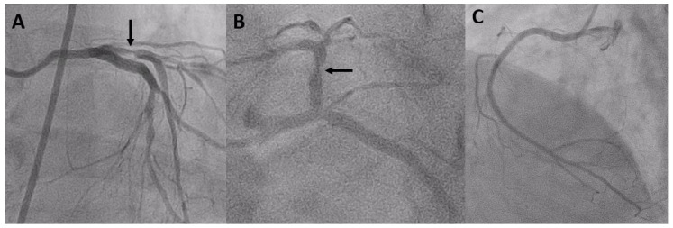 Figure 1
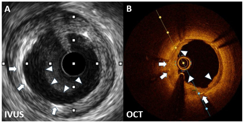 Figure 2
Peer Reviews
No public reviews on file for this paper yet. If you reviewed it on a platform where reviews are public (OpenReview, ICLR, NeurIPS, ICML), you can paste yours below so the community can read it here.
Videos
No videos yet. Explain this paper in a talk, walkthrough, or lecture? Add one.
Taxonomy
TopicsCoronary Interventions and Diagnostics · Cardiac Imaging and Diagnostics · Cerebrovascular and Carotid Artery Diseases
In-stent restenosis (ISR) is a common complication that can occur following the placement of a coronary stent [1,2]. In recent years, the use of drug-eluting stents (DESs) has reduced ISR and major adverse cardiac events [3,4]. However, ISR after DES implantation still occurs [5,6]. The black hole (BH) phenomenon is when an intraluminal restenotic lesion image is obtained by intravascular ultrasound (IVUS) and optical coherence tomography (OCT) [7,8,9,10,11,12]. The development of ISR involves multiple factors, including biological, mechanical, and patient- and operator-related factors [13,14,15]. Intracoronary imaging is crucial for determining the specific mechanism of ISR, enabling tailored treatment strategies based on the identified cause. Therefore, we present a case of an ISR lesion with a suspected BH phenomenon on IVUS confirmed by OCT.
A 72-year-old man presented to our outpatient department with incremental exertional dyspnea and chest pain over the past 2 weeks. He had no history of cardiovascular risk factors, such as smoking and alcohol consumption, but had hypertension and type 2 diabetes (T2D). Additionally, he had a history of ischemic heart disease (chronic coronary syndrome) and underwent his first percutaneous coronary intervention (PCI) on 21 June 2022, which involved sirolimus-eluting stents (SESs) being placed at the proximal portion of the left anterior descending (LAD) artery and the proximal to the middle portion of the left circumflex artery. His ongoing medications included aspirin, clopidogrel, bisoprolol, atorvastatin, ezetimibe, furosemide, and hypoglycemic agents (pioglitazone, glimepiride, and dulaglutide).
During the cardiovascular outpatient department visit, his symptoms subsided, and ECGs displayed sinus rhythms without significant ST/T wave changes for 9 months. According to the history and positive findings of a treadmill exercise test, he was admitted for coronary angiography. On admission, his laboratory data revealed poor diabetic control (hemoglobin A1C of 8.3%) and controlled hyperlipidemia (total cholesterol, 132 mg/dL; triglycerides, 54 mg/dL; low-density lipoprotein, 55 mg/dL; and high-density lipoprotein, 59 mg/dL). Blood counts, serum electrolytes, and renal and liver function tests were within normal limits.
Coronary angiography on 27 April 2023 revealed severe ISR of the proximal LAD (Figure 1). A 60 MHz high-definition IVUS was performed, which revealed the homogenous echolucent appearance of ISR of the SES (Figure 2A) and a suspected BH phenomenon.
Given the significant constriction of the lumen area of the ISR lesion, obtaining a high-quality OCT image without balloon predilatation was challenging. Hence, we chose a 2.0 mm balloon to minimize the effect on the original lesion. OCT revealed a layered pattern with a superficial high signal-intensity band adjacent to the luminal surface and a signal-poor region near the stent (Figure 2B). According to the above imaging findings, the patient was diagnosed with a BH phenomenon through OCT.
A DES was implanted and subsequently post dilated using a noncompliant balloon. After PCI, the lumen surface appeared smooth, with no evidence of tissue prolapse or residual intraluminal thrombus, with a well-deployed stent. Thrombolysis in myocardial infarction III distal blood flow was achieved using angiography.
Following the placement of a coronary stent, ISR can commonly emerge as a complication [1,2]. Despite advances in technology, the occurrence rate of ISR has remained relatively consistent, affecting approximately 10% of patients after DES implantation [5,6].
The cause of ISR is multifactorial and can be attributed to various biological, mechanical, and procedural factors [13,14,15]. The mechanical factors primarily include stent expansion or fracture, whereas the biological factors involve localized inflammation leading to excessive neointimal growth and late neoatherosclerosis.
Previous epidemiological studies have revealed T2D as an important risk factor for developing ISR in patients following DES implantation [16,17]. Another study showed that the presence of T2D with elevated hemoglobin A1C levels is associated with ISR development in patients with DES [18]. The findings from this case highlight an increased ISR risk in patients with T2D and high hemoglobin A1C levels. Therefore, maintaining satisfactory sugar control in these patients may help prevent ISR following DES implantation.
The “BH” phenomenon, which has been characterized as an intraluminal restenotic lesion with a homogeneous black appearance (echolucent) on IVUS, was initially observed in patients following brachytherapy [7]. This phenomenon has also been identified in patients who have received SES implants [8].
DES improved BMS limitations but raised concerns about complications such as late restenosis and thrombosis. Late DES thrombosis is marked by delayed healing with impaired reendothelialization and persistent fibrin deposition. The exact cause of late restenosis remains unclear but may involve a delayed healing response to stent polymers and drugs.
Although the BH phenomenon is unusual in DES restenosis, establishing a universal mechanism is challenging. Tissue analysis of the BH revealed a primarily hypocellular matrix with areas rich in proteoglycans, possibly due to delayed vascular wound healing following SES implantation. The echolucent appearance was likely attributed to this hypocellular matrix and its high water content [9,10].
OCT revealed a layered structure with an inner layer with high signal intensity and outer layers with low signal intensity. The low OCT signal intensity was a result of restenotic tissue rich in proteoglycan and poor in collagen matrix or fibrin-rich thrombus formation. Although various restenotic tissues can exhibit low OCT signal intensity, the consistent border and structure in this case suggest that the BH phenomenon in OCT may have been due to the lack of organized mature connective tissue elements in the restenotic tissue [11,12].
Assessments by IVUS or OCT before DES restenosis treatment can help identify restenotic tissue characteristics and guide the development of an optimal strategy to prevent recurrent ISR. However, OCT is superior to IVUS for the diagnosis of the BH phenomenon after DES implantation.
We conclude that a BH phenomenon can occur after SES implantation. The described case suggests the superiority of OCT over IVUS rather than further research being necessary.
The reference list from the paper itself. Each links out to its DOI / PubMed record.
- 1Kyaw H. Johal G. Gedela M. Barman N. Kini A. Sharma S.K. Is coronary brachytherapy staging a Comeback for the treatment of in-stent sestenosis?Curr. Cardiol. Rep.20212315610.1007/s 11886-021-01582-434599432 · doi ↗ · pubmed ↗
- 2Huang C.W. Huang M.S. Su P.F. Chao T.H. Lee C.H. Liu P.Y. Management of restenosis after stenting in left main coronary artery disease Acta Cardiol. Sin.2023392772863691155110.6515/ACS.202303_39(2).20220821 APMC 9999179 · doi ↗ · pubmed ↗
- 3Liou K. Jepson N. Cao C. Luo R. Pala S. Ooi S.Y. Drug-eluting balloon versus second generation drug eluting stents in the treatment of in-stent restenosis: A systematic review and meta-analysis Heart Lung Circ.2016251184119410.1016/j.hlc.2016.04.00127180214 · doi ↗ · pubmed ↗
- 4Macovei L. Magopet R. Campo G. Drug-eluting stents: New presumed effects over in-stent restenosis prevention Minerva Cardiol. Angiol.20216914114310.23736/S 2724-5683.20.05372-432975393 · doi ↗ · pubmed ↗
- 5Fu G. Yu Z. Chen Y. Chen Y. Tian F. Yang X. Direct adsorption of anti-CD 34 antibodies on the Nano-Porous stent surface to enhance endothelialization Acta Cardiol. Sin.2016322732802727416710.6515/ACS 20150813 APMC 4884754 · doi ↗ · pubmed ↗
- 6Parfrey S. Siu V. Graham J.J. Vijayaraghavan R. Li C. Pang J. Kalra S. Džavík V. Wijeysundera H.C. Bagai A. from the University of Toronto CTO Collaborative. Evaluation and management of drug-eluting stent in-stent restenosis Curr. Opin. Cardiol.20233843344010.1097/HCO.000000000000107337477129 · doi ↗ · pubmed ↗
- 7Castagna M.T. Mintz G.S. Weissman N. Maehara A. Finet G. Waksman R. "Black hole": Echolucent restenotic tissue after brachytherapy Circulation 200110377810.1161/01.CIR.103.5.77811156893 · doi ↗ · pubmed ↗
- 8Hirose M. Kobayashi Y. Leon M.B. Echolucent neointimal hyperplasia "dark wall" after sirolimus eluting stent implantation Heart 200490114310.1136/hrt.2003.01102315367508 PMC 1768498 · doi ↗ · pubmed ↗