Non-Invasive High-Resolution Imaging of In Vivo Human Myelinated Axons
Marco Lombardo, Massimo Cesareo, Benedetto Falsini, Andrea Cusumano

TL;DR
This study uses a new imaging technique to visualize myelinated nerve fibers in the human eye with high resolution, offering insights into nerve anatomy and disease.
Contribution
The first non-invasive in vivo imaging of human myelinated axons at microscopic resolution using transscleral optical phase imaging with adaptive optics.
Findings
Human myelinated retinal nerve fibers were imaged in vivo at 2–3 micrometer resolution.
The method provided better detail than traditional ocular imaging techniques like optical coherence tomography.
The microscopic appearance of human myelin and myelinated axons was documented for the first time.
Abstract
This work aims to reveal the microscopic (2–3 micrometer resolution) appearance of human myelinated nerve fibers in vivo for the first time. We analyzed the myelinated retinal nerve fibers of a male patient without other neurological disorders in a non-invasive way using the transscleral optical phase imaging method with adaptive optics. We also analyzed the fellow eye with non-myelinated nerve fibers and compared the results with traditional ocular imaging methods such as optical coherence tomography. We documented the microscopic appearance of human myelin and myelinated axons in vivo. This method allowed us to obtain better details than through traditional ocular imaging methods. We hope these findings will be useful to the scientific community to evaluate neuro-retinal structures through new imaging techniques and more accurately document nerve anatomy and the pathophysiology of…
Genes, proteins, chemicals, diseases, species, mutations and cell lines named across the full text — each resolved to its canonical identifier and authoritative record.
Click any figure to enlarge with its caption.
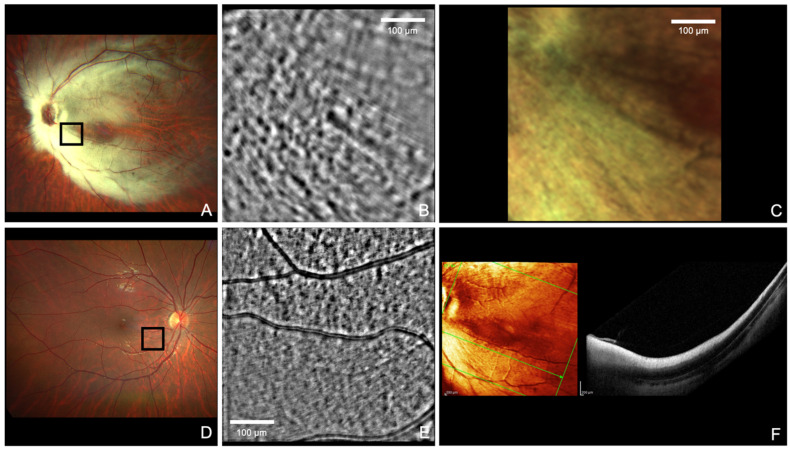 Figure 1
Peer Reviews
No public reviews on file for this paper yet. If you reviewed it on a platform where reviews are public (OpenReview, ICLR, NeurIPS, ICML), you can paste yours below so the community can read it here.
Videos
No videos yet. Explain this paper in a talk, walkthrough, or lecture? Add one.
Taxonomy
TopicsOptical Coherence Tomography Applications · Glaucoma and retinal disorders · Retinal Development and Disorders