Notes on species of Talaus Simon, 1886 (Araneae, Thomisidae) from China, with descriptions of two new species

Abstract
Genes, proteins, chemicals, diseases, species, mutations and cell lines named across the full text — each resolved to its canonical identifier and authoritative record.
Click any figure to enlarge with its caption.
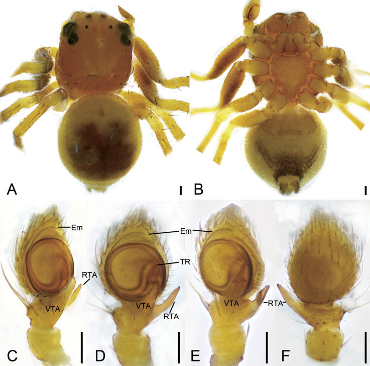 Figure 1
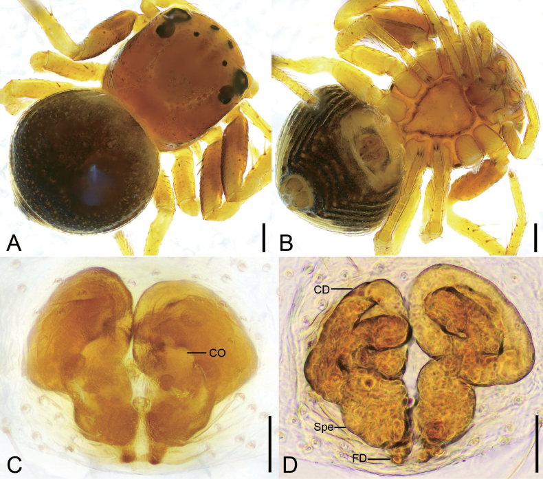 Figure 2
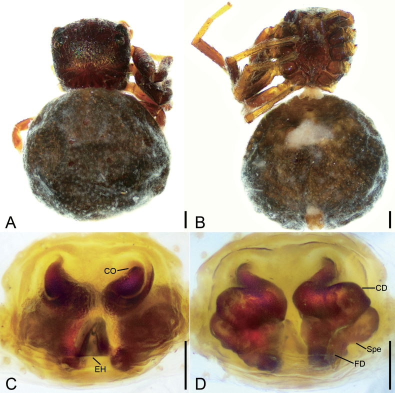 Figure 3
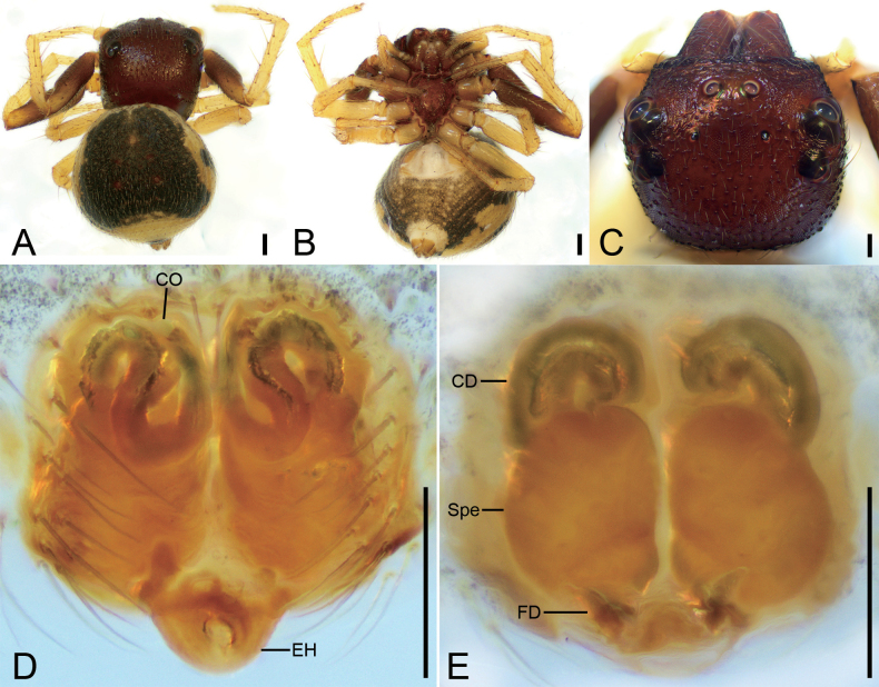 Figure 4
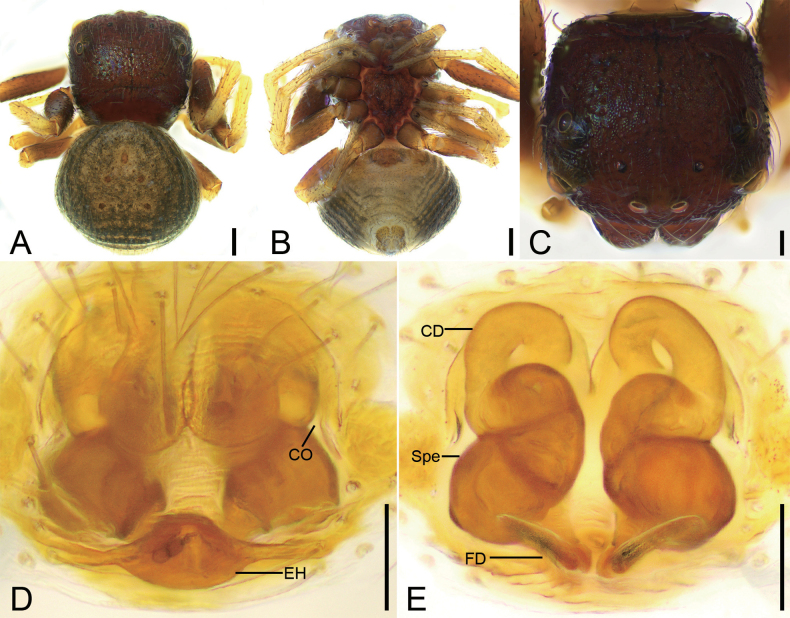 Figure 5
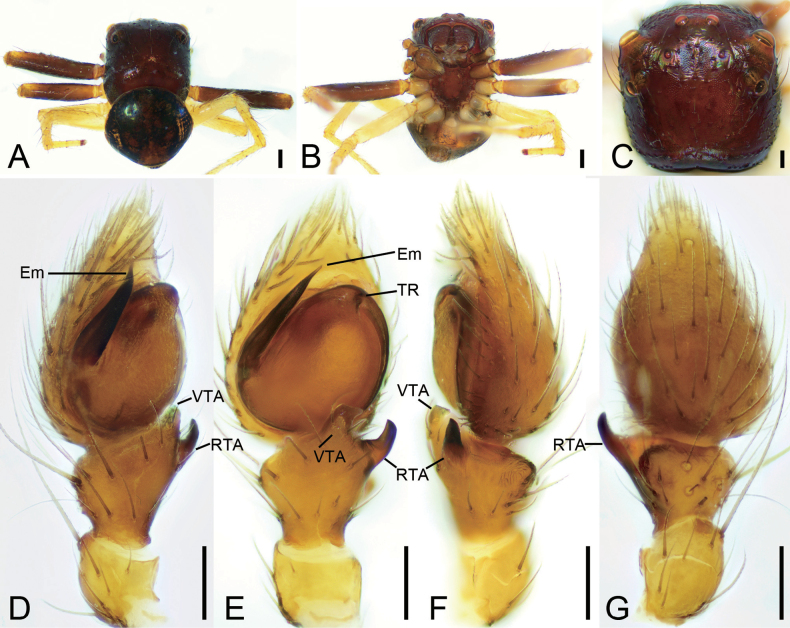 Figure 6
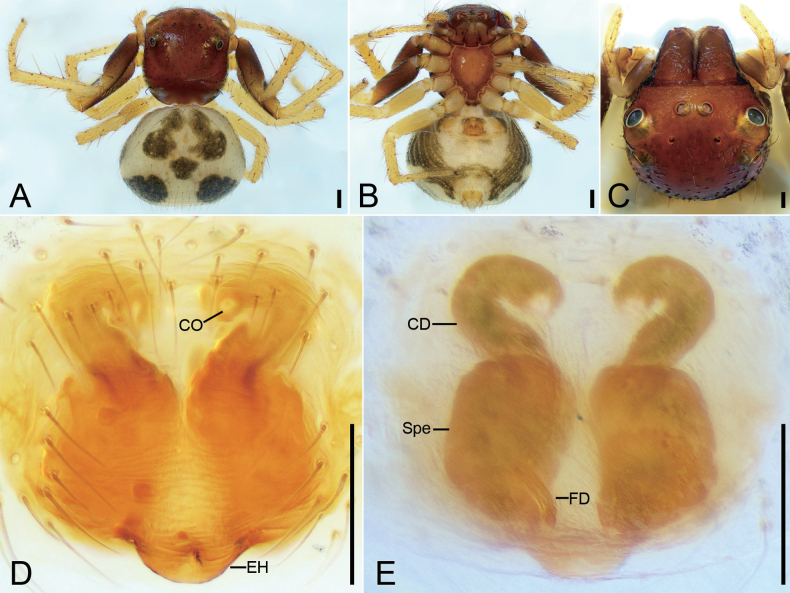 Figure 7
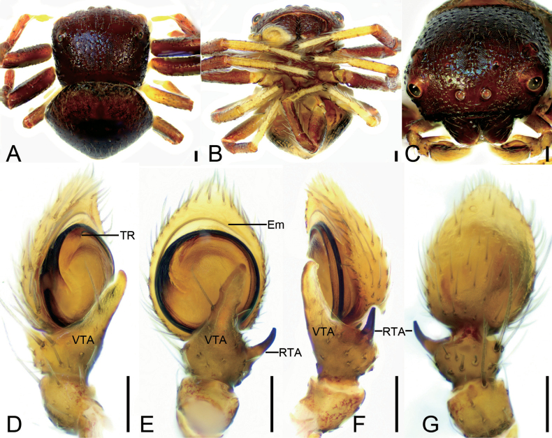 Figure 8
 Figure 9
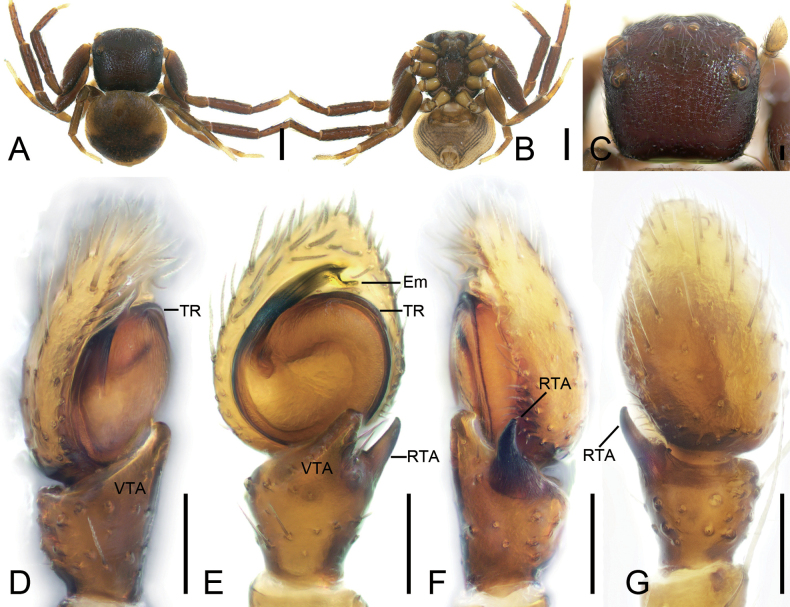 Figure 10
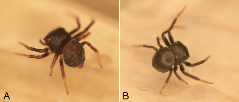 Figure 11
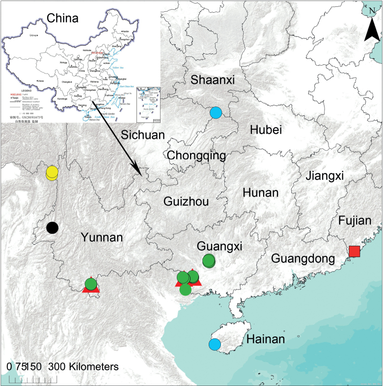 Figure 12
Peer Reviews
No public reviews on file for this paper yet. If you reviewed it on a platform where reviews are public (OpenReview, ICLR, NeurIPS, ICML), you can paste yours below so the community can read it here.
Videos
No videos yet. Explain this paper in a talk, walkthrough, or lecture? Add one.
Taxonomy
TopicsSpider Taxonomy and Behavior Studies · Lepidoptera: Biology and Taxonomy · Genetic diversity and population structure
Introduction
The crab spider genus Talaus Simon, 1886 comprises 13 species, of which 12 are endemic in Southeast Asia (WSC 2023); the validity of the other species, T.limbatus Simon, 1895 recorded from South Africa based on a juvenile specimen (Simon 1895), was doubted by Benjamin (2020). The genus has been neglected for the past ten years and only four species are known from Yunnan, Guangxi and Hainan in southwest and south China (Li and Lin 2016).
Talaus is currently non-monotypic with its type species, Talaustriangulifer Simon, 1886, described from Sumatra, Indonesia based on a female specimen (Benjamin et al. 2008; WSC 2023). Based on the combination of morphological characters of T.beccarii Benjamin, 2020, T.nanus Thorell, 1890, T.oblitus O. Pickard-Cambridge, 1899, T.opportunus (O. Pickard-Cambridge, 1873) and T.xiphosus Zhu & Ono, 2007, Benjamin (2020) re-defined the genus with the following features: the male palp with a retrolateral tibial apophysis and a ventral tibial apophysis, with a dorsal tibial apophysis in some species, and a short or long spiniform embolus with a broad base; the epigyne lacks a median septum, has a short copulatory duct, and the spermathecae are rounded to irregular in shape. Furthermore, Benjamin (2020) highlighted questions about the species to be included in this genus, and suggested that T.limbatus from South Africa was probably misplaced, that T.xiphosus might be a synonym of the type species T.triangulifer; and that T.elegans Thorell, 1890 might also be a junior synonym of T.triangulifer. When these problems are resolved, this genus will be unambiguous in the spider family Thomisidae Sundevall, 1833.
The present paper describes six species based on ample material (including some paratypes) from China which have been collected by spider enthusiasts, our colleagues, and the authors. Two new Talaus species are identified as undescribed and examination of these specimens reveals that T.xiphosus is a junior synonym of T.triangulifer.
Material and methods
The new materials were collected in the bush with the malaise trap, fogging and beating method. Specimens were examined using a SZ6100 stereomicroscope. Both male and female copulatory organs were dissected and examined in 80% ethanol using an Olympus CX43 compound microscope with a KUY NICE CCD camera (Beijing Tiannuoxiang Scientific Instrument Co., Ltd, China). Epigynes were cleared with pancreatin solution (Álvarez-Padilla and Hormiga 2007).
The measurements were taken using a stereomicroscope (AxioVision SE64 Rel. 4.8.3) and are given in millimeters. The body lengths of all specimens exclude the chelicerae and spinnerets. Terminology of the male and female copulatory organs follows Benjamin (2020). Leg measurements are given as total length (femur, patella, tibia, metatarsus, tarsus). The abbreviations used in the figures are as follows:
ALE anterior lateral eye;
AME anterior median eye;
CD copulatory duct;
CO copulatory opening;
d dorsal;
Em embolus;
EH epigynal hood;
FD fertilization duct;
MOA median ocular area;
p prolateral;
PLE posterior lateral eye;
PME posterior median eye;
r retrolateral;
RTA retrolateral tibial apophysis;
Spe spermatheca;
TR tegular ridge;
v ventral;
VTA ventral tibial apophysis.
Depositories of all specimens examined are abbreviated as:
ASM-JGSU Animal Specimen Museum, College of Life Science, Jinggangshan University, Ji’an, China;
CASCalifornia Academy of Sciences, San Francisco, USA;
HNUHunan Normal University, Changsha, China.
Taxonomy
Family Thomisidae Sundevall, 1833
Talaus
Taxon classificationAnimaliaAraneaeThomisidae
Genus
Simon, 1886
D2A4F311-21A3-5354-AD39-C5EBA256E015
Type species.
Talaustriangulifer Simon, 1886
Notes.
Before this study, the genus included 13 species (WSC 2023). One-third of these species are recorded from southern China; the validity of one species, T.xiphosus was doubted by Benjamin (2020) and is confirmed as a synonym in this study; therefore, only 12 Talaus species are restricted to Southeast Asia and the Indomalayan Realm (Bhutan, China, India, Indonesia, Myanmar, Sri Lanka, Vietnam; WSC 2023). The 13^th^ species, T.limbatus from South Africa, is probably misplaced.
Talaus
dulongjiang
Taxon classificationAnimaliaAraneaeThomisidae
Tang, Yin, Ubick & Peng, 2008
D3BB4F92-54B5-5A24-9A18-EAAA8E02EF7C
Talaus dulongjiang
Tang et al., 2008: 63, figs 1–12.
Type material examined.
Paratypes. 2 ♂ (DHK-2004-068): China, Yunnan. Province, Gongshan County, Dulongjiang Township, 2.3–3.3 km south of Longyuan Village along Dulongjiang, 28.00532°N, 98.32145°E, 1685 m, 2 November 2004, David Kavanaugh leg. (Tho-159, paratypes examined, HNU); 4 ♀ (Tang-04-08), Mokewang Bridge, 27.83827°N, 98.32103°E, 1455 m, 6–7 November 2004, Guo Tang leg., other data same as previous (Tho-159). Holotype not examined.
Diagnosis.
The male of this species is similar to T.niger Tang, Yin, Ubick & Peng, 2008 (Tang et al. 2008: 65, figs 16–18) in having the same position of the tegular ridge and the filiform embolus, but can be separated from it by the retrolateral tibial apophysis strongly bending forward (vs slightly) and the longer ventral tibial apophysis (vs relatively short) (Fig. 1C–F). The females can be easily separated from T.niger (Tang et al. 2008: 65, fig. 20) by the helical copulatory duct (vs S-shaped) and the slightly separated swollen spermathecae (vs clearly separated) (Fig. 2D).
Talausdulongjiang Tang, Yin, Ubick & Peng, 2008, male A habitus, dorsal view B habitus, ventral view C palp, prolatero-ventral view D palp, ventral view E palp, ventro-retrolateral view F palp, dorsal view. Abbreviations: Em – embolus, RTA – retrolateral tibial apophysis, TR – tegular ridge, VTA – ventral tibial apophysis. Scale bars: 0.1 mm.
Talausdulongjiang Tang, Yin, Ubick & Peng, 2008, female A habitus, dorsal view B habitus, ventral view C epigyne, ventral view D epigyne, dorsal view. Abbreviations: CD – copulatory duct, CO – copulatory opening, FD – fertilization duct, Spe – spermatheca. Scale bars: 0.2 mm (A, B); 0.05 mm (C, D).
Description.
Male habitus as in Fig. 1A, B, palp in Fig. 1C–F. Palp: tibia with two apophyses, the ventral one slightly shorter than retrolateral one, but longer than tibia, with a strongly curved apex directed prolaterally; retrolateral one relatively thin, basally slightly curved antero-retrolaterally in ventral view, longer than tibia; tegular ridge long, basally arising from ~ 12 o’clock position of the tegulum; embolus filiform, spiraling nearly 3/4 coil, arising from 3 o’clock and ending at ~ 1 o’clock on tegulum. Female habitus as in Fig. 2A, B, epigyne in Fig. 2C, D. Epigyne: copulatory openings relatively large, sub-antero-laterally located; copulatory ducts broad and long, looping 1.75 coils; spermathecae swollen, slightly separated by ~ 1/7 of their width; fertilization ducts located posteriorly, directed anterolaterally.
Distribution.
Known from Yunnan Province, China (Fig. 12).
Remarks.
According to Tang et al. (2008) the original material consisted of three males and seven females; two males and four females were deposited in HNU and one male and three females in CAS. However, only two males and four females (but not the holotype) were found in HNU and there are no striking markings on them: the label data match the localities of the paratypes recorded by Tang et al. (2008: 63).
Talaus
niger
Taxon classificationAnimaliaAraneaeThomisidae
Tang, Yin, Ubick & Peng, 2008
83943FE9-595B-5569-AC5E-3DC31173A7FB
Talaus niger
Tang et al., 2008c: 65, figs 13–23.
Type material examined.
Paratype. 2 ♀ (GKJ020): CHINA, Yunnan Province, Tengchong County, Wuhe Township, Tongjiazhuang Village, Longchuanjiang River (Longjiang Bridge), along river, 24.89284°N, 98.67439°E, 1210 m, 24 May 2005, Heng-mei Yan & Ke-ji Guo leg. (Tho-202). Holotype not examined.
Diagnosis.
The female can easily be recognized by the S-shaped copulatory ducts with a swollen median part (Fig. 3D).
Description.
Female habitus as in Fig. 3A, B, epigyne in Fig. 3C, D. Epigyne: copulatory openings relatively large, antero-medially located, with sclerotized and round margins; epigynal hood posteriorly located, thumb-like; copulatory ducts S-shaped, anterior part relative narrow, L-shaped, medial and posterior part swollen, extending transversally; spermathecae triangular, widely separated by approximately half of their width; fertilization ducts located posteriorly, directed anteriorly.
Talausniger Tang, Yin, Ubick & Peng, 2008, female A habitus, dorsal view B habitus, ventral view C epigyne, ventral view D epigyne, dorsal view. Abbreviations: CD – copulatory duct, CO – copulatory opening, EH – epigynal hood, FD – fertilization duct, Spe – spermatheca. Scale bars: 0.2 mm (A, B); 0.05 mm (C, D).
Distribution.
Known only from the type locality in Yunnan Province, China (Fig. 12).
Remarks.
According to Tang et al. (2008: 65), the original materials consisted of a male and two females, one female deposited in HNU and one female in CAS. However, two females and no male holotype were found in HNU and there are no labels designating the paratypes. Based on the locality information and the original illustration given in their paper, these specimens can be confirmed as the paratypes of T.niger and have been labelled as such. They are somewhat shriveled after alcohol evaporated during storage, but their epigynes still can be clearly recognized after pancreatin digestion and are now placed on labelled slides.
Talaus
sulcus
Taxon classificationAnimaliaAraneaeThomisidae
Tang & Li, 2010
8D5EA179-6D30-5DA9-8557-0E4E2A01D271
Talaus sulcus Tang & Li, 2010b: 93, f. 71A–D, 72A–F, 73A–D.
Other material examined.
2 ♀: China, Guangxi Zhuang Autonomous Region, Chongzuo City, Jiangzhou District, Zuozhou Town, Nongxue Village, 22°36.024'N, 107°24.93'E, 252 m, 5 September 2015, Bing Zhou, Wang Liu, Ji-he Liu, Qu Cai, Xian-feng Huang & Da Li leg. (Tho-334, HNU); 1 ♀, Guanghe Village, Pairutun, Nongzui, 22°32.556'N, 107°26.970'E, 311 m, 11 September 2015, other data same as previous (Tho-334, HNU); 1 ♀, Quxi Village, Nongqiong, 22°34.208'N, 107°25.003'E, 276 m, 31 August 2015, other data same as previous (Tho-334, HNU); 1 ♀, Longzhou County, Nonggang National Nature Reserve, Longjiang Station, Checkpoints, 22.4777°N, 106.9092°E, 204 m, 28 October 2017, Ai-lan He, Ke-ke Liu, Qu Cai, Ji-he Liu, Jin-xin Liu & Zong-guang Huang leg. (Tho-335, HNU).
Diagnosis.
Female resembles Talaustriangulifer (Fig. 7C, D) in having a bean-shaped spermathecae, but can be easily recognized by the copulatory openings located antero-medially and directed posteriorly (vs located antero-medially and directed anteriorly, or located latero-medially) and the S-shaped or spiral copulatory ducts (vs C-shaped) (Figs 4D, E, 5D, E).
Talaussulcus Tang & Li, 2010, female A habitus, dorsal view B habitus, ventral view C prosoma, dorso-frontal view D epigyne, ventral view E epigyne, dorsal view. Abbreviations: CD – copulatory duct, CO – copulatory opening, EH – epigynal hood, FD – fertilization duct, Spe – spermatheca. Scale bars: 0.2 mm (A, B); 0.1 mm (C–E).
Talaussulcus Tang & Li, 2010, female A habitus, dorsal view B habitus, ventral view C prosoma, dorsal view D epigyne, ventral view E epigyne, dorsal view. Abbreviations: CD – copulatory duct, CO – copulatory opening, EH – epigynal hood, FD – fertilization duct, Spe – spermatheca. Scale bars: 0.2 mm (A, B); 0.1 mm (C); 0.05 mm (D, E).
Description.
Female habitus as in Figs 4A, B, 5A, B, eyes as in Figs 4C, 5C, epigyne as in Figs 4D, E, 5D, E. Epigyne: copulatory openings small, antero-medially located, separated by half of width of epigynal hood; epigynal hood postero-medially located, looks like a semi-circular extrusion; copulatory ducts long, S-shaped in ventral view, C-shaped in dorsal view; spermathecae ovoid, swollen, slightly separated from each other; fertilization ducts postero-medially located, directed anterolaterally. The epigynes also include the other variations: copulatory openings small, slit-like, latero-medially located; epigynal hood postero-medially located, triangular, curved forward; copulatory ducts long, like an oval ring in dorsal view; spermathecae sac-shaped, with obvious constrictions, nearly sac-shaped, anterior slightly separated from each other anteriorly; fertilization ducts postero-medially located, touching their bases, directed anterolaterally.
Distribution.
Known from Yunnan (Tang and Li 2010) and Guangxi Province (present records), China (Fig. 12).
Remarks.
The detailed study of newly collected specimens from Guangxi revealed that they have two variants of the epigyne conformation as shown for paratype specimens from Yunnan by Tang and Li (2010). Specimens from Jiangzhou District have S-shaped (in ventral view) and C-shaped (in dorsal view) copulatory ducts and ovoid spermathecae as illustrated in Tang and Li (2010: fig. 72C, D); the specimens from Longzhou County have loop-like copulatory ducts and nearly C-shaped spermathecae similar to the illustration in Tang and Li (2010: fig. 72E, F). However, our new female specimens do not have a yellow abdomen dorsally bearing a large subtriangular spot subposteriorly, which is an acceptable color-variation proposed by Tang and Li (2010: 93).
Talaus
triangulifer
Taxon classificationAnimaliaAraneaeThomisidae
Simon, 1886
EE15D876-64B4-5D18-BC35-4649845C5619
Talaus triangulifer Simon, 1886: 172); Benjamin 2020: 414, figs 2C, H–J, 4E, 7A–B. Talaus xiphosus Zhu & Ono, 2007: 81, figs 1–5; Benjamin 2020: 415 (“probably a synonym of T.triangulifer”). syn. nov.
Other material examined.
3 ♂: China, Guangxi Zhuang Autonomous Region, Chongzuo City, Longzhou County, Nonggang National Nature Reserve, Sanlian Station, Longdan, 22.53470°N, 106.83697°E, 307 m, 31 October 2017, Ai-lan He, Ke-ke Liu, Qu Cai, Ji-he Liu, Jin-xin Liu & Zong-guang Huang leg. (Tho-029, HNU); 1 ♀, 30 October 2017, other data as previous; 14 ♂ 11 ♀, Nonggang Station, Boarded-up Houses, 22.46444°N, 106.92359°E, 188 m, 28 October 2017, other data as previous; 5 ♂ 11 ♀, Core Area, 22.46415°N, 106.93238°E, 228 m, 26 October 2017, other data as previous; 4 ♂ 3 ♀, 27 October 2017, other data as previous; 6 ♂ 6 ♀, Longjiang Ligatures, 22.4770°N, 106.90921°E 204 m, 28 October 2017, other data as previous; 12 ♂ 1 ♀, 27 October 2017, other data as previous; 12 ♂ 8 ♀, Longhengtun, 22.47450°N, 106.98307°E, 270 m, 29 October 2017, other data as previous; 1 ♂ 3 ♀, 22.47166°N, 106.97051°E, 163 m, other data as previous; 1 ♂ 1 ♀, Nanning City, Wuming County, Damingshan National Nature Reserve, Sanbao Station, Chaoyang, 23°31'13.679"N, 108°23'4.560"E, 3 November 2018, 593 m, Ai-lan He, Ke-ke Liu, Hui-juan Sheng, Ji-he Liu, Jin-xin Liu & Zong-guang Huang leg.; 9 ♂ 1 ♀, Ganlan Station, 23°34'15.380"N, 108°25'16.284"E, 7 November 2018, 485 m, other data as previous; 1 ♂ 2 ♀, Shanglin County, Zhaojiang Station, 23°27'1.8"N, 108°23'32.639"E, 6 November 2018, 263 m, other data as previous; 1 ♂ 1 ♀, Jilong Station 23°26'5.279"N, 108°26'32.639"E, 591 m, 5 November 2018, other data as previous; 17 ♂ 28 ♀, Chongzuo City, Jiangzhou District, Zuozhou Town, Guanghe Village, Hecuntun, Nongyao, 22°36.318'N, 107°25.677'E, 224 m, 9 September 2015, Bing Zhou, Wang Liu, Ji-he Liu, Qu Cai, Xian-feng Huang & Da Li leg.; 8 ♂ 7 ♀, Longmitun, Nongxing, 22°34.190'N, 107°26.283'E, 272 m, 7 September 2015, other data as previous; 11 ♀, Hecuntun, Nongteng, 22°35.074'N, 107°25.430'E, 235 m, 4 September 2015, other data as previous; 1 ♂ 11 ♀, Nongdan, 22°34.054'N, 107°24.295'E, 296 m, 30 August 2015, other data as previous; 2 ♂, 12 ♀, Pairutun, 22°34.911'N, 107°25.684'E, 226 m, 3 September 2015, other data as previous; 7 ♂, 26 ♀, Nongheng, along the mountain road, 22°34.740'N, 107°24.915'E, 271 m, 29 August 2015, other data as previous; 2 ♂ 4 ♀, Duolu Town, Duobai Village, Longquantun, Nongquan, 22°32.392'N, 107°27.221'E, 145 m, 10 September 2015, other data as previous.
Diagnosis.
The species can be easily differentiated from other Talaus species by the long straight xiphoid embolus [vs curved in T.beccarii (Benjamin, 2020: 406, fig. 1A); short in T.opportunus (Benjamin 2020: 411, fig. 6A, B); flagelliform and curved in other species]. Females resemble those of T.opportunus (Benjamin 2020: 411, fig. 6C, D) in having a pair of question-mark-like copulatory ducts, but can be easily distinguished from it by the oval spermathecae (vs irregular in T.opportunus). The females are similar to those of T.sulcus (Tang and Li 2010: 93, fig. 72C–F) in having a semi-circular epigynal scape and the swollen spermathecae, but can be separated from it by the question-mark-like copulatory ducts (vs S-shaped or spiral in T.sulcus).
Talaustriangulifer Simon, 1886, male A habitus, dorsal view B habitus, ventral view C prosoma, dorsal view D palp, prolatero-ventral view E palp, ventral view F palp, retrolateral view G palp, dorsal view. Abbreviations: Em – embolus, RTA – retrolateral tibial apophysis, TR – tegular ridge, VTA – ventral tibial apophysis. Scale bars: 0.2 mm (A, B); 0.1 mm (C–G).
Description.
Male habitus as in Fig. 6A, B, eyes as in Fig. 6C, palp as in Fig. 6D–G. Palp: tibia with two apophyses: the ventral one bird-head-like in ventral view, with a sharp, narrowed apex directed retrolaterally; retrolateral one horn-like, well sclerotized, longer than ventral one; tegular ridge arising from ~ 1 o’clock position; embolus (Em) xiphoid, arising from 8 o’clock and ending at ~ 12 o’clock. Female habitus as in Fig. 7A, B, epigyne as in Fig. 7D, E. Epigyne: copulatory openings small, directed backwards, separated by half width of spermathecae; epigynal hood located posteriorly, semi-circular; copulatory ducts question-mark-like; spermathecae nearly oval, swollen, anterior part nearly touching, posterior part separated by less than half of spermathecal width.
Talaustriangulifer Simon, 1886, female A habitus, dorsal view B habitus, ventral view C prosoma, dorso-frontal view D epigyne, ventral view E epigyne, dorsal view. Abbreviations: CD – copulatory duct, CO – copulatory opening, EH – epigynal hood, FD – fertilization duct, Spe – spermatheca. Scale bars: 0.2 mm (A, B); 0.1 mm (C–E).
Distribution.
Known from Indonesia (Borneo, Sumatra; WSC 2023), Malaysia (Sabah; Benjamin 2020), and China (new records, Yunnan and Guangxi provinces; Fig. 12).
Remarks.
According to Zhu and Ono (2007), the original materials of Talausxiphosus were collected from Longzhou County, Chongzuo City in Guangxi Zhuang Autonomous Region. Of these specimens, some were collected from Chongzuo City, the others from Nanning City. Although there is 100 km from the locality of the holotype (Ningming County in Chongzuo City) to the nearest point of the locality of our specimens, and there is intraspecific variation in the abdomen color in the specimens from Zhu and Ono (2007), we did not find any noticeable differences in the male palps or in the female epigynes and therefore confirm Benjamin’s (2020) proposal that T.xiphosus is a junior synonym of T.triangulifer. The results presented here suggest that this species has a wide distribution in Southeast Asia.
Talaus
yuyang
Taxon classificationAnimaliaAraneaeThomisidae
Yao & Liu sp. nov.
E13AC877-4A9D-5E9C-88D6-B81B8AF3D45A
https://zoobank.org/0B1D782D-8049-473F-9399-E05EE564F269
Type material.
Holotype ♂, China: Chongqing Municipality: Chengkou County, Hongjun Park, 31°56'56.89"N, 108°40'15.84"E, late October 2022, Y.Y. Zhou leg. (Tho-322, ASM-JGSU). Paratypes: 1 ♂ 7 ♀, same data as holotype; 1 ♂, 1 ♀; Ledong County, Jianfengling National Natural Reserve, Mingfenggu scenic spot, 18°44'25.87"N, 108°50'47.83"E, 1–31 May 2021, Yun-hu Mo leg. (Tho-322, ASM-JGSU).
Etymology.
The specific name is taken from the first name of Mr Yuyang Zhou, who collected the specimens at Hongjun Park; noun in apposition.
Diagnosis.
The male is similar to that of Talaussulcus (Tang and Li 2010: 93, fig. 71B–D) in having the horn-like retrolateral tibial apophysis and the filariform embolus, but can be easily distinguished from it by the tegular ridge arising from ~ 8 o’clock position (vs 9 o’clock) and the very long finger-like ventral tibial apophysis (vs short, hump-like). The females of the new species can be easily recognized by the tunnel-like copulatory openings located anterolaterally, the very long copulatory ducts, and the spermathecae with many constrictions.
Description.
Male (holotype). Habitus (Figs 8A, B, 11). Total length 1.68. Carapace (Fig. 8A) red brown, length 0.71, width 1.0, with densely short setae. Eyes (Fig. 8C) diameters and interdistances: AME 0.05, ALE 0.11, PME 0.05, PLE 0.07; AME–AME 0.13, ALE−AME 0.26, PME–PME 0.27, PLE−PME 0.26, AME−PME 0.14, AME−PLE 0.42, ALE−ALE 0.76, PLE−PLE 0.79, ALE−PLE 0.18. MOA 0.24 long, front width 0.24, back width 0.38. Chelicerae red brown, straight, robust, without retromarginal or promarginal teeth. Endites yellow brown, longer than wide. Labium yellow brown, longer than wide. Sternum red brown, longer than wide. Legs red brown except yellow metatarsi and tarsi (Fig. 8A, B); measurements: I 2.33 (0.71, 0.29, 0.51, 0.5, 0.32); II 2.41 (0.71, 0.29, 0.54, 0.53, 0.34); III 1.63 (0.48, 0.23, 0.35, 0.31, 0.26); IV 1.62 (0.49, 0.24, 0.38, 0.29, 0.22); spination: I Pa: d1; Ti: d2, p2, r2, v2; Mt: d4, p2, r2, v4; II Pa: d1; Ti: d2, p2, r2, v2; Mt: d4, p1, r2, v4; III Pa: d1; Ti: d2, p1; Mt: d2; IV: Pa: d1; Ti: r1; Mt: d1, p1, r1. Abdomen (Fig. 8A, B) 0.98 long, 1.12 wide, ovoid, black brown; venter yellow.
Talausyuyang sp. nov., male holotype A habitus, dorsal view B habitus, ventral view C prosoma, dorso-frontal view D palp, prolatero-ventral view E palp, ventral view F palp, retrolateral view G palp, dorsal view. Abbreviations: Em – embolus, RTA – retrolateral tibial apophysis, TR – tegular ridge, VTA – ventral tibial apophysis. Scale bars: 0.1 mm.
Palp (Fig. 8D−G). Ventral tibial apophysis digitiform, longer than tibia, apex slightly curved retrolaterally. Retrolateral tibial apophysis slightly shorter than tibia, horn-like, slightly curved in ventral view. Tegular ridge located at ~ 10 o’clock. Embolus filiform, arising from 9 o’clock and ending at ~ 3 o’clock, spiraling 1.5 coils.
Female (paratype). Habitus (Fig. 9A, B). As in male except as follows. Total length 1.84. Carapace (Fig. 9A) broadly square, length 0.83, width 0.95, with densely short setae. Eye (Fig. 9C) diameters and interdistances: AME 0.05, ALE 0.09, PME 0.05, PLE 0.07; AME–AME 0.11, ALE−AME 0.26, PME–PME 0.24, PLE−PME 0.26, AME−PME 0.16, AME−PLE 0.41, ALE−ALE 0.72, PLE−PLE 0.71, ALE−PLE 0.19. MOA 0.24 long, front width 0.22, back width 0.33. Chelicerae yellow, with abundant thick setae on frontal surface. Legs measurements: I 1.98 (0.65, 0.27, 0.41, 0.36, 0.29); II 2.09 (0.71, 0.31, 0.45, 0.34, 0.28); III 1.32 (0.33, 0.24, 0.28, 0.23, 0.24); IV 1.51 (0.48, 0.22, 0.35, 0.27, 0.19); spination: I Fe: d2, p2; Ti: p2, v2; Mt: d4, p2, v2; II Ti: p2; Mt: d4, p2, v2; III Fe: d1; Pa: d1; Mt: d2, p2; IV: Fe: d1; Pa: d1; Mt: d2, p1. Abdomen (Fig. 9A, B) ovoid, 1.01 long, 1.05 wide, yellow to black brown, with yellow margin dorsally; venter yellow.
Talausyuyang sp. nov., female paratype A habitus, dorsal view B same, ventral view C prosoma, dorso-frontal view D epigyne, ventral view E same, dorsal view. Abbreviations: CD – copulatory duct, CO – copulatory opening, EH – epigynal hood, FD – fertilization duct, Spe – spermatheca. Scale bars: 0.2 mm (A–C); 0.05 mm (D, E).
Epigyne (Fig. 9D, E). Copulatory openings located at antero-lateral part of atrium, transversal, tunnel-shaped. Copulatory ducts very long, convoluted, double S-shaped in ventral view, splay in dorsal view. Spermathecae sac-shaped, with many constrictions its surface. Fertilization ducts blade-like, directed anterolaterally.
Distribution.
Known only from the Chongqing Municipality and Hainan Province of China (Fig. 12).
Talaus
zhangjiangkou
Taxon classificationAnimaliaAraneaeThomisidae
Yao & Liu sp. nov.
20D5D046-05A6-5B0D-B06D-36F39D58C638
https://zoobank.org/6945DACB-01F3-472D-9459-95C013CD83D1
Type material.
Holotype ♂, China: Fujian Province: Zhangzhou City, Yunxiao County, Dongxia Town, Fujian Zhangjiangkou National Mangrove Nature Reserve, 23°55'38.08"N, 117°24'52.91"E, 4 March 2023, H.T. Song, Z.H. Qi, R.X. Su, and B. Ding leg. (Tho-347, ASM-JGSU).
Etymology.
The specific name is taken from the type locality; noun in apposition.
Diagnosis.
This new species is similar to that of Talaustriangulifer (Benjamin 2020: 414, figs 2I, J, 7A, B) in having the longer, broad-based embolus and the configuration of the tibial apophyses, but can be easily distinguished from it by the retrolateral tibial apophysis with a distinctly curved tip directed mostly dorsally as seen in retrolateral view (vs directed mostly ventrally) and the embolus with a furcate tip (vs pointed) in ventral view (Fig. 10E–G).
Talauszhangjiangkou sp. nov., male holotype A habitus, dorsal view B habitus, ventral view C prosoma, dorsal view D palp, prolatero-ventral view E palp, ventral view F palp, retrolateral view G palp, dorsal view. Abbreviations: Em – embolus, RTA – retrolateral tibial apophysis, TR – tegular ridge, VTA – ventral tibial apophysis. Scale bars: 0.1 mm (A, B); 0.1 mm (C–G).
Description.
Habitus (Fig. 10A, B). Total length 2.24. Carapace (Fig. 10A) red brown, length 1.07, width 1.14, with densely short setae. Eyes (Fig. 10C) diameters and interdistances: AME 0.07, ALE 0.12, PME 0.05, PLE 0.08; AME–AME 0.12, ALE−AME 0.31, PME–PME 0.32, PLE−PME 0.3, AME−PME 0.17, AME−PLE 0.5, ALE−ALE 0.88, PLE−PLE 0.88, ALE−PLE 0.21. MOA 0.29 long, front width 0.25, back width 0.41. Chelicerae red brown, straight, robust, without retromarginal or promarginal teeth. Endites yellow brown, longer than wide. Labium yellow brown, longer than wide. Sternum red brown, longer than wide, with long dense setae. Legs red brown except yellow metatarsi and tarsi (Fig. 10A, B); measurements: I 3.24 (1, 0.42, 0.77, 0.61, 0.44); II 3.49 (1.04, 0.48, 0.81, 0.7, 0.46); III 2.23 (0.68, 0.32, 0.54, 0.38, 0.31); IV 2.08 (0.67, 0.3, 0.48, 0.37, 0.26); spination: I Pa: d1, p1; Ti: d3, p2, r3, v2; Mt: d3, p1, r1, v4; II Ti: d3, p3, r3, v2; Mt: d3, p2, r1, v3; III Pa: d1; Ti: d1, r1, v1; Mt: d2, p1, r1, v1; IV: Ti: d1, r1; Mt: d1, p1, r1, v1. Abdomen (Fig. 10A, B) 1.17 long, 1.37 wide, ovoid, with a round and a fan-shaped black-brown spots dorsally; venter yellow, with black sloping stripes.
Palp (Fig. 10D−G). Ventral tibial apophysis digitiform, shorter than tibia, apex slightly curved retrolaterally. Retrolateral tibial apophysis nearly as long as tibia, horn-like, slightly curved in retrolateral view, directed mostly dorsally. Tegular ridge arising from ~ 12 o’clock. Embolus stout, with broad base, apically furcate, arising from 9 o’clock and ending at ~ 1 o’clock.
Female. Unknown.
Distribution.
Known only from the Fujian Province of China (Fig. 12).
Photographs of live specimen from China A, BTalausyuyang sp. nov., male.
Distributional records of Talausdulongjiang Tang, Yin, Ubick & Peng, 2008 (yellow circles), T.niger Tang, Yin, Ubick & Peng, 2008 (black circle), T.sulcus Tang & Li, 2010 (red triangles), T.triangulifer Simon, 1886 (green circles), T.yuyang sp. nov. (blue circles) and T.zhangjiangkou sp. nov. (red square) from China.
Supplementary Material
XML Treatment for Talaus
XML Treatment for Talaus dulongjiang
XML Treatment for Talaus niger
XML Treatment for Talaus sulcus
XML Treatment for Talaus triangulifer
XML Treatment for Talaus yuyang
XML Treatment for Talaus zhangjiangkou
The reference list from the paper itself. Each links out to its DOI / PubMed record.
- 1Álvarez-Padilla F Hormiga G (2007) A protocol for digesting internal soft tissues and mounting spiders for scanning electron microscopy.The Journal of Arachnology 35(3): 538–542. 10.1636/Sh 06-55.1 · doi ↗
- 2Benjamin SP (2020) Distributional and taxonomic notes on the crab spider genus Talaus Simon, 1886 with description of a new species (Araneae: Thomisidae).Zootaxa 4858(3): 405–416. 10.11646/zootaxa.4858.3.633056222 · doi ↗ · pubmed ↗
- 3Benjamin SP Dimitrov D Hormiga G Gillespie RG (2008) Family ties: molecular phylogeny of crab spiders (Araneae: Thomisidae).Cladistics 24(5): 708–722. 10.1111/j.1096-0031.2008.00202.x · doi ↗
- 4Li SQ Lin YC (2016) Species Catalogue of China (Vol. 2). Animals. Invertebrates (1). Arachnida: Araneae.Science Press, Beijing, 549 pp.
- 5Simon E (1895) Descriptions d’arachnides nouveaux de la famille des Thomisidae.Annales de la Société Entomologique de Belgique 39: 432–443.
- 6Tang G Li SQ (2010) Crab spiders from Xishuangbanna, Yunnan Province, China (Araneae, Thomisidae).Zootaxa 2703(1): 1–105. 10.11646/zootaxa.2703.1.1 · doi ↗
- 7Tang G Yin CM Ubick D Peng XJ (2008) Two new species of the crab spider genus Talaus (Araneae: Thomisidae) from Yunnan Province, China.Zootaxa 1815(1): 62–68. 10.11646/zootaxa.1815.1.6 · doi ↗
- 8WSC (2023) World Spider Catalog. Natural History Museum Bern. Version 24. https://wsc.nmbe.ch/ [Accessed 22 April 2023]