Coexistence of meningioma and craniofacial fibrous dysplasia: a case series of clinicopathological study and literature review
Xiaowen Song, Zhi Li

TL;DR
This study examines rare cases where meningioma and craniofacial fibrous dysplasia coexist, highlighting the challenges in diagnosis and treatment.
Contribution
The paper presents a case series and literature review on the coexistence of meningioma and CFD, emphasizing diagnostic and therapeutic challenges.
Findings
CFD most commonly involved the sphenoid bone, and meningiomas were predominantly located at the skull base.
Simpson grade I-II resection was achieved in 12 out of 14 resected meningiomas, with most classified as WHO I grade.
GNAS variants were not detected, but Gαs protein was positively expressed in varying degrees in the samples.
Abstract
The co-existence of meningioma and craniofacial fibrous dysplasia (CFD) is rare. Due to the similar radiological characteristics, it is challenging to differentiate such co-existence from solitary hyperostotic meningioma resulting in a dilemma of prompt diagnosis and appropriate intervention. We conducted a retrospective review of the data from 21 patients with concomitant meningioma and CFD who were treated at Beijing Tiantan Hospital from 2003 to 2021. We summarized their clinicopathological features and performed a comprehensive literature review. Additionally, we tested the characteristic pathogenic variants in exon 8 and 9 of GNAS gene and the expression of corresponding α-subunit of the stimulatory G protein (Gαs) related to CFD to explore the potential interactions between these two diseases. The cohort comprised 4 men and 17 women (mean age, 45.14 years). CFD most commonly…
Genes, proteins, chemicals, diseases, species, mutations and cell lines named across the full text — each resolved to its canonical identifier and authoritative record.
Click any figure to enlarge with its caption.
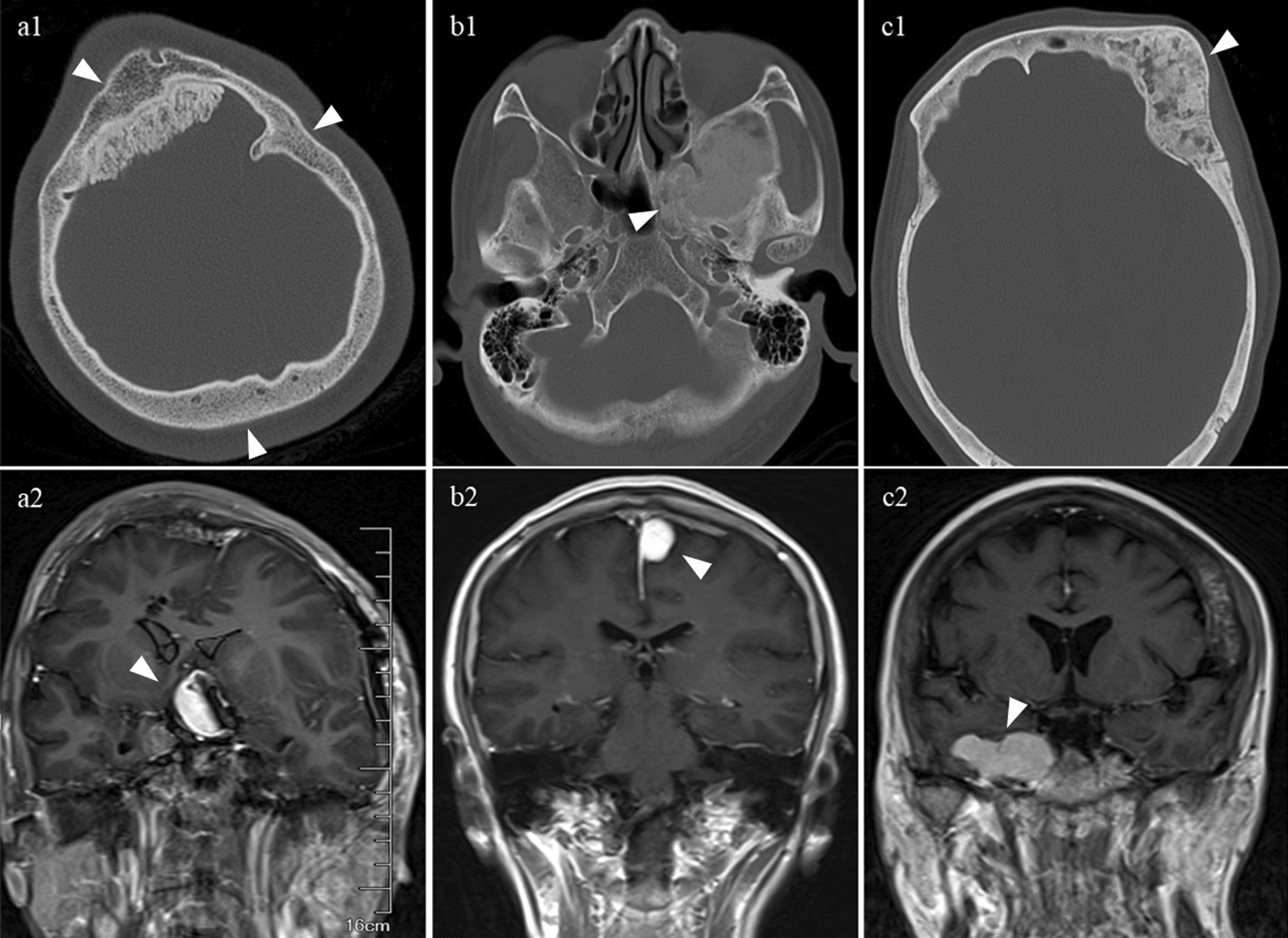 Figure 1
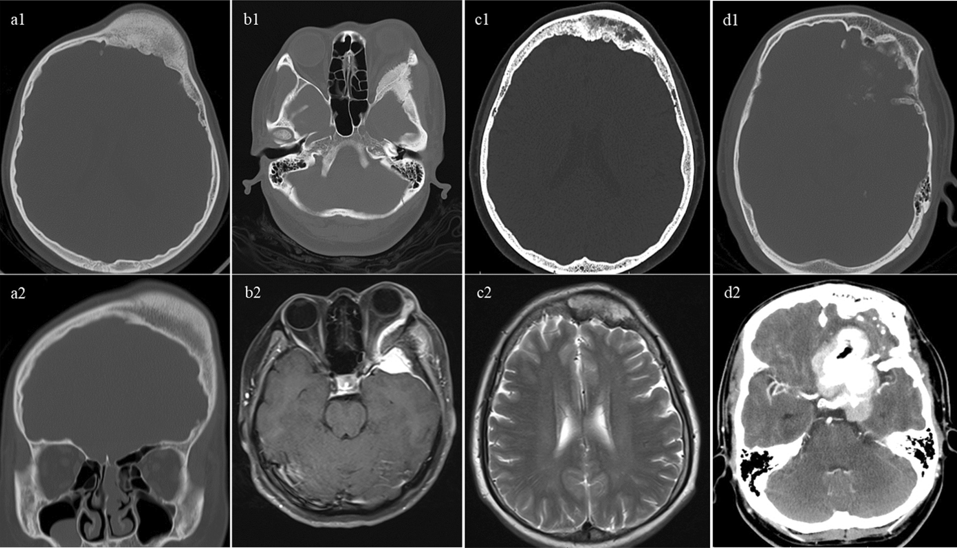 Figure 2
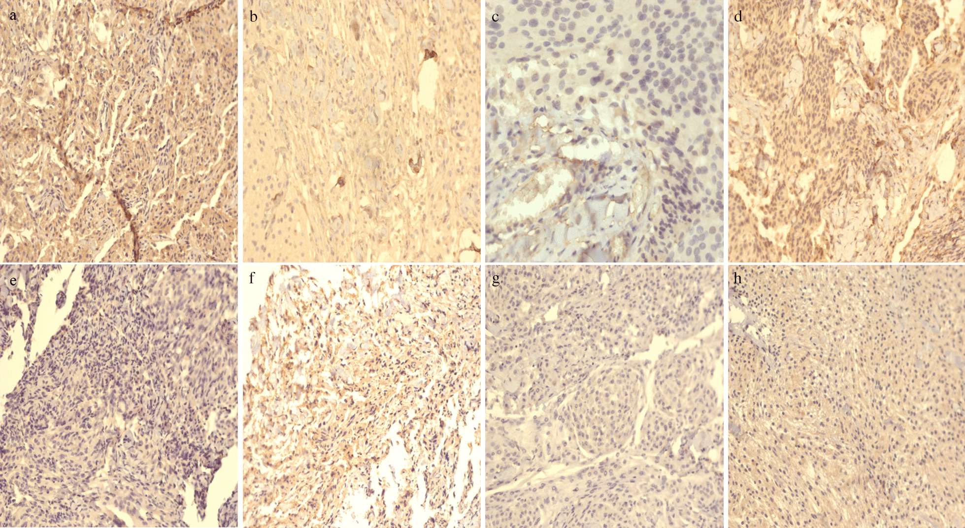 Figure 3
Peer Reviews
No public reviews on file for this paper yet. If you reviewed it on a platform where reviews are public (OpenReview, ICLR, NeurIPS, ICML), you can paste yours below so the community can read it here.
Videos
No videos yet. Explain this paper in a talk, walkthrough, or lecture? Add one.
Taxonomy
TopicsBone Tumor Diagnosis and Treatments · Meningioma and schwannoma management · Head and Neck Surgical Oncology
Background
Meningiomas, primarily arising from meningothelial arachnoid cells, are the most common intracranial tumors at present, accounting for almost one third of all primary central nervous system tumors [1]. Its incidence rate varies from 1.28 to 8.81 per 100,000 persons in different studies around the world [2, 3]. Fibrous dysplasia (FD) is an uncommon mosaic disorder resulting in replacement of normal bone with fibro-osseous tissue. The actual incidence of FD is once reported to be 10–30 in 1,000,000 persons, representing as many as 7% of benign bone tumors [4, 5]. It may occur in one single bone (monostotic FD), in multiple bones (polyostotic FD) or in combination with extra-skeletal disease. Craniofacial bones are the most common location affecting as many as 87% of patients with polyostotic FD [6–9].
The co-existence of meningiomas and craniofacial fibrous dysplasia (CFD) is a fairly uncommon condition which has only been described in a few case reports [10–16]. However, the clinical and radiological characteristics of this condition have not been well-demonstrated and the actual interactions between these two entities still remain unclear. Sporadic activating variants in the GNAS locus not only result in replacement of normal bone with fibro-osseous tissue in CFD lesions [4], but also is mutationally activated in various cancer types, such as growth hormone-secreting pituitary tumors, pancreatic cancer and colorectal cancer [17, 18]. It remains highly concerned whether GNAS gene is the common genetic predisposition between CFD and meningiomas.
Craniofacial FD typically demonstrates dense and sclerotic lesions or appears as an area of radiolucent ground glass matrix. Relevant differential diagnoses of CFD should consider meningiomas, Paget’s disease of the skull bone, and benign osteosclerotic lesions like osteoma [19]. Since meningioma itself could inflict the adjacent bones resulting in bone destruction with similar radiological manifestations to CFD [5, 20], differential diagnosis between bone-invasive meningiomas and concomitant meningiomas and CFD is clinically problematic.
This article was designed to describe a seldom seen series of coexisting meningiomas and CFD, demonstrate their clinical characteristics, explore the underlying interactions and pathogenesis, and discuss the difference between concomitant meningiomas and CFD and single hyperostotic meningiomas in order to facilitate diagnosis and improve treatment.
Methods
Patients’ selection
In the period 2003–2021, a total of 1176 patients diagnosed with CFD at Beijing Tiantan Hospital were retrospectively screened. The study finally enrolled 21 cases that were reported to have concomitant CFD and cerebral meningiomas. All patients underwent computed tomography (CT) and magnetic resonance imaging (MRI) for diagnosis and evaluation. CFD and meningiomas were diagnosed in accordance with histological examinations in patients managed with surgery, while for patients received conservative treatment, diagnoses were made according to typical radiological characteristics. Demographic characteristics, clinical manifestations, radiological and pathological features, treatment procedures and outcomes were recorded.
Follow-up was accomplished via telephone or at the clinic. CT and MRI was carefully evaluated and whether there was disease recurrence or progression was recorded.
This study was approved by the Institutional Review Board. Due to the retrospective nature of our study, the board waived the need for written consent.
Immunohistochemistry and genetic analysis
Immunohistochemistry was performed to detect α-subunit of the stimulatory G-protein (Gα_s_) protein expression in meningioma specimens. The tissue sections were incubated with primary Gα_s_ antibody (1:100, sc-365855, Santa Cruz). Each stained slide was individually reviewed and independently scored by two neuropathologists. Genomic DNA was extracted from paraffin-embedded meningioma specimens using the Wizard Genomic DNA Purification kit following the manufacturer’s instructions (Promega, Madison, WI). The Gα_s_ encoding exons 8 and 9 of GNAS were amplified by PCR and sequenced by conventional Sanger sequencing (BigDye Terminator Cycle Sequencing Ready reaction kit, Applied Biosystems).
Literature review
In addition, we searched 3 medical database, PubMed, EMBASE and Cochrane Library up to 2021 for published studies focusing on the coexistence of CFD and meningioma. The following combined terms ([MESH] “fibrous dysplasia” AND [MESH] “meningioma”) were used. A manual researching on the reference of identified studies was performed for more related studies.
Results
Clinical and radiological characteristics
Among the 1176 CFD patients evaluated, concurrent meningiomas were found in 21 patients (17 females, mean age 45.14 years old). Only 1 patient was adolescent. The teenage boy had a heavy disease burden of CFD (Fig. 1a1) and a meningioma located at tuberculum sellae (Fig. 1a2). Tables 1 and 2 presented the distribution of CFD and meningiomas. The majority (57.14%) of meningiomas located at the skull base and most (47.62%) of the CFD lesions affected the sphenoid bone.Fig. 1. Radiological manifestations. a1, a2 radiology of case 1 shows diffuse CFD in right maxilla, ethmoid sinus and sphenoid bone and tuberculum sellae meningioma; b1–b2 radiology of case 19 shows CFD in left sphenoid bone and left frontal-parietal parafalx meningioma; c1, c2 radiology of case 9 shows CFD in bilateral sphenoid, temporal and occipital bones and left frontal parasagittal meningiomaTable 1Demographical, radiological, therapeutic and prognosis characteristics of the 21 patientsVariablesAge (mean)45.14 years oldGender (%) Male4 (19.05%) Female17 (80.95%)Symptom Craniofacial deformity2 Neurological dysfunction8 Seizure1 Headache/dizziness7 Symptom free4Meningioma Location Convexity2 Para-falx/para-sinus4 Skull base12 Ventricle3 Treatment Resection14 Gamma knife1 Watchful waiting6 Pathology Transitional6 Mixed1 Meningothelial4 Fibrous2 Metaplastic1 Follow-up (mean) Lost2 All patients56.89 months Surgical patients56.64 months Watchful waiting patients57.60 months Prognosis Recurrence after resection2 Favorable outcome19Cranial fibrous dysplasia Location Sphenoid bone10 Maxilla2 Frontal bone5 Temporal bone5 Parietal bone2 Occipital bone2 Ethmoid sinus2 Clivus2 Treatment Surgery4 Watchful waiting17 Follow-up (mean) Lost2 All patients56.89 months Surgical patients47.00 months Watchful waiting patients59.53 months Prognosis Progression0 Favorable outcome19Table 2Detailed clinical information of the 21 patients included in this studyNoAge /yGenderOnset symptomLesion locationManagementSimpson gradePrognosisCFDMeningiomaCFDMeningiomaFollow-up/ monthCFDMeningioma115MaleCraniofacial deformityRight maxilla, ethmoid sinus and sphenoid boneSuprasellar regionSurgerySurgeryIII80UnchangedNo recurrence231FemaleHeadacheSphenoid boneLeft parietal-occipital lobe (parafalx)Watchful waitingSurgeryI118UnchangedNo recurrence339FemaleHearing lossSphenoid boneRight cerebellopontine angleWatchful waitingSurgeryI57UnchangedRecurrence447FemaleVision lossRight clivusRight parasellar regionWatchful waitingSurgeryII65UnchangedNo recurrence554FemaleVision loss and double visionLeft orbit and frontal boneRight parasellar regionWatchful waitingSurgeryI120UnchangedNo recurrence658MaleSeizureLeft maxillaLeft olfactory sulcusWatchful waitingSurgeryI77UnchangedNo recurrence737MaleVision lossLeft frontal boneLeft sphenoid ridgeSurgerySurgeryII84UnchangedNo recurrence837FemaleHeadacheLeft orbit, frontal, sphenoid and temporal bonesLeft tentorium and left frontal lobe (convexity)SurgerySurgeryI12UnchangedNo recurrence936FemaleSymptom freeBilateral sphenoid, temporal and occipital bonesLeft frontal lobe (parasagittal)Watchful waitingSurgeryI10UnchangedNo recurrence1056FemaleVision lossRight temporal boneRight anterior clinoid processWatchful waitingSurgeryI118UnchangedNo recurrence1161FemaleDizzinessLeft sphenoidLeft frontal lobe (parafalx)Watchful waitingSurgeryI8UnchangedNo recurrence1226MaleHeadacheBilateral parietal bonesRight petroclival regionWatchful waitingSurgeryIII24UnchangedRecurrence1366FemaleHeadacheLeft sphenoid bonePosterior part of third ventricleWatchful waitingSurgeryII8UnchangedNo recurrence1451FemaleDouble visionLeft frontal–temporal bonesLeft cavernous sinus and sphenoid ridgeSurgerySurgeryII12UnchangedNo recurrence1538FemaleHeadacheLeft sphenoid boneRight lateral ventricleWatchful waitingGamma Knife radiosurgery24UnchangedNo progression1618FemaleCraniofacial deformity and vision lossLeft frontal boneLeft parasellar regionWatchful waitingWatchful waiting72UnchangedNo progression1754FemaleHearing lossRight clivus and temporal boneRight frontal-parietal lobe (convexity)Watchful waitingWatchful waiting36UnchangedNo progression1860FemaleDizzinessSphenoid bone and sphenoid sinusLeft lateral ventricleWatchful waitingWatchful waiting72UnchangedNo progression1960FemaleSymptom freeLeft sphenoid boneLeft frontal-parietal lobe (parafalx)Watchful waitingWatchful waiting84UnchangedNo progression2061FemaleSymptom freeLeft ethmoid sinusRight petroclival regionWatchful waitingWatchful waitingLost2143FemaleSymptom freeRight parietal boneRight sphenoid ridgeWatchful waitingWatchful waitingLostCFD cranial fibrous dysplasia, NA not available
Surgical intervention was executed in 14 meningiomas. The extent of resection was considered gross-total in 12 patients (Simpson grade I-II) and subtotal in 2 patients (Simpson grade III). No postoperative complications was noticed in the 14 surgically treated patients and none of them received any adjuvant therapy postoperatively. During the average 56.64-month follow-up after surgery, the radiological examinations showed 2 recurrences. Among the 7 unresected meningiomas, 6 opted for watchful waiting, while 1 was treated with Gamma Knife radiosurgery. For instance, case 19 was a 60-year-old female with CFD involving the left sphenoid bone led to the diagnosis of CFD (Fig. 1b1) and a parafalx meningioma at left frontal-parietal lobe (Fig. 1b2). The meningioma showed any sign of progression during the 84 months of “watchful waiting”. These unresected meningiomas showed no progression during the mean 57.60-month follow-up. Referring to the included 21 CFD lesions, only 4 were managed with operation. The mean follow-up time was 56.89 months, and no recurrence or progression was observed in any of the CFD lesions. For example, case 5 who were diagnosed with CFD involving the left frontal bone and left orbit (Fig. 1c1) and meningioma in the right parasellar region (Fig. 1c2). The unresected CFD lesion stayed stable during 120-month follow-up.
CFD typically demonstrated dense, sclerotic lesions and was often associated with the term “ground glass bone matrix”. However, a smooth outer cortical contour always maintained (Fig. 2a). Although meningioma related hyperostosis (Fig. 2b) and intraosseous meningiomas (Fig. 2c) were also evaluated as sclerotic lesions, these lesions exhibited irregular and spiculated borders. Figure 2d demonstrated a co-existing meningioma adjacent to the CFD lesion, the involvement of the lamina interna cranii caused by the meningioma could complicate and interfere with the identification of CFD, making it challenging to differentiate the co-occurrence from the bone-invasive meningioma.Fig. 2. Differential diagnosis between bone invasive meningioma and concomitant meningioma and CFD. a1, 2 typical CFD showing asymmetric expansive lesion at the left frontal bone with typical ground-glass matrix; b1, b2 hyperostosis caused by meningioma revealing a sclerotic lesion of the left greater sphenoid wing with spiculated margins; c1, c2 hyperostotic intraosseous meningioma with irregular inner table; d1, d2 concomitant meningioma and CFD
Pathological characteristics and genetic results
Among the 14 surgically resected and pathologically examined meningiomas, transitional meningiomas were the most common type (6, 42.86%), and almost all the meningiomas (92.86%) were reported to be WHO I grade (Table 3) (Additional file 1). DNA sequencing was accomplished in 7 cases with no GNAS variant detected. In addition, 8 meningiomas were immunohistochemically examined and Gα_s_ expression was positive (grade 1 and grade 2) in 6 specimens (Fig. 3).Table 3. Pathological characteristics of the resected meningioma specimensNoWHO gradeHistological diagnosisGα_s_ proteinGNAS gene1WHO ITransitional2Negative2WHO IMixed1Negative3WHO IMeningothelial1Negative4WHO IMeningothelial1Negative5WHO IFibrous0Negative6WHO ITransitional2Negative7WHO I-IIMeningothelial0NA8WHO ITransitional1Negative9WHO IMetaplasticNANA10WHO IMeningothelialNANA11WHO ITransitionalNANA12WHO ITransitionalNANA13WHO IFibrousNANA14WHO ITransitionalNANANA not availableFig. 3Immunohistochemical analysis of the Gα_s_ expression of the meningiomas in patients 1–8. Gα_s_ is strongly expressed (grade 2) in case 1 (a) and case 6 (f), moderately expressed (grade 1) in case 2–4 (b–d) and case 8 (h) and mildly expressed (grade 0) in case 5 (e) and case 7 (g)
Both of the meningioma samples with grade 2 Gα_s_ expression were transitional meningiomas (case 1 and case 6). Case 1 was a suprasellar meningioma co-existed with diffuse CFD involving right maxilla, ethmoid sinus and sphenoid bone and case 6 was a left olfactory groove meningioma concomitant with a CFD lesion inflicting left maxilla (Additional file 2). Although these 2 CFD lesions showing strongly positive Gα_s_ expression both involved maxilla, no definite correlation between the level of Gα_s_ expression and the location of CFD lesions could be drawn because of the limited sample size.
The two recurrent meningiomas (case 3 and case 12) were both WHO I grade. Case 3 was a meningothelial meningioma in the right cerebellopontine angle co-diagnosed with sphenoid bone CFD. Although without GNAS mutation, the meningioma revealed grade 1 Gα_s_ expression. Co-occurrence of CFD inflicting the parietal bone and a transitional meningioma in the right petroclival region was seen in case 12 (Additional file 2). Immunohistochemistry and genetic analysis was missing for this recurrent meningioma. No relation was found between recurrence and pathological characteristics.
Literature review
Only 8 studies met the inclusion criteria and all of them were case reports. Detailed information of these 4 articles were described in Table 4. The mean age of the included patients was 29.38 years old with 6 males and only 2 females. Operations were reported in 5 meningiomas and 4 FD lesions. None of them explored GNAS variant and Gα_s_ expression in meningioma specimens and no hypothesis was put forward to explain the co-occurrence.
Discussion
This article reported an infrequent series of concomitant meningiomas and CFD. To the best of our knowledge, this study was the largest case series highlighting the clinicopathologic features, treatment modalities and prognosis. In addition, we also reviewed the literature and discussed the challenges to differentiate such co-existence with solitary bone-invasive meningioma.
The actual mechanism of co-existed meningiomas and CFD remains unclear. There are several possible explanations: (1) genetic predisposition; (2) a purely coincidental event; (3) environmental influence as an irritating agent for the local proliferation and growth of the other [21–23]. Sporadic CFD is reported to be most common in children and adolescents and barely have any gender difference [24], which varies a lot from the age (mean age 45.14 years old) and gender profile (marked female: male ratio up to 4:1) of the included 21 CFD co-diagnosed with meningiomas. What’s more, the incidence rate of meningioma in the general population varies from 1.28 to 8.81 per 100,000 persons [2, 3, 25], however, the present study found 21 meningiomas in the 1176 CFD patients indicating much higher incidence of meningioma (1.8%). Therefore, it is reasonable to hypothesize a possible link between meningiomas and CFD. However, radiological imaging is more regularly performed in CFD patients increasing the chance of incidental findings of other intracranial lesions including meningiomas, which should also be taken into account when evaluating the actual mechanism of the co-existence. Additionally, previous case reports show quite different demographic and radiological characteristics. There were 8 cases included in the literature review. When compared with regular meningioma patients, their mean age was much younger (29.38 years old) and the female predominance was absent since male patients outnumbered female by a ratio of 3:1 [10–16, 26]. And inconsistent with the present findings showing only two meningiomas in the same side of CFD, most of meningiomas included in the previous reports were found to be adjacent to CFD lesions. Such random demographic and radiological profile also provides further evidence for the possibility that the co-existence might be coincidental.
FD is caused by a mosaic activating pathogenic variant in GNAS gene [27, 28], and the development of sporadic meningiomas also has genetic predisposition including NF2, TRAF7, KLF4, AKT1 and TERT [29–31]. GNAS pathogenic variants have been previously found in various systems and has been reported to be associated with many extra-skeletal diseases such as thyroid hyperfunction, hormone-secreting pituitary tumors, pancreatic cancer, breast cancer and colorectal cancer [6–9, 17, 18, 32, 33].Furthermore, GNAS pathogenic variant is also detected in an endothelial meningioma with multiple recurrences recently [34]. However, the present results did not find any pathogenic GNAS variant in the 7 meningiomas analyzed, consistent with the study of Eun who examined 13 meningioma samples [35]. To date there is no evidence concerning the definite role of GNAS variants in the co-occurrence of meningioma and CFD. The current study also tested the Gα_s_ protein encoded by GNAS. Though there was no association between Gα_s_ expression and the histology of meningiomas, the different expression levels of Gα_s_ in the meningioma specimens delineated the possibility that the development of these two diseases might share a common molecular pathway. Case 1 showed a 15-year-old transitional meningioma with strong positive Gα_s_ expression. The relatively young age of meningioma onset and the multiple surgeries of CFD provided some evidence for the hypothesis that CFD might have environmental influence as an irritating agent on the occurrence and development of meningiomas.
Bone involvement is a major concern in meningioma [36], which is documented in 20–68% of meningiomas by histopathological studies [37] and is proved to influence tumor recurrence and prognosis [38]. Bone invasive meningiomas are associated with NF2 and TRAF7 variants [39]. Radiographic evidence of bone involvement includes hyperostosis, bone sclerosis and osteolytic lesions [40]. Both characterized by an increased bone density involving the craniofacial bones, meningioma associated hyperostosis and CFD can be confounded easily resulting in the dilemma to differentiate concomitant meningioma and CFD from meningioma with hyperostotic bone involvement. Seen in 25–49% of meningiomas [41], meningioma associated hyperostosis most frequently affects the convexity and sphenoid wing [5, 42] and is featured by irregular inner surface margins and diffuse “hairy spicules” trabecular hyperostosis without the destruction of trabecular structures [43–45]. Additionally, as a special condition of meningioma restricted in bone (accounting for about 2%) [46, 47], intraosseous meningiomas are readily evaluated as sclerotic lesions with irregular and spiculated borders [48–50]. However, CFD prototypically appears as an area of radiolucent homogeneous ground glass matrix with a smooth cortical contour [51–53]. Therefore, it can be inferred that the key to diagnose CFD is the regular contours of cortical table, but when the co-exist meningioma was adjacent to CFD, the intact lamina interna cranii could be destroyed, making the differential diagnosis more complicated.
Misdiagnosis may influence the treatment preferences and patients’ prognosis. The management strategy should be based on the accurate diagnosis. If it is considered to be meningioma with reactive hyperostosis or intraosseous meningioma, complete resection might be recommended to reduce recurrence and improve prognosis [39, 54]. However, if the patient is diagnosed with co-existed meningioma and CFD, “watchful waiting” treatment of the bone lesion may be acceptable especially when there is no CFD related symptom since FD turns to be stable after adolescence. This managemeng strategy is further proved by the current series of co-existed meningioma and CFD. Although only 4 CFD was surgically resected, most patients had favorable prognosis without any obvious CFD progression suggesting that the co-existence of CFD may not influence the prognosis of meningiomas. However, if important structures are compressed causing complaints, surgical resection should be considered. In addition, when meningioma is located in close juxtaposition of CFD, the bone lesions caused by CFD will make the exposure laborious for the resection of meningioma. In this situation, surgical resection of CFD can be recommended. However, whether these two diseases should be managed at one session ought to be evaluated carefully [55, 56]. Therefore, interdisciplinary and more personalized management should be adopted for patients diagnosed with concomitant CFD and meningioma.
Our study has some limitations. Firstly, due to the rarity, the sample size of qualified cases is limited. More cases are needed to strengthen the reliability. Secondly, no definite mechanism concerning the coexisting meningioma and CFD is clarified which still needs further exploration and verification. Technologies such as whole exome sequencing can be considered to study the common molecular pathway of meningioma and CFD in future researches.Table 4. Literature review of co-existed meningiomas and CFDAuthorPublication yearGenderAge/yOnset symptomsType of CFDLocationTreatmentHistological examinationCFDMeningiomaCFDMeningiomaSettecase et al. [16]2016Male13Enlarging lump on right foreheadMASInvolving the entire skullMultiple: right frontal and right posterior falxUntreatedUntreatedNAAlves et al. [15]2009Male35Growing massCFDFrontal boneRight frontalSurgerySurgeryMeningothelialGhosal et al. [13]2007Male25Diminishing vision and seizuresCFDSphenoid sinus and boneRight frontalSurgerySurgeryAtypical lymphoplasmacyte-rich (WHO grade II)Tasar et al. [14]2004Male20Exophthalmus and lost vision of right eyeCFDRight frontal and temporal bonesMultiple: sphenoidal-temporoparietalNANANAGao et al. [26]2002Female37Headache, dizziness and seizureCFDRight sphenoid boneRight middle cranial fossaSurgerySurgerySyncytialBayas et al. [57]1999Female38MyelopathyMASLeft maxilla, left ramus and right corpus mandibulaeT3-T4UntreatedSurgeryPsammomatousFehlow et al. [58]1992Male17Psychic maldevelopmentMASRight frontal and temporal bonesRight temporalNANANAFrankel J, et al. [59]1988Male50Skull swelling and double visionCFDFrontal, parietal and sphenoid bonesLeft parietalSurgerySurgeryMeningothelial (infiltrating bone)CFD cranial fibrous dysplasia, MAS McCune-Albright syndrome, NA not available
Conclusion
We reported a seldom seen case series of co-diagnosed meningioma and CFD and provided a detailed description of their clinicopathological features, treatment strategy and prognosis. Although a definite causative relationship is still undefined, possible genetic or environmental interplay between these two diseases cannot be excluded and requires further investigations. It can be quite intriguing to be differentiated from bone invasive meningiomas. The comprehensive assessment of this seldom seen and challenging condition in the present study can provide more profound understanding of this co-occurrence thus facilitating the diagnosis and helping with the determination of the appropriate treatment strategy.
Supplementary Information
Additional file 1: Fig. 1. Representative images of meningioma pathology. (a) Pathological hematoxylin–eosin staining of case 1 tumor specimen indicating transitional meningioma (WHO I grade); (b) Pathological hematoxylin–eosin staining of case 5 tumor specimen showing fibrous meningioma (WHO I grade); (c) Pathological hematoxylin–eosin staining of case 9 tumor specimen reporting metaplastic meningioma with a Ki-67 label index of 3% (WHO I grade).Additional file 2: Clinical descriptions and radiological presentations of 21 included cases.
The reference list from the paper itself. Each links out to its DOI / PubMed record.
- 1Ostrom QT Price M Neff C Cioffi G Waite KA Kruchko CCBTRUS statistical report: primary brain and other central nervous system tumors diagnosed in the United States in 2015–2019 Neuro-Oncol 202224 v 1v 9510.1093/neuonc/noac 20236196752 PMC 9533228 · doi ↗ · pubmed ↗
- 2Ogasawara C Philbrick BD Adamson DC Meningioma: a review of epidemiology, pathology, diagnosis, treatment, and future directions Biomedicines.202110.3390/biomedicines 903031933801089 PMC 8004084 · doi ↗ · pubmed ↗
- 3Huntoon K Toland AMS Dahiya S Meningioma: a review of clinicopathological and molecular aspects Front Oncol 20201057959910.3389/fonc.2020.57959933194703 PMC 7645220 · doi ↗ · pubmed ↗
- 4Javaid MK Boyce A Appelman-Dijkstra N Ong J Defabianis P Offiah A Best practice management guidelines for fibrous dysplasia/Mc Cune-Albright syndrome: a consensus statement from the FD/MAS international consortium Orphanet J Rare Dis 20191413910.1186/s 13023-019-1102-931196103 PMC 6567644 · doi ↗ · pubmed ↗
- 5Van de Voorde N Mortier GR Vanhoenacker FM Fibrous dysplasia, paget's disease of bone, and other uncommon sclerotic bone lesions of the craniofacial bones Semin Musculoskelet Radiol.20202457057810.1055/s-0039-340029233036044 · doi ↗ · pubmed ↗
- 6Belsuzarri TA Araujo JF Melro CA Neves MW Navarro JN Brito LG Mc Cune-Albright syndrome with craniofacial dysplasia: clinical review and surgical management Surg Neurol Int 20167 S 165S 16910.4103/2152-7806.17856727057395 PMC 4804399 · doi ↗ · pubmed ↗
- 7Collins MT Singer FR Eugster E Mc Cune-Albright syndrome and the extraskeletal manifestations of fibrous dysplasia Orphanet J Rare Dis 20127 Suppl 1S 410.1186/1750-1172-7-S 1-S 422640971 PMC 3359955 · doi ↗ · pubmed ↗
- 8Boyce AM Chong WH Shawker TH Pinto PA Linehan WM Bhattacharryya N Characterization and management of testicular pathology in Mc Cune-Albright syndrome J Clin Endocrinol Metab 201297 E 1782 E 179010.1210/jc.2012-179122745241 PMC 3431566 · doi ↗ · pubmed ↗