Atypical Wassel type VI thumb duplication treated with on-top plasty
Krešimir Bulić, Goran Tintor

TL;DR
This paper presents a rare case of thumb duplication treated with a specific surgical technique to achieve optimal function and appearance.
Contribution
The paper introduces the use of on-top plasty in treating Wassel type VI thumb polydactyly based on combined clinical and radiological assessments.
Findings
Preoperative X-rays showed a well-formed radial thumb and an underdeveloped ulnar thumb.
On-top plasty combined the best parts of both thumbs to achieve functional and aesthetic results.
Clinical and radiological assessments are crucial for determining treatment strategies in thumb duplication cases.
Abstract
Congenital thumb duplication is estimated to occur between 0.08 and 7.6 times per 1,000 live births; however its cause is still undetermined. In this report, we present a case of Wassel type VI thumb polydactyly. clinical examination revealed an optimal functional position and an aesthetically pleasing shape of the ulnar thumb as well as a superior nail and pulp. However, preoperative X-ray indicated a well formed carpometacarpal joint of the radial thumb compared to an underdeveloped CMC joint of the ulnar thumb. Through surgical procedure we combined the best parts of both thumbs with on-top plasty to achieve the most optimal outcome. In conclusion, it is important to determine an adequate treatment strategy for a patient based on both clinical and radiological assessments.
Genes, proteins, chemicals, diseases, species, mutations and cell lines named across the full text — each resolved to its canonical identifier and authoritative record.
Click any figure to enlarge with its caption.
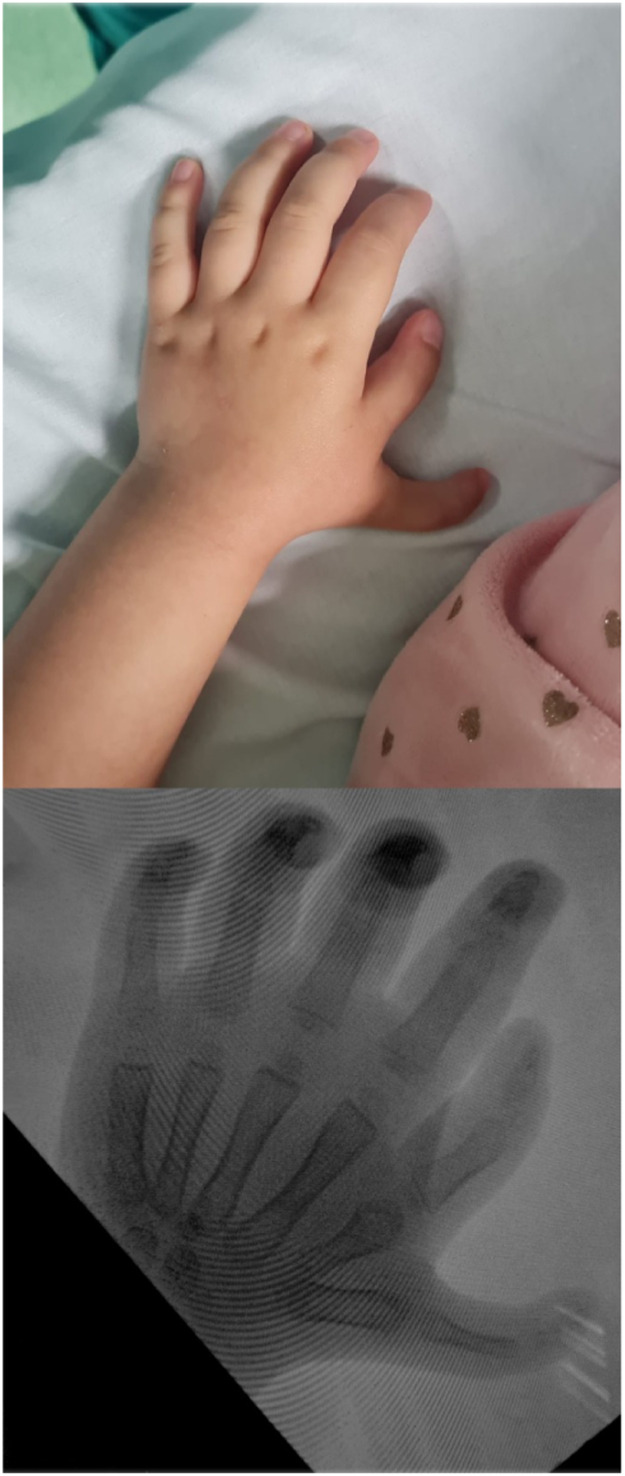 Figure 1
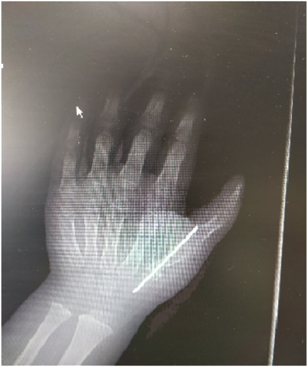 Figure 2
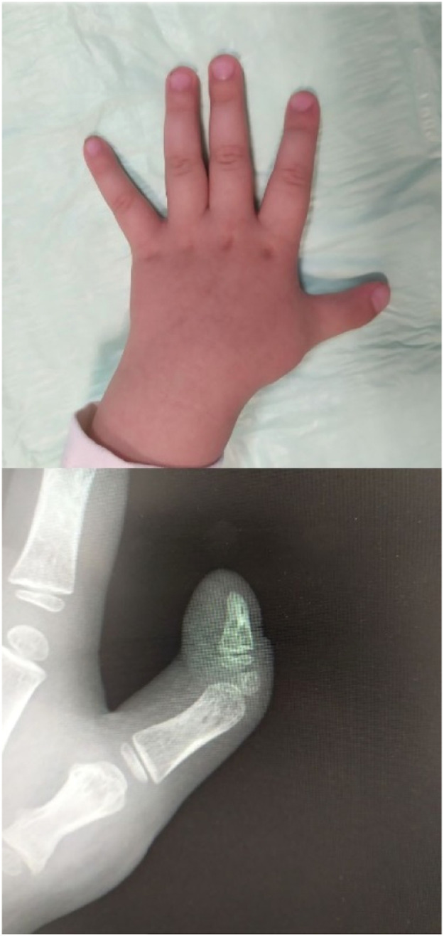 Figure 3
Peer Reviews
No public reviews on file for this paper yet. If you reviewed it on a platform where reviews are public (OpenReview, ICLR, NeurIPS, ICML), you can paste yours below so the community can read it here.
Videos
No videos yet. Explain this paper in a talk, walkthrough, or lecture? Add one.
Taxonomy
TopicsCongenital limb and hand anomalies · Genetic and rare skin diseases.
Congenital thumb duplication is estimated to occur between 0.08 and 7.6 times per 1,000 live births; however its cause is still undetermined.1, 2, 3 Furthermore,only a few studies have objectively and quantitatively examined the outcomes of surgical treatment.4^,^5 The most widely used Wassel classification of thumb duplications in seven types is practical and based on the level of duplication, but it doesn't provide a knowledge of soft-tissue anatomy, stability and mobility of the joints or which thumb possess appropriate size and shape.6
In this report, we present a case of Wassel type VI thumb polydactyly. During the assessment of the patient's condition, clinical examination revealed an optimal functional position and an aesthetically pleasing shape of the ulnar thumb as well as a superior nail and pulp. However, preoperative X-ray indicated a well formed carpometacarpal joint of the radial thumb compared to an underdeveloped CMC joint of the ulnar thumb (Figure 1).Figure 1. Wassel VI of the thumb is evident on a preoperative plain radiograph.Figure 1
Following the excision of the skin and subcutaneous tissue around the radial thumb, dissection was extended up to the metacarpophalangeal joint. The abductor pollicis brevis (APB) and extensor pollicis longus (EPL) tendons were divided at their insertions. An amputation of the radial thumb was performed at the level of the lower third of the metacarpal bone to preserve the capsular-periosteal attachment of the CMC joint. A transverse osteotomy of the ulnar thumb was performed to remove articular surface of the metacarpal base and the distal part of the ulnar thumb was transposed onto the proximal part of the radial thumb. The bones were fixed with a longitudinal K-wire and the reconstruction was completed with a transposition of the APB and EPL tendons to the newly created insertions on the CMC joint and extensor apparatus (Figure 2).Figure 2. Intraoperative image after fixation of the remaining bone fragments with Kirschner wire.Figure 2
Finally, the excision was closed with an absorbable suture. A long-arm splint was applied for 6 weeks, after which time the K-wire was removed. Although the option of excising the rudimentary accessory phalanx causing a 15-degree ulnar deviation was presented to the parents, they decided not to have it performed at this time due to a fully functional and aesthetically pleasing thumb (Figure 3).Figure 3. Images taken six weeks after surgery exhibiting the thenar eminence's contour and the adequate CMC joint of the thumb.Figure 3
The majority of research on surgical interventions for thumb duplication is focused on the two most prevalent forms, the Wassel II and Wassel IV. However, the incidence and optimal treatment of infrequent variants are not as well documented. Shen et al. demonstrated their on-top reconstructive procedure for Wassel VI radial polydactyly with triphalangeal thumb deformity. The study concentrated on osseous restructuring and simplified surgical technique without taking into consideration the soft-tissue reconstruction due to an uncommon and challenging variant – a mixture of Wassel VI and Wassel VII subtypes.7 In our case report, we incorporated the description of the surgical approach, which puts emphasis not just on the bone reconstruction but also on the tendon transfer and capsular-periosteal flap preservation.
Ogino et al. revealed that insufficient results have been prevalent in Wassel types III, V, and VI, as well as triphalangeal-type thumb polydactyly. This outcome was primarily attributed to the adduction contracture of the thumb.8 In contrast to these findings, our case demonstrated a cosmetically pleasing postoperative result as well as the maintenance of optimal function.
In conclusion, it is important to determine an adequate treatment strategy for a patient based on both clinical and radiological assessments.
Declaration of Competing Interest
None declared.
The reference list from the paper itself. Each links out to its DOI / PubMed record.
- 1Wu J Shi W Lin X Epidemiological characteristics and distribution of congenital thumb duplication in south China: An analysis of 2,300 thumbs in 2,108 children Front Pediatr 102022102724310.3389/fped.2022.1027243 PMC 966668936405832 · doi ↗ · pubmed ↗
- 2Van Wyhe RD Trost JG Koshy JC The duplicated thumb: A review Semin Plast Surg 3020161811882789554110.1055/s-0036-1593736 PMC 5115921 · doi ↗ · pubmed ↗
- 3Lin S Tong K Zhang G Clinical characteristics and distribution of thumb polydactyly in South China: A retrospective analysis of 483 hands J Hand Surg Am 4520209389463247383510.1016/j.jhsa.2020.04.003 · doi ↗ · pubmed ↗
- 4Maillet M Fron D Martinot-Duquennoy V Results after surgical treatment of thumb duplication: A retrospective review of 33 thumbs J Child Orthop 120071351411930848610.1007/s 11832-007-0019-3PMC 2656706 · doi ↗ · pubmed ↗
- 5Ozalp T Coskunol E Ozdemir O Thumb duplication: An analysis of 72 thumbs Acta Orthop Traumatol Turc 40200638839117220648 · pubmed ↗
- 6Wassel HD The results of surgery for polydactyly of the thumb. A review Clin Orthop Relat Res 6419691751934894526 · pubmed ↗
- 7Shen K Wang Z Xu Y Reconstruction of Wassel type VI radial polydactyly with triphalangeal thumb using an on-top osteotomy Plast Reconstr Surg Glob Open 52017 e 12162828066110.1097/GOX.0000000000001216 PMC 5340476 · doi ↗ · pubmed ↗
- 8Ogino T Ishii S Takahata S Long-term results of surgical treatment of thumb polydactyly J Hand Surg Am 211996478486872448310.1016/S 0363-5023(96)80366-2 · doi ↗ · pubmed ↗