Fast reconstruction of single-shot wide-angle diffraction images through deep learning
Thomas Stielow, Robin Schmidt, Christian Peltz, Thomas Fennel, Stefan, Scheel

TL;DR
This paper introduces a deep learning approach for rapid and accurate reconstruction of nanoparticle structures from single-shot wide-angle diffraction images, enabling real-time analysis in high-throughput nanocrystallography.
Contribution
The authors develop a deep learning method trained on simulated data that significantly accelerates the reconstruction of nanoparticle shapes and orientations from experimental diffraction images.
Findings
Deep learning achieves fast reconstruction times.
High accuracy in shape and orientation determination.
Potential for real-time nanostructure analysis.
Abstract
Single-shot X-ray imaging of short-lived nanostructures such as clusters and nanoparticles near a phase transition or non-crystalizing objects such as large proteins and viruses is currently the most elegant method for characterizing their structure. Using hard X-ray radiation provides scattering images that encode two-dimensional projections, which can be combined to identify the full three-dimensional object structure from multiple identical samples. Wide-angle scattering using XUV or soft X-rays, despite yielding lower resolution, provides three-dimensional structural information in a single shot and has opened routes towards the characterization of non-reproducible objects in the gas phase. The retrieval of the structural information contained in wide-angle scattering images is highly non-trivial, and currently no efficient rigorous algorithm is known. Here we show that deep…
Click any figure to enlarge with its caption.
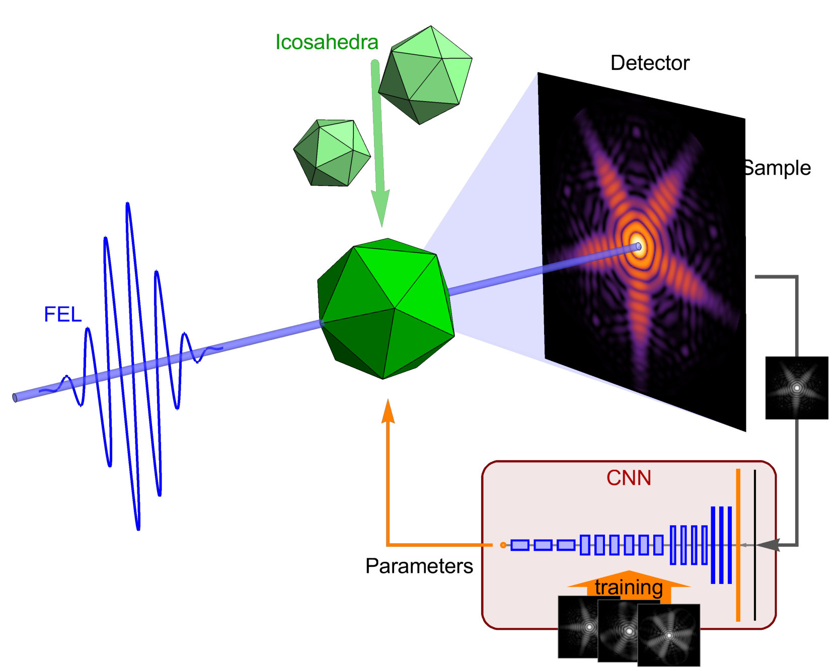 Figure 1
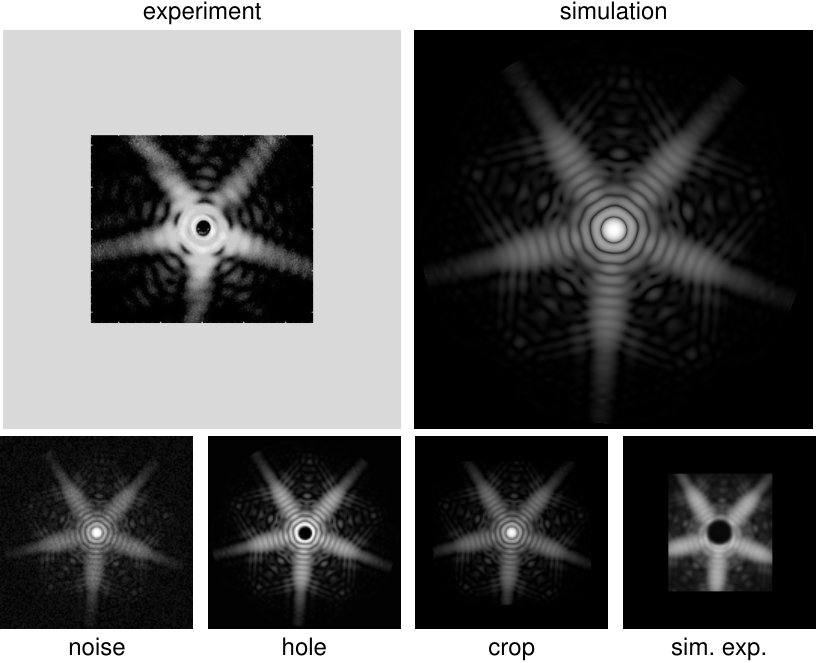 Figure 2
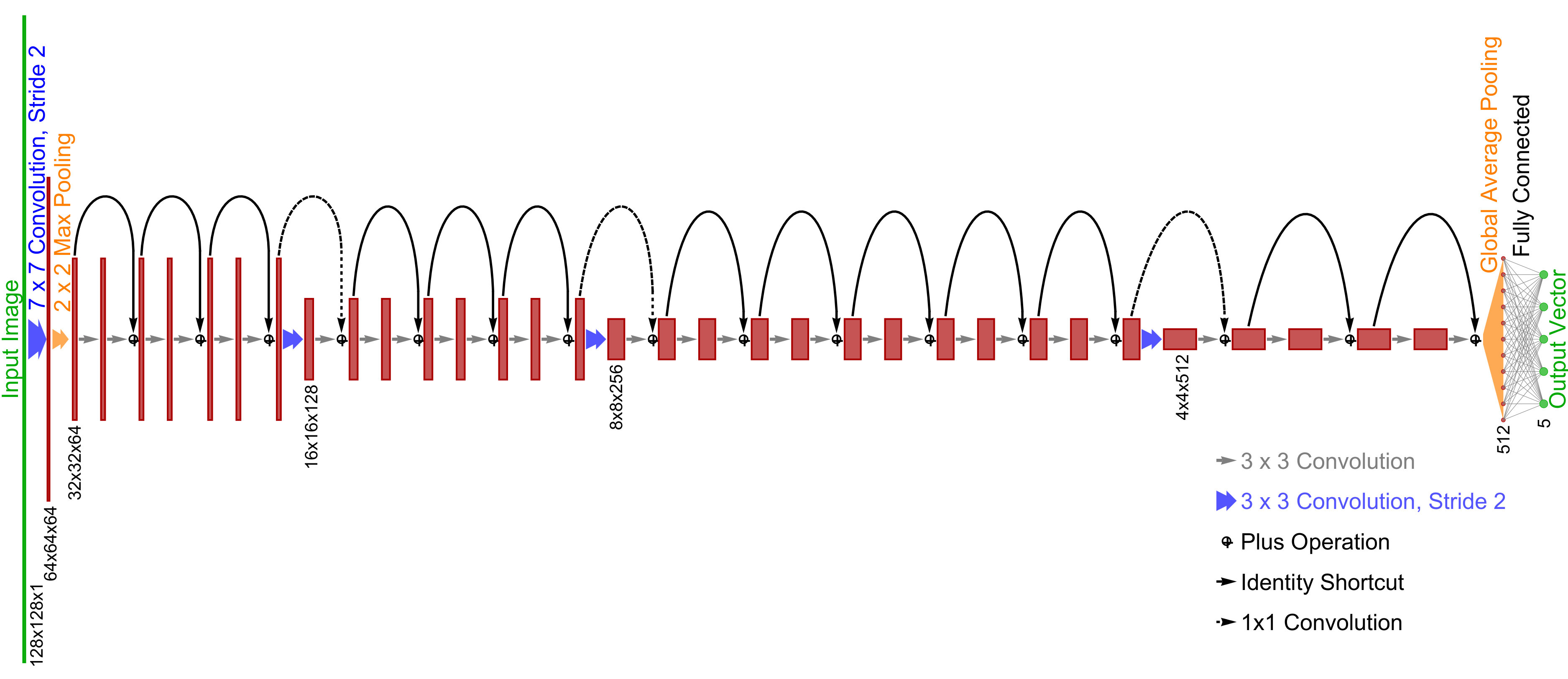 Figure 3
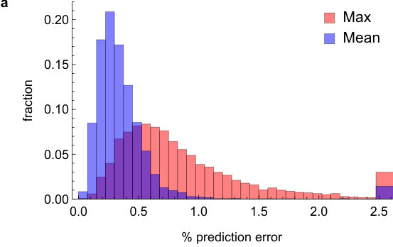 Figure 4
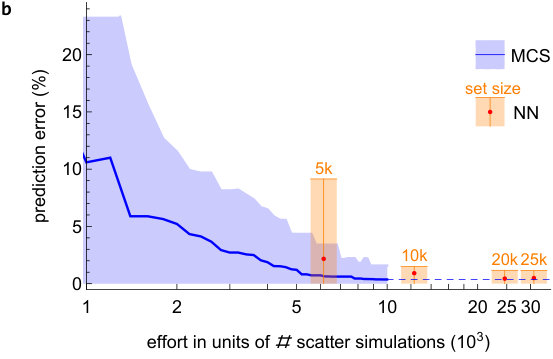 Figure 4
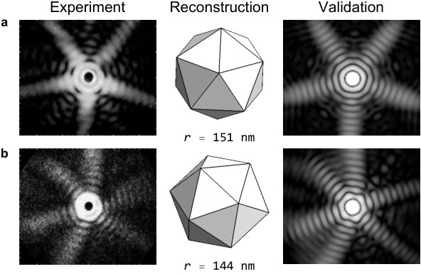 Figure 5
Peer Reviews
No public reviews on file for this paper yet. If you reviewed it on a platform where reviews are public (OpenReview, ICLR, NeurIPS, ICML), you can paste yours below so the community can read it here.
Videos
No videos yet. Explain this paper in a talk, walkthrough, or lecture? Add one.
Fast reconstruction of single-shot wide-angle diffraction images through
deep learning
T Stielow, R Schmidt, C Peltz, T Fennel and S Scheel
Institut für Physik, Universität Rostock, Albert-Einstein-Straße 23–24, D-18059 Rostock, Germany
Abstract
Single-shot X-ray imaging of short-lived nanostructures such as clusters and nanoparticles near a phase transition or non-crystalizing objects such as large proteins and viruses is currently the most elegant method for characterizing their structure. Using hard X-ray radiation provides scattering images that encode two-dimensional projections, which can be combined to identify the full three-dimensional object structure from multiple identical samples. Wide-angle scattering using XUV or soft X-rays, despite yielding lower resolution, provides three-dimensional structural information in a single shot and has opened routes towards the characterization of non-reproducible objects in the gas phase. The retrieval of the structural information contained in wide-angle scattering images is highly non-trivial, and currently no efficient rigorous algorithm is known. Here we show that deep learning networks, trained with simulated scattering data, allow for fast and accurate reconstruction of shape and orientation of nanoparticles from experimental images. The gain in speed compared to conventional retrieval techniques opens the route for automated structure reconstruction algorithms capable of real-time discrimination and pre-identification of nanostructures in scattering experiments with high repetition rate – thus representing the enabling technology for fast femtosecond nanocrystallography.
††: Machine Learning: Science and Technology
1 Introduction
Sources of soft and hard X-rays with large photon flux such as free electron lasers [1, 2] have enabled the high-resolution imaging of unsupported nanosystems such as viruses [3], helium droplets [4, 5, 6], rare-gas clusters [7], or metallic nanoparticles [8]. For reproducible samples, a set of scattering images taken for different orientations in the small-angle scattering limit, each delivering a two-dimensional projection of the object’s density, can be used to retrieve its three-dimensional structure using conventional reconstruction algorithms [9, 10]. Short-lived and non-reproducible objects, however, elude the repeated acquisition of several images required for the tomographic reconstruction from small-angle scattering. The partial three-dimensional information contained in wide-angle scattering enables to overcome this main deficiency, for the prize of an even more complicated inversion problem [5, 8, 11]. Finding a fast reconstruction method thus remains the major obstacle for exploiting the potential of wide-angle scattering for genuine single-shot structure characterization.
Two aspects distinguish wide-angle from small-angle scattering. First, the projection approximation is no longer valid due to substantial contributions of the longitudinal component of the wavevector, such that the curvature of the Ewald sphere plays an important role. Second, for the wavelength range for which wide-angle scattering is realized, the refractive index of most materials deviates substantially from unity, and hence multiple scattering, absorption, backpropagating waves, and refraction all have to be accounted for. Currently, all these constraints can only be met by solving the full three-dimensional scattering problem by, e.g., finite-difference time-domain (FDTD) methods, gridless discrete-dipole approximation (DDA) techniques, or appropriate approximate solutions based on multislice Fourier transform (MSFT) techniques [6, 12].
These methods allow, for an assumed geometry model of the nanoparticle, to describe their wide-angle scattering patterns. However, the determination of the geometry from those patterns is highly nontrivial, as there exists no rigorous inversion method. Consequently, the existing applications of wide-angle scattering had to be based on a parametrized geometry model whose parameters can be determined by an iterative forward fit, e.g. by an ensemble of optimization trajectories in phase space as employed in the simplex Monte Carlo approach in [6]. Because for every iteration step, at least one forward simulation has to be performed, this method is only applicable to a small data set and for a sufficiently simple geometry model [6]. Hence, there is an urgent need for efficient reconstruction methods that can be used in real time for a large data set. Here we present a proof-of-principle study that shows, by considering icosahedra, that a neural network, trained with simulated scattering images, establishes a high-quality reconstruction method of particle size and orientation with unprecedented speed.
Machine learning using neural networks, and deep learning in particular, are ideally suited for the extraction of structural parameters from scattering images, as this is equivalent to the retrieval of a small number of parameters or classes from high-dimensional spaces [13, 14]. Originally conceived for analyzing big data, deep learning has already had significant impact in natural sciences, ranging from analyzing phase transitions and properties of matter [15, 16, 17, 18, 19] and simulations of many-body quantum systems [20] to quantum state reconstruction [21, 22]. In X-ray imaging, neural networks have been introduced only recently. In the small-angle regime, it has been demonstrated that both the tasks of phase retrieval and reconstruction of theoretically generated binary two-dimensional density distributions can be solved also with neural networks [23]. From transmission scattering patterns of thin materials lattice cell orientation maps were extracted [24], as a parameter representation of the material structure. In a next step, using the computational power of the world’s largest supercomputer, full density projection images have been recovered [25]. In addition, the task of pre-classifying scattering patterns has been successfully tackled with neural networks [6], and reinforcement learning techniques have provided further insights into experimental features of X-ray scattering patterns [26]. In these previous applications, the neural networks were either trained and tested solely on theoretical data or were used for feature extraction from experimental diffraction images. In our work we take the decisive step by training a neural network on augmented theoretical data and use it for predictions on experimental scattering data.
2 Experimental and theoretical framework
The choice of icosahedra as test objects was motivated by their ubiquity in nature, ranging from viruses [3, 9, 27, 10] to rare-gas [28] and metal clusters [8]. Focussing on the last example, which already constitutes a wide-angle scenario (see figure 1a), we compute scattering images of icosahedral silver clusters with a range of sizes and spatial orientations using an MSFT algorithm [6], representing the training data. The employed generalized multi-sliced Fourier transform (MSFT) algorithm includes an effective treatment of absorption [8].
We numerically generated individual scattering images for clusters with a uniform size distribution () and random orientations in the fundamental domain of the icosahedron, which represent perfect theoretical data. When representing spatial rotations by unit quaternions (see A for details), the fundamental domain of the icosahedron has the shape of a dodecahedron in imaginary space [29], which is simply connected and possesses a natural metric, unlike other lower dimensional representations such as Euler angles. Furthermore, any arbitrary rotation in the axis-angle representation may be projected into this domain by determining the distance to the closest quaternion associated to one of the symmetry rotations.
The ultimate goal is to analyze realistic scattering data that are obtained from experiments with various imperfections. Therefore, the neural network should not be trained solely using the ideal theoretical data, but also with appropriately augmented data [30, 31, 32]. In that way, the network will be trained to focus on physically relevant features. Here, we augment our data by adding noise, blur, spatial offsets, a central hole, as well as blind spots and cropping of the images. These augmentation features address common experimental imperfections associated with photon noise, limited detector resolution, source-point and beam-pointing jitter, transmission of the high-intensity primary beam, and detector segmentation and finite size (see figure 2). These augmentations (see D for details) increase the training set 11-fold.
3 Network Design and Training
Based upon the quaternion representation of rotations, we can find a unique, bijective parametrization of arbitrary icosahedra of uniform density by a vector with five scalar entries: the four components of the quaternions and the radius of the circumsphere. On the other hand, the associated scattering patterns can be understood as two-dimensional single-channel (or grayscale) images with a size of 128x128 pixels. For the regression task of assigning a parameter vector to an image, we utilize the ResNet architecture of a convolutional neural network with 34 layers. This architecture was found to both offer the complexity needed for learning the specified task while also keeping the number of free parameters as low as possible to counteract overfitting and minimizing training times.
The exact network structure is visualized in Figure 3. It is composed of an initial 7x7 convolution layer with stride 2 and 64 filters, followed by a 2x2 max pooling for lateral dimension reductions, feeding into four stacks of 3, 4, 6 and 4 consecutive residual blocks as defined in [33] with 64, 128, 256 and 512 filters each, respectively. The first convolution of each stack has stride 2 and consequently the corresponding identity shortcut is implemented by 1x1 convolutions. Behind the final residual layer follows a global average pooling operation, reducing the tensor size to a 512-vector from which the terminal 5-vector, composed of the four quaternion components and the radius, is computed by a fully connected layer. All activation functions are set to tanh.
Upon training, the network parameters were optimized to minimize the mean-squared deviation of the predicted parameters compared to their target values. The training was performed on an Nvidia GTX 1060 consumer graphics card with the Wolfram language neural network framework, which was completed within approximately . During training, the generalization capability is supervised by checking the prediction on the validation set after each iteration on the training set.
After training, the network’s predictive capacity has been benchmarked on a separate test set containing 5000 scattering patterns. The measure of interest is the mean relative prediction error (normalized to the possible parameter range). In addition, we also specified the maximal relative prediction error out of the five parameters (for further information regarding error definitions see C). These two errors (mean and maximum) are computed for each of the test patterns. Figure 4a displays the resulting histograms for the mean (blue bins) and the maximum prediction error (red bins). The reconstruction of the relevant physical parameters is highly accurate, with prediction errors well below .
For comparison with established forward fitting methods, we also reconstructed 30 images of the test set with a state-of-the-art Monte Carlo simplex procedure, as used in [6]. For each image, the reconstruction started from 50 random initial points in parameter space, and 50 simplex iteration steps have been taken. The convergence of the reconstruction error as function of required image calculations can be seen in figure 4b, where we estimated that, on average, four scattering patterns need to be calculated in each iteration step. The solid line marks the median best approximation, while the shaded area marks the quantile. In comparison, the red bars denote the same measures for individual neural networks trained on different sized portions of the complete training set. It can be seen that both methods eventually achieve the same level of accuracy. The optimized scattering code requires per image on a hexa-core Intel Xeon E5, resulting in a generation time of for the complete training set. The subsequent training of the network on the complete data set takes additional , which result in a total of or the equivalent of 31k scattering image calculations to yield the ready-to-use neural network. After successful training, the evaluation of a single image takes only , which is a negligible numerical effort compared to that required during forward fitting. Hence, already for a small number of to be reconstructed images, the computational overhead for training data set generation and actual network training can be compensated by the exceptional reconstruction speed whilst still providing reconstruction results of comparable accuracy (see figure 4b).
We demonstrate the network’s ability in recognizing structures in imperfect experimental images by applying it to data taken from [8], where two icosahedral clusters have been identified among the images (left column in figure 5). The reconstructed size and spatial orientation (central column in figure 5) are validated to reproduce the experimental scattering images (right column in figure 5). Our results match the reconstructed data published in [8], with the exception of one of the radii which we attribute to the reduced visibility of the radial fringes which complicates an accurate radius determination with any method.
4 Summary
We have shown that, using a deep-learning technique based on augmented theoretical scattering data, neural networks enable the accurate and fast reconstruction of wide-angle scattering images of individual icosahedral nanostructures. Our results demonstrate that a network, which has only been trained on theoretical data, can be employed for the analysis of experimental scattering data, with image processing times on the millisecond time scale. The neural network reaches the same level of accuracy as established forward fitting methods based on Monte Carlo Simplex algorithms. Although the reconstruction of a single image using the neural network is orders of magnitude faster than the direct optimization, the generation of the training data and subsequent training of the network requires a substantial constant overhead. However, the reconstruction speed of the network compensates the extra effort after only a few scattering images.
Motivated by the performance of this method, we anticipate that a generalization to a wide range of particle morphologies will be feasible. Combined with pre-selection algorithms as utilized in [6], this may evolve into a classification tool for archimedean bodies. The envisaged combination of identification of arbitrary three-dimensional shapes with short processing times is anticipated to represent the enabling technology for a fully automated analysis of scattering data and real-time reconstruction of ultrafast nanoscale dynamics probed at the next generation of X-ray light sources with high repetition rate — with major implications for a broad range of physical, chemical and biological applications.
T.S. gratefully acknowledges a scholarship from “Evangelisches Studienwerk Villigst”. S.S. acknowledges financial support from Deutsche Forschungsgemeinschaft (DFG) via the SPP 1929 “Giant interactions in Rydberg systems”. T.F. acknowledges financial support from the DFG via the Heisenberg program (No. 398382624) and the BMBF (project 05K16HRB). This work was partially supported by the NEISS project of the European Social Fund (ESF) (reference ESF/14-BM-A55-0007/19) and the Ministry of Education, Science and Culture of Mecklenburg-Vorpommern, Germany. The authors thank Dr. Katharina Sander for sharing her insights into the MSFT technique and Dr. Ingo Barke for providing information on the experimental data aquisition.
Data Availability
The data that support the findings of this study are available from the corresponding author upon reasonable request.
Appendix A Icosahedral Symmetry
The icosahedron is one of the five platonic solids and is spanned by 20 equilateral triangle faces, intersecting with 30 edges and twelve corners. It possesses three-fold rotation symmetry axes about the center-of-mass of each triangle, two-fold axes about the center of each edge and five-fold axes about each corner, which together form the icosahedral rotation group . The 60 symmetry rotations imply that any rotation of a body with icosahedral symmetry is 60-fold degenerate. Hence, the mapping of three-dimensional rotation representations, such as Euler-angle or axis-angle representations, to icosahedral orientations are not unique, but have to be constrained in their parameter range. The fundamental domain of rotations has an exceptionally simple form in quaternion representation of rotations, where it forms a dodecahedron in imaginary space [29].
Quaternions are the four-dimensional extension of the complex numbers with three imaginary units and fulfilling the relations and . With real coefficients , any quaternion may be written as . Imaginary quaternions () are isomorphic to the space , implying that all vectors can be represented by quaternions as . The sum of two vectors then translates into the sum of two quaternions, whereas the quaternion product contains both the scalar product of two cartesian vectors (in its real part) and their cross product (in the imaginary part). The rotation by an angle of any vector about a unit vector can thus be expressed by the product of the quaternion with the unit quaternion . Hence, any rotation can be projected into the fundamental domain by applying all inverse symmetry rotations and selecting the one yielding the smallest rotation angle. For the training of a neural network, the quaternion representation has the additional advantage of providing a useful metric for the distance between rotations.
Appendix B Dataset Generation
The scattering patterns used for training are created by using the MSFT algorithm described in [8]. In accordance with the experiment described therein, we simulate the scattering of ultra-short XUV pulses with wavelength and femto-second duration on nano-sized silver clusters. The material parameters are assumed to be equal to bulk silver, with absorption length . For the calculations, the electron density of the cluster is discretized on a cuboid grid, chosen to contain a depth of 192 pixels. The outgoing scattered field is determined by the phase-coherent summation of the scattered field of each slice, which can be obtained by Fourier transformation. Before transformation, each slice is zero-padded to a width of 512 pixels. The computed scattered field is then reduced to an logarithmic intensity profile of pixels with random background noise, which is stored as a grayscale image. The rotation quaternions are sampled uniformly from the fundamental domain, while the size of the clusters range from to . With this procedure, a dataset of images has been generated, one fifth of which has been reserved for validation during training. Another set of is generated for final testing indepentently from the training process.
Appendix C Error Measures
The five parameters of our icosahedron representation reconstructed by the neural network cover very different parameter ranges. For training purposes, both the parameters of the radius and the real part of the rotation quaternion are linearly scaled to the interval . However, when testing the prediction quality of the network it is more useful to have a uniform error measure, that gives the same weight to each parameter. Hence, all error measures within this work are calculated from the relative errors of each prediction parameter, obtained by normalization to the allowed parameter range. All four components of the rotation quaternion are normalized to the maximal span of the fundamental domain in each direction. The deviation of the radius is normalized to the range defined in the previous section. These five individual relative errors, normalized to the parameter range allowed in our model, now weigth each parameter equally. From these, further error measures can be defined. More precisely, the mean error, taking the mean values of all five relative errors, and the maximum error, picking the largest of all five relative errors.
Appendix D Image Augmentation
Prior to training the neural network, image augmentation is applied to the dataset. The augmentation is performed by applying eleven different filters to each ideal scattering pattern, and randomly adding the newly generated images to the training set. These filters can be divided into five groups: trivial, noise, blur, cropping and successive application. The trivial filter is the identity mapping, leaving the image unchanged. Noise is applied both with uniform distribution with a randomly chosen intensity upto half the maximum signal, changing every pixel by a random margin as well as salt-and-pepper statistics, where random pixels are set to either minimal or maximal signal. Blurring is performed by convoluting with a Gaussian kernel with randomly chosen radius of upto five pixels, and by jitter distortion. Cropping filters delete different parts of the image, mainly to account for the characteristics of real detectors. Images are either center-cropped for limited detector size, a central hole of random radius is deleted to simulate the shadow of a beam dump, images are shifted or uneven detector sensitivity is simulated by attenuating parts of the image. Finally, we both randomly combine all image effects, and in addition apply them in a well-defined order so as to generate images that closely resemble experimental results (see figure 2).
References
- [1]
Chapman H N, Barty A, Bogan M J, Boutet S, Frank M, Hau-Riege S P, Marchesini S, Woods B W, Bajt S, Benner W H et al. 2006 Nature Physics 2 839
- [2]
Gaffney K and Chapman H 2007 Science 316 1444–1448
- [3]
Seibert M M, Ekeberg T, Maia F R, Svenda M, Andreasson J, Jönsson O, Odić D, Iwan B, Rocker A, Westphal D et al. 2011 Nature 470 78
- [4]
Gomez L F, Ferguson K R, Cryan J P, Bacellar C, Tanyag R M P, Jones C, Schorb S, Anielski D, Belkacem A, Bernando C et al. 2014 Science 345 906–909
- [5]
Rupp D, Monserud N, Langbehn B, Sauppe M, Zimmermann J, Ovcharenko Y, Möller T, Frassetto F, Poletto L, Trabattoni A et al. 2017 Nature communications 8 493
- [6]
Langbehn B, Sander K, Ovcharenko Y, Peltz C, Clark A, Coreno M, Cucini R, Drabbels M, Finetti P, Di Fraia M et al. 2018 Physical review letters 121 255301
- [7]
Rupp D, Adolph M, Gorkhover T, Schorb S, Wolter D, Hartmann R, Kimmel N, Reich C, Feigl T, De Castro A et al. 2012 New Journal of Physics 14 055016
- [8]
Barke I, Hartmut H, Rupp D, Flückiger L, Sauppe M, Adolph M, Schorb S, Bostedt C, Treusch R, Peltz C, Bartling S, Fennel T, Meiwes-Broes K H and Möller T 2015 Nature communications 6 6187
- [9]
Ekeberg T, Svenda M, Abergel C, Maia F R, Seltzer V, Claverie J M, Hantke M, Jönsson O, Nettelblad C, Van Der Schot G et al. 2015 Physical review letters 114 098102
- [10]
Ayyer K, Morgan A J, Aquila A, DeMirci H, Hogue B G, Kirian R A, Xavier P L, Yoon C H, Chapman H N and Barty A 2019 Optics Express 27 37816–37833
- [11]
Raines K S, Salha S, Sandberg R L, Jiang H, Rodríguez J A, Fahimian B P, Kapteyn H C, Du J and Miao J 2010 Nature 463 214
- [12]
Gessner O and Vilesov A 2019 Annual Review of Physical Chemistry 70
- [13]
Hinton G E and Salakhutdinov R R 2006 Science 313 504–507
- [14]
LeCun Y, Bengio Y and Hinton G 2015 Nature 521 436
- [15]
Carleo G, Cirac I, Cranmer K, Daudet L, Schuld M, Tishby N, Vogt-Maranto L and Zdeborová L 2019 arXiv preprint arXiv:1903.10563
- [16]
Wang L 2016 Physical Review B 94 195105
- [17]
Carrasquilla J and Melko R G 2017 Nature Physics 13 431
- [18]
Van Nieuwenburg E P, Liu Y H and Huber S D 2017 Nature Physics 13 435
- [19]
Venderley J, Khemani V and Kim E A 2018 Physical Review Letters 120 257204
- [20]
Carleo G and Troyer M 2017 Science 355 602–606
- [21]
Torlai G, Mazzola G, Carrasquilla J, Troyer M, Melko R and Carleo G 2018 Nature Physics 14 447
- [22]
Xin T, Lu S, Cao N, Anikeeva G, Lu D, Li J, Long G and Zeng B 2019 npj Quantum Information 5 1–8
- [23]
Cherukara M J, Nashed Y S and Harder R J 2018 Scientific reports 8 1–8
- [24]
Laanait N, He Q and Borisevich A Y 2019 arXiv preprint arXiv:1902.06876
- [25]
Laanait N, Romero J, Yin J, Young M T, Treichler S, Starchenko V, Borisevich A, Sergeev A and Matheson M 2019 arXiv preprint arXiv:1909.11150
- [26]
Zimmermann J, Langbehn B, Cucini R, Di Fraia M, Finetti P, LaForge A C, Nishiyama T, Ovcharenko Y, Piseri P, Plekan O et al. 2019 Physical Review E 99 063309
- [27]
Gorkhover T, Ulmer A, Ferguson K, Bucher M, Maia F R, Bielecki J, Ekeberg T, Hantke M F, Daurer B J, Nettelblad C et al. 2018 Nature Photonics 12 150
- [28]
Miehle W, Kandler O, Leisner T and Echt O 1989 The Journal of chemical physics 91 5940–5952
- [29]
Frank F C 1986 Le Journal de Physique Colloques 47 C3–165
- [30]
Krizhevsky A, Sutskever I and Hinton G E 2012 Imagenet classification with deep convolutional neural networks Advances in neural information processing systems pp 1097–1105
- [31]
Dosovitskiy A, Springenberg J T, Riedmiller M and Brox T 2014 Discriminative unsupervised feature learning with convolutional neural networks Advances in Neural Information Processing Systems pp 766–774
- [32]
Perez L and Wang J 2017 arXiv preprint arXiv:1712.04621
- [33]
He K, Zhang X, Ren S and Sun J 2016 Deep residual learning for image recognition Proceedings of the IEEE conference on computer vision and pattern recognition pp 770–778
The reference list from the paper itself. Each links out to its DOI / PubMed record.
- 1[1] Chapman H N, Barty A, Bogan M J, Boutet S, Frank M, Hau-Riege S P, Marchesini S, Woods B W, Bajt S, Benner W H et al. 2006 Nature Physics 2 839
- 2[2] Gaffney K and Chapman H 2007 Science 316 1444–1448
- 3[3] Seibert M M, Ekeberg T, Maia F R, Svenda M, Andreasson J, Jönsson O, Odić D, Iwan B, Rocker A, Westphal D et al. 2011 Nature 470 78
- 4[4] Gomez L F, Ferguson K R, Cryan J P, Bacellar C, Tanyag R M P, Jones C, Schorb S, Anielski D, Belkacem A, Bernando C et al. 2014 Science 345 906–909
- 5[5] Rupp D, Monserud N, Langbehn B, Sauppe M, Zimmermann J, Ovcharenko Y, Möller T, Frassetto F, Poletto L, Trabattoni A et al. 2017 Nature communications 8 493
- 6[6] Langbehn B, Sander K, Ovcharenko Y, Peltz C, Clark A, Coreno M, Cucini R, Drabbels M, Finetti P, Di Fraia M et al. 2018 Physical review letters 121 255301
- 7[7] Rupp D, Adolph M, Gorkhover T, Schorb S, Wolter D, Hartmann R, Kimmel N, Reich C, Feigl T, De Castro A et al. 2012 New Journal of Physics 14 055016
- 8[8] Barke I, Hartmut H, Rupp D, Flückiger L, Sauppe M, Adolph M, Schorb S, Bostedt C, Treusch R, Peltz C, Bartling S, Fennel T, Meiwes-Broes K H and Möller T 2015 Nature communications 6 6187