TL;DR
This paper introduces a novel deep volumetric network that integrates multiple spatial priors to improve MRI head anatomy segmentation, especially in cases with abnormalities like lesions.
Contribution
The study presents the Multiprior network, combining spatial, morphological, and contextual priors, enhancing segmentation accuracy over existing methods for abnormal brain anatomies.
Findings
Multiprior network outperforms existing segmentation tools like SPM, FSL, and DeepMedic.
Adding a tissue probability map (TPM) significantly boosts segmentation performance.
The approach generalizes well to clinical cases with disorders of consciousness.
Abstract
Purpose: Conventional automated segmentation of the head anatomy in MRI distinguishes different brain and non-brain tissues based on image intensities and prior tissue probability maps (TPM). This works well for normal head anatomies, but fails in the presence of unexpected lesions. Deep convolutional neural networks leverage instead spatial patterns and can learn to segment lesions, but often ignore prior probabilities. Approach: We add three sources of prior information to a three-dimensional convolutional network, namely, spatial priors with a TPM, morphological priors with conditional random fields, and spatial context with a wider field-of-view at lower resolution. We train and test these networks on 3D images of 43 stroke patients and 4 healthy individuals which have been manually segmented. Results: We demonstrate the benefits of each sources of prior information, and we show…
Click any figure to enlarge with its caption.
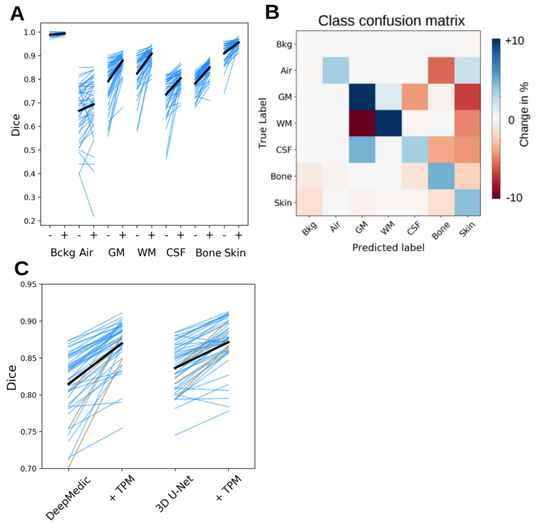 Figure 1
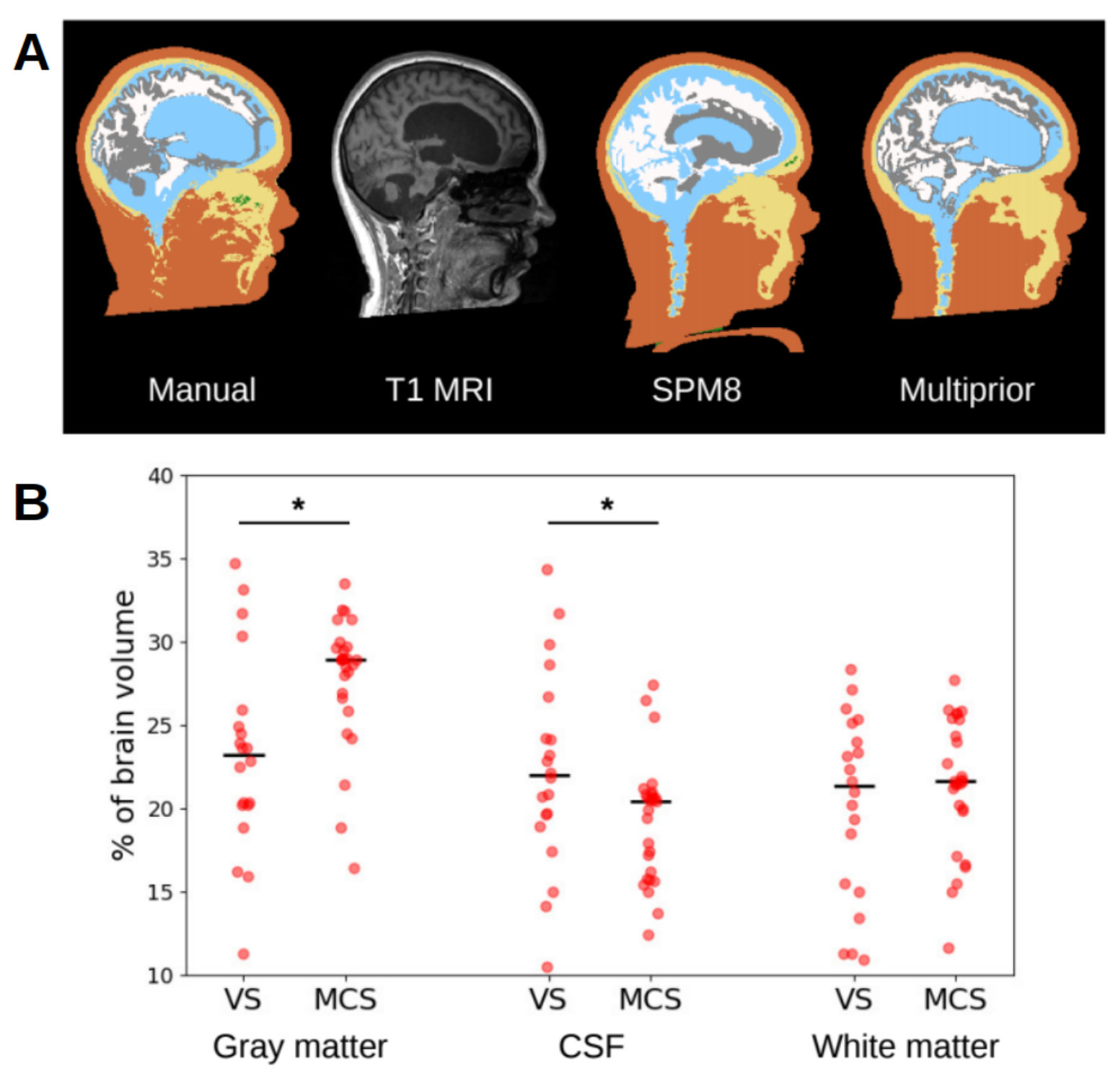 Figure 2
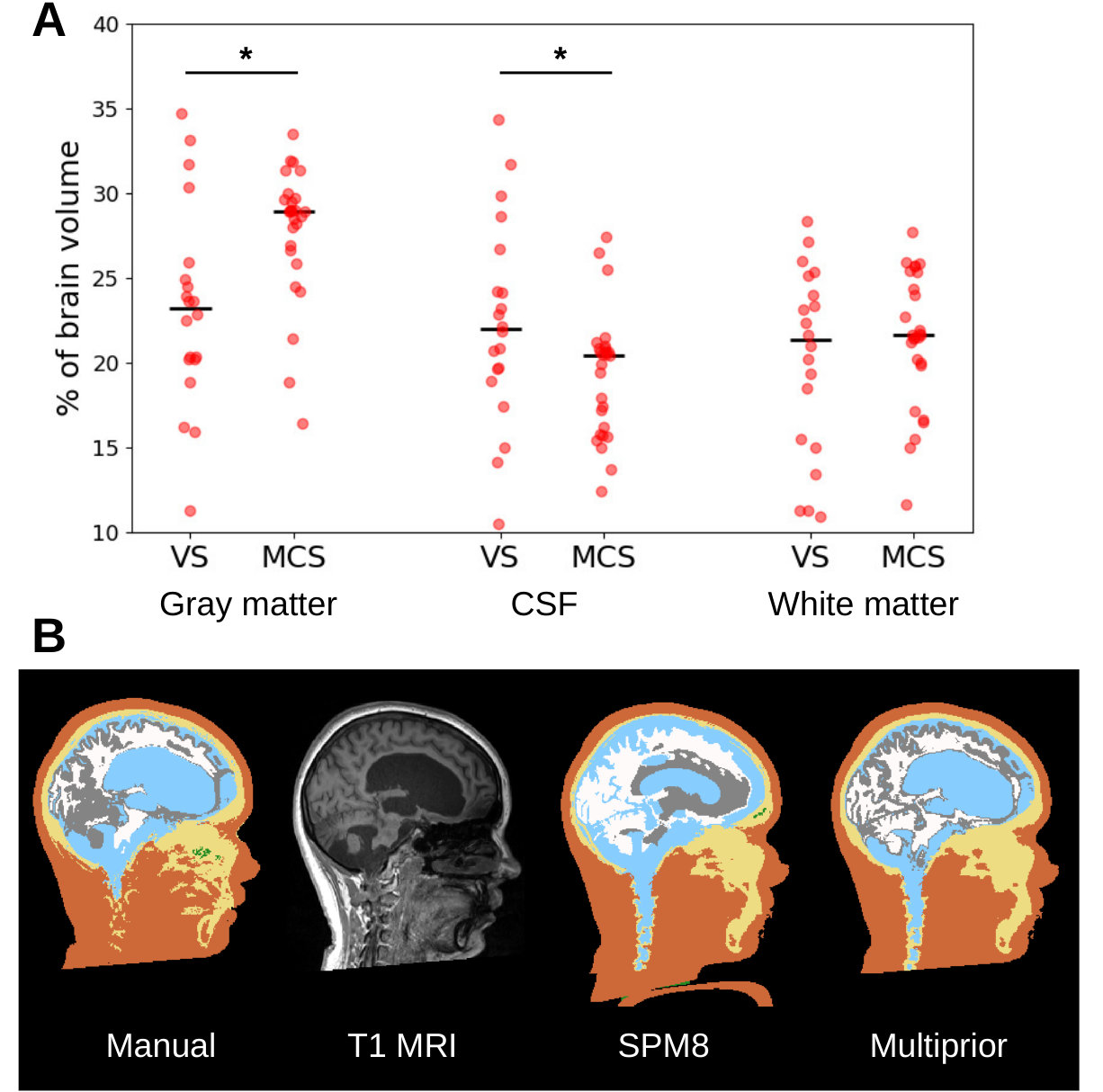 Figure 3
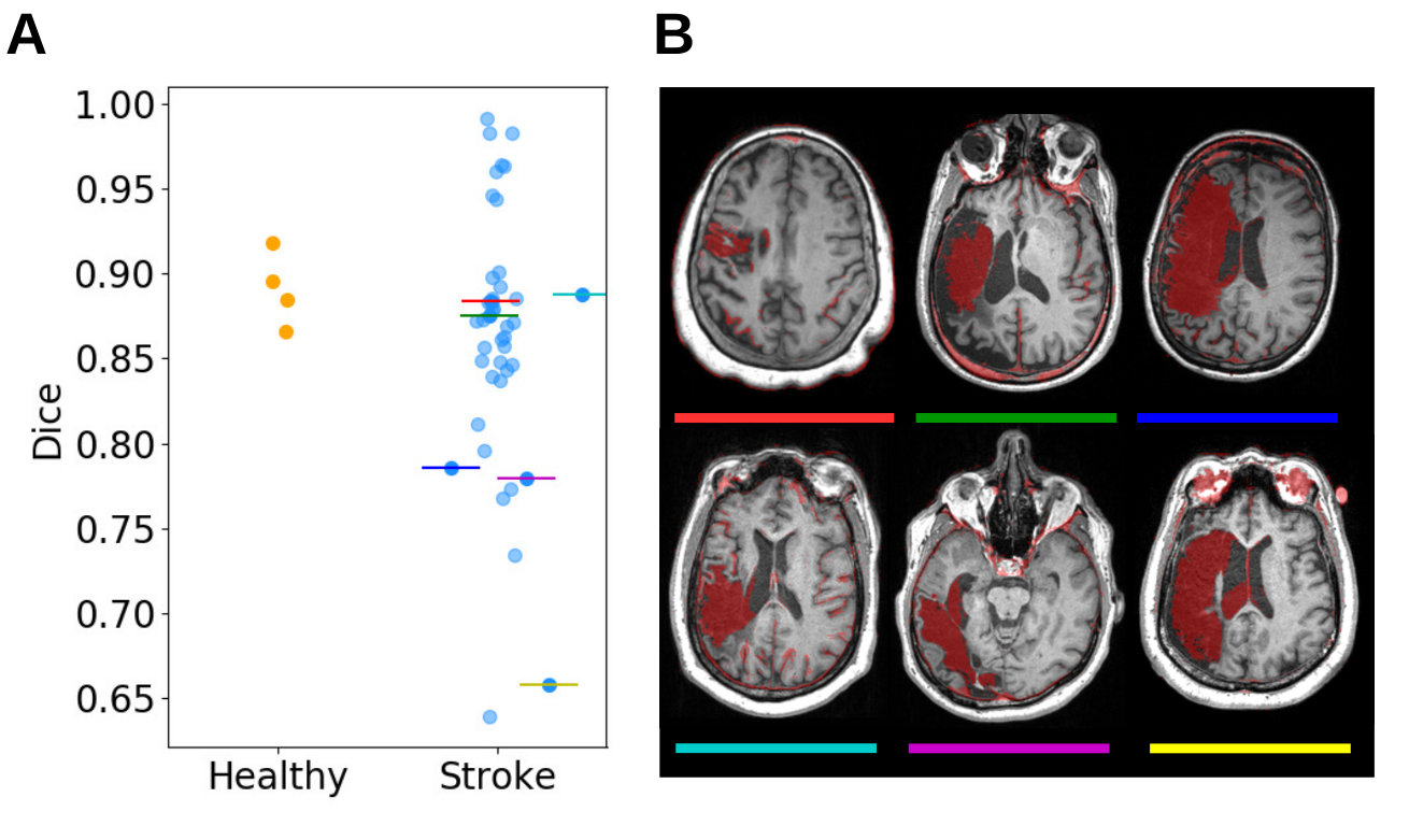 Figure 4
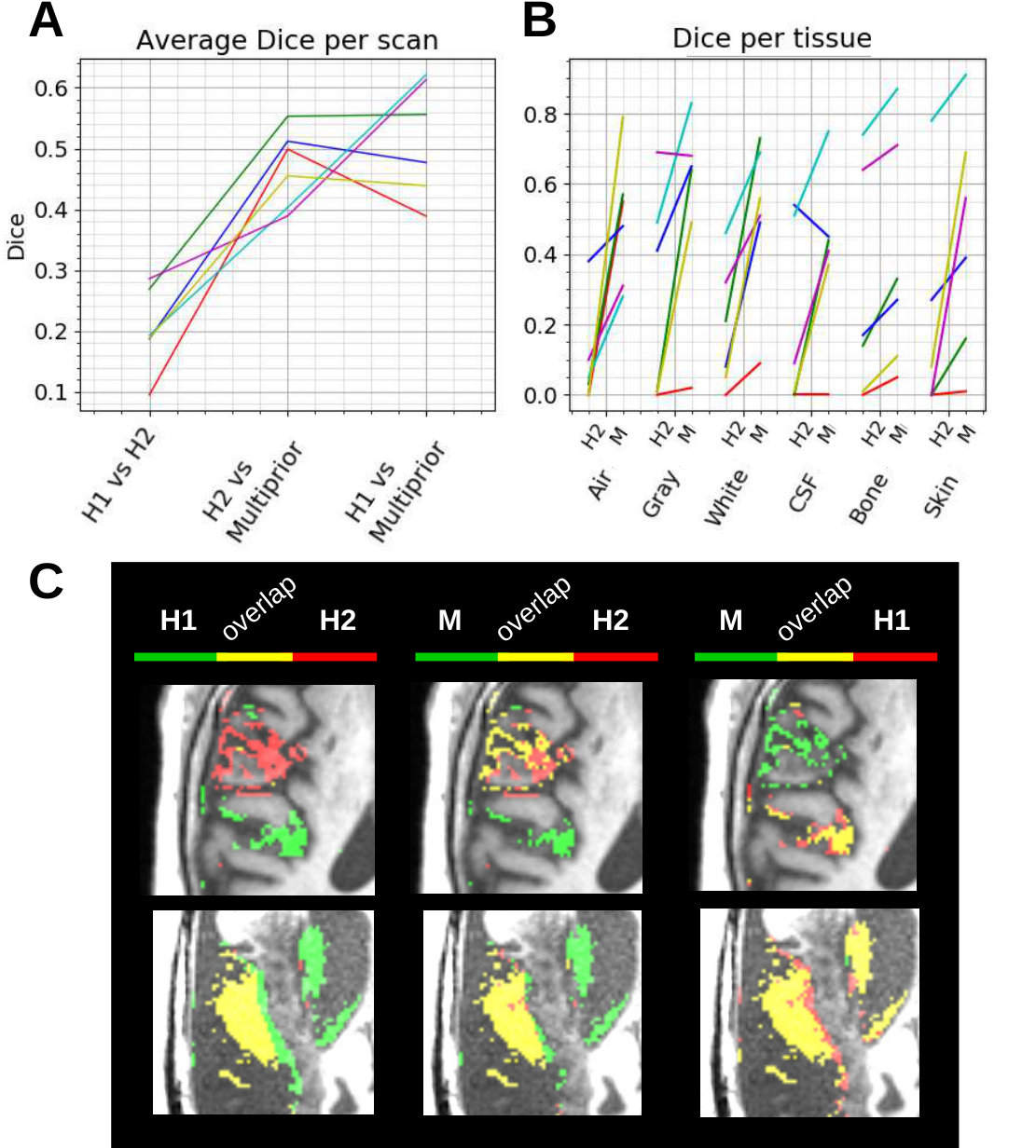 Figure 5
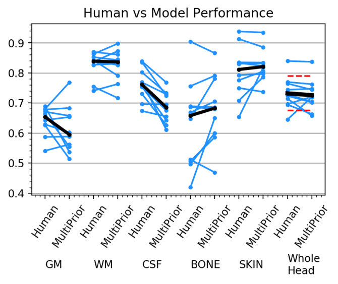 Figure 6
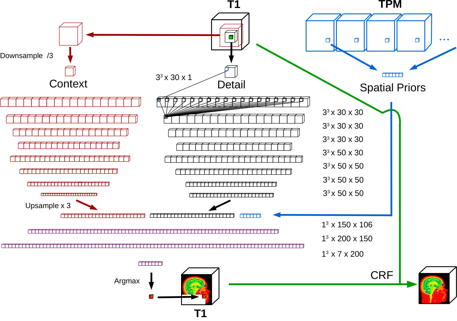 Figure 7
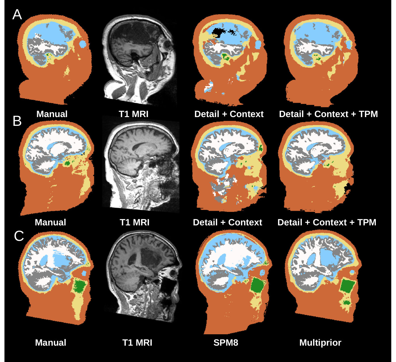 Figure 8
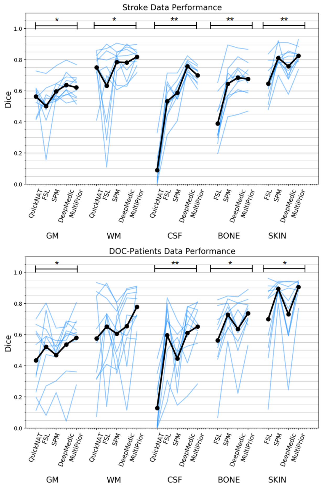 Figure 9
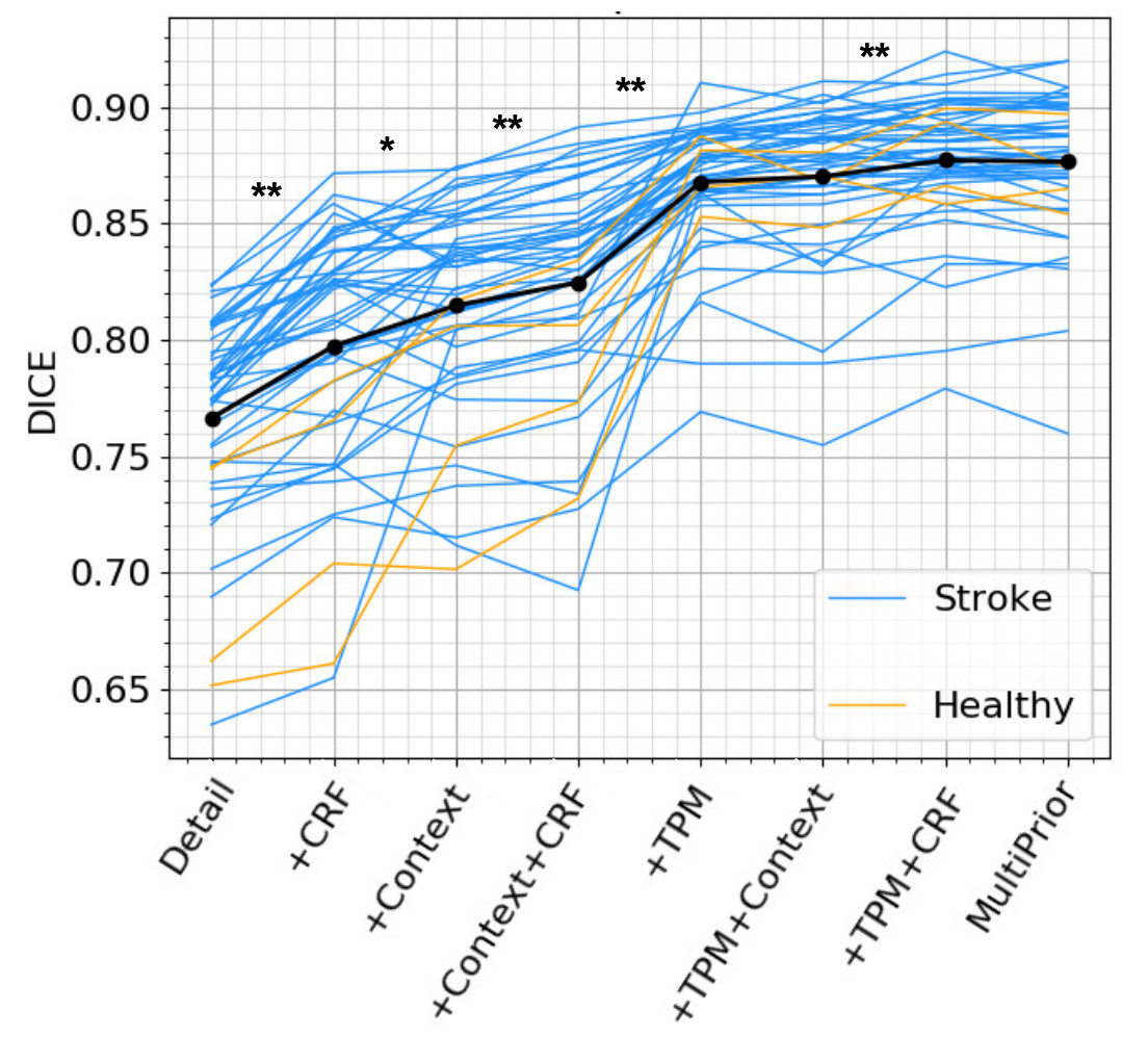 Figure 10
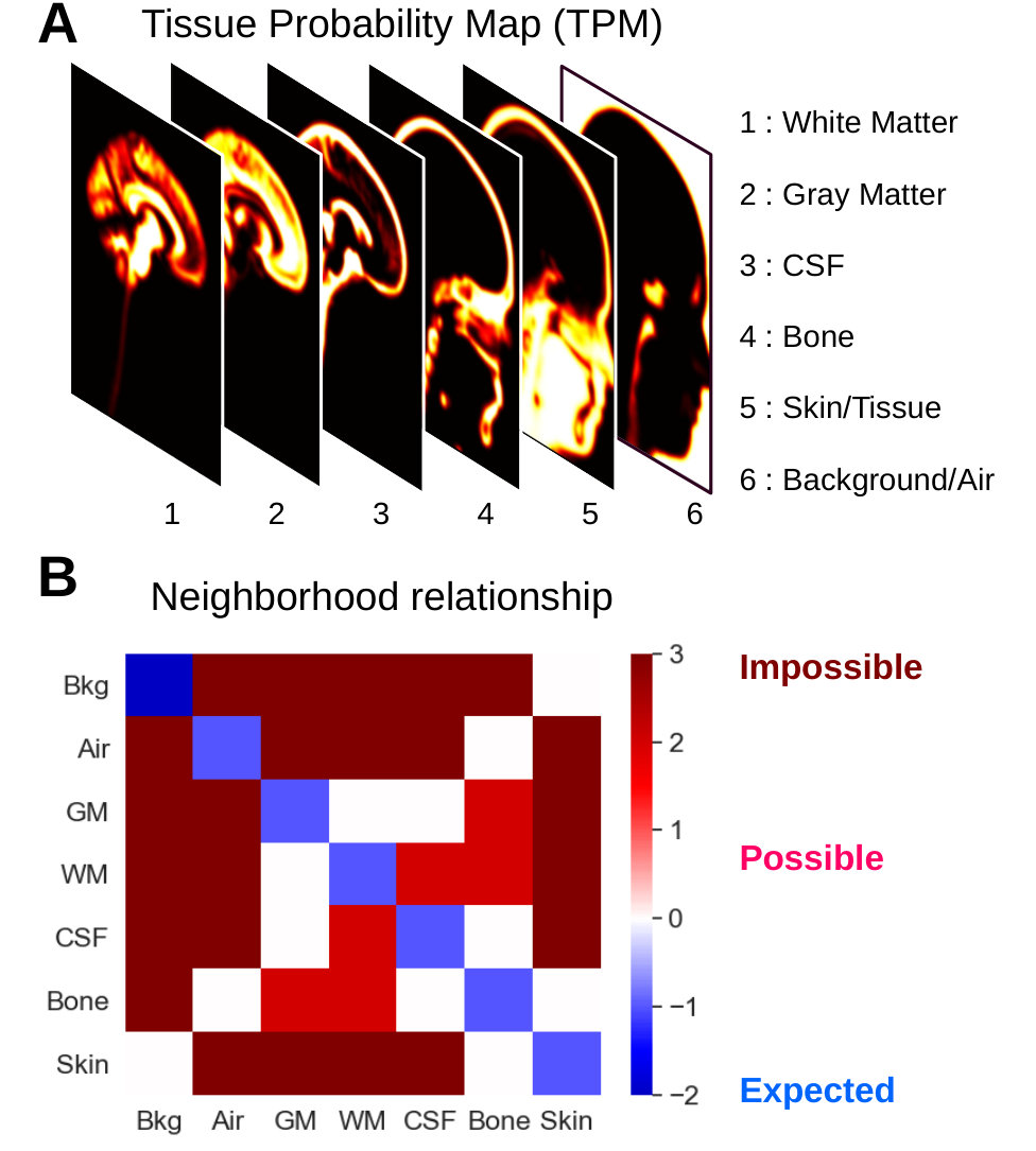 Figure 11
Peer Reviews
No public reviews on file for this paper yet. If you reviewed it on a platform where reviews are public (OpenReview, ICLR, NeurIPS, ICML), you can paste yours below so the community can read it here.
Code & Models
Videos
No videos yet. Explain this paper in a talk, walkthrough, or lecture? Add one.
