Multiple Fourier Component Analysis of X-ray Second Harmonic Generation in Diamond
P.Chakraborti, B.Senfftleben, B.Kettle, S.W.Teitelbaum, P.H.Bucksbaum,, S.Ghimire, J.B.Hastings, H.Liu, S.Nelson, T.Sato, S. Shwartz, Y.Sun,, C.Weninger, D.Zhu, D.A.Reis, M.Fuchs

TL;DR
This paper investigates multiple Fourier component second harmonic generation in diamond using high-intensity X-ray interactions, revealing insights into nonlinear processes beyond the free-electron approximation.
Contribution
It extends previous studies by analyzing multiple Fourier components of XSHG in diamond with higher statistics, determining the nonlinear structure factor and source contributions.
Findings
Good agreement with the quasi-free electron model
Extended analysis to multiple Fourier components
Measured efficiency and angular dependence of XSHG
Abstract
The unprecedented brilliance of X-ray free-electron lasers (XFELs) [1, 2] has enabled first studies of nonlinear interactions in the hard X-ray range. In particular, X-ray-optical mixing [3], X-ray second harmonic generation (XSHG) [4] and nonlinear Compton scattering (NLCS) [5] have been recently observed for the first time using XFELs. The former two experiments as well as X-ray parametric downconversion (XPDC)[6, 7] are well explained by nonlinearities in the impulse approximation[8], where electrons in a solid target are assumed to be quasi free for X-ray interactions far from atomic resonances. However, the energy of the photons generated in NLCS at intensities reaching up to 4 x 10²⁰ W/cm2 exhibit an anomalous red-shift that is in violation with the free-electron model. Here we investigate the underlying physics of X-ray nonlinear interactions at intensities on order of 1016…
Click any figure to enlarge with its caption.
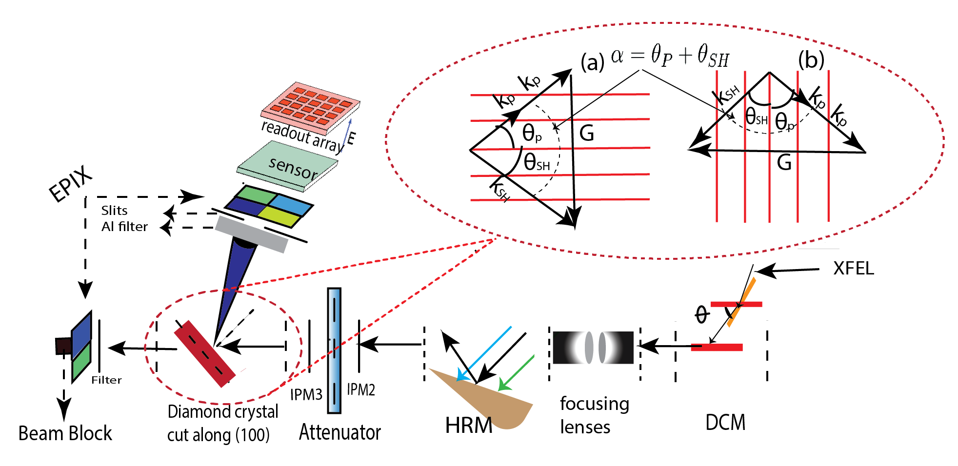 Figure 1
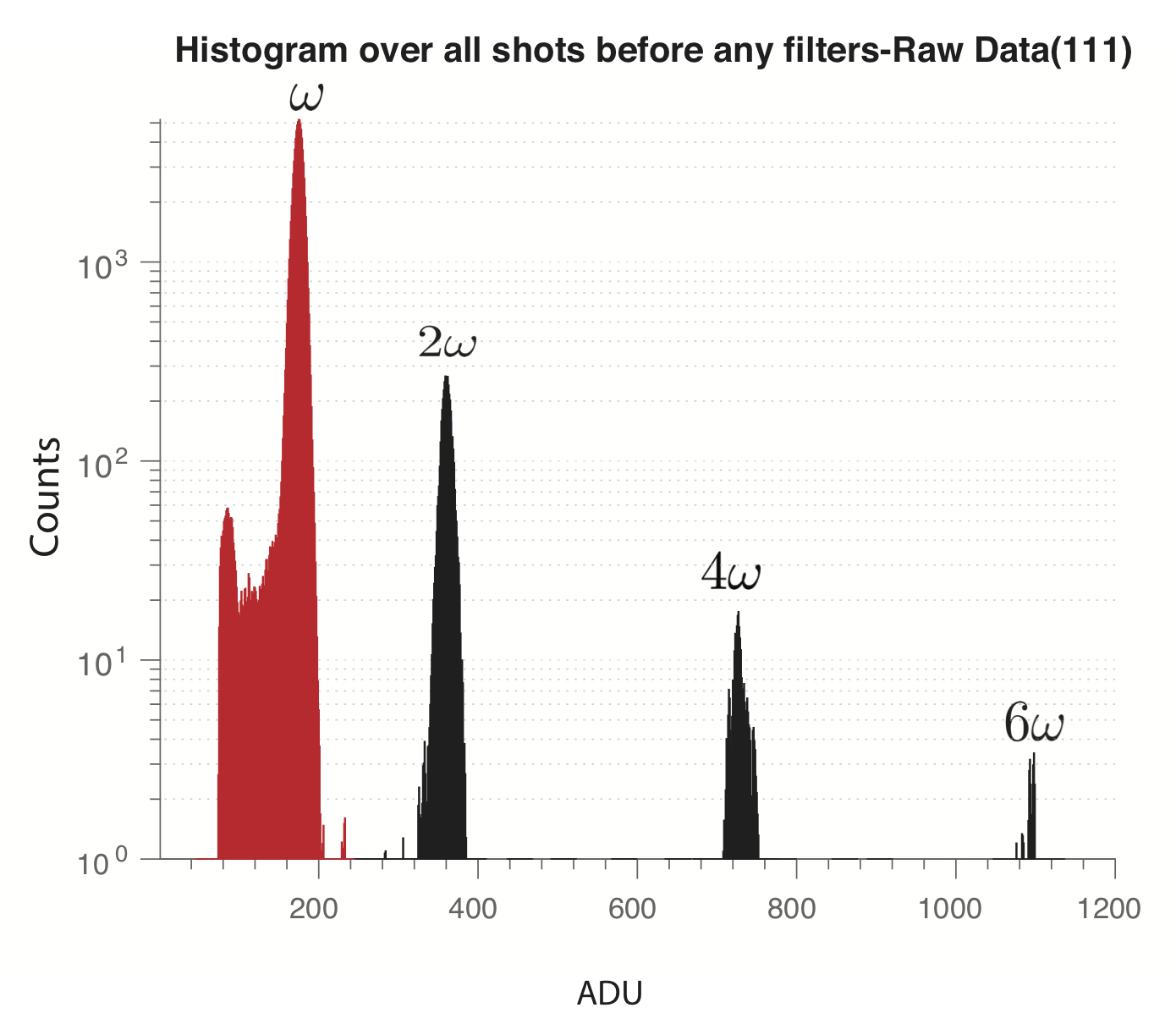 Figure 2
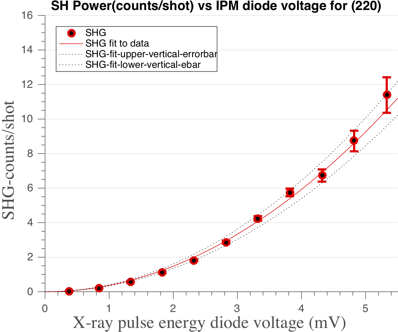 Figure 3
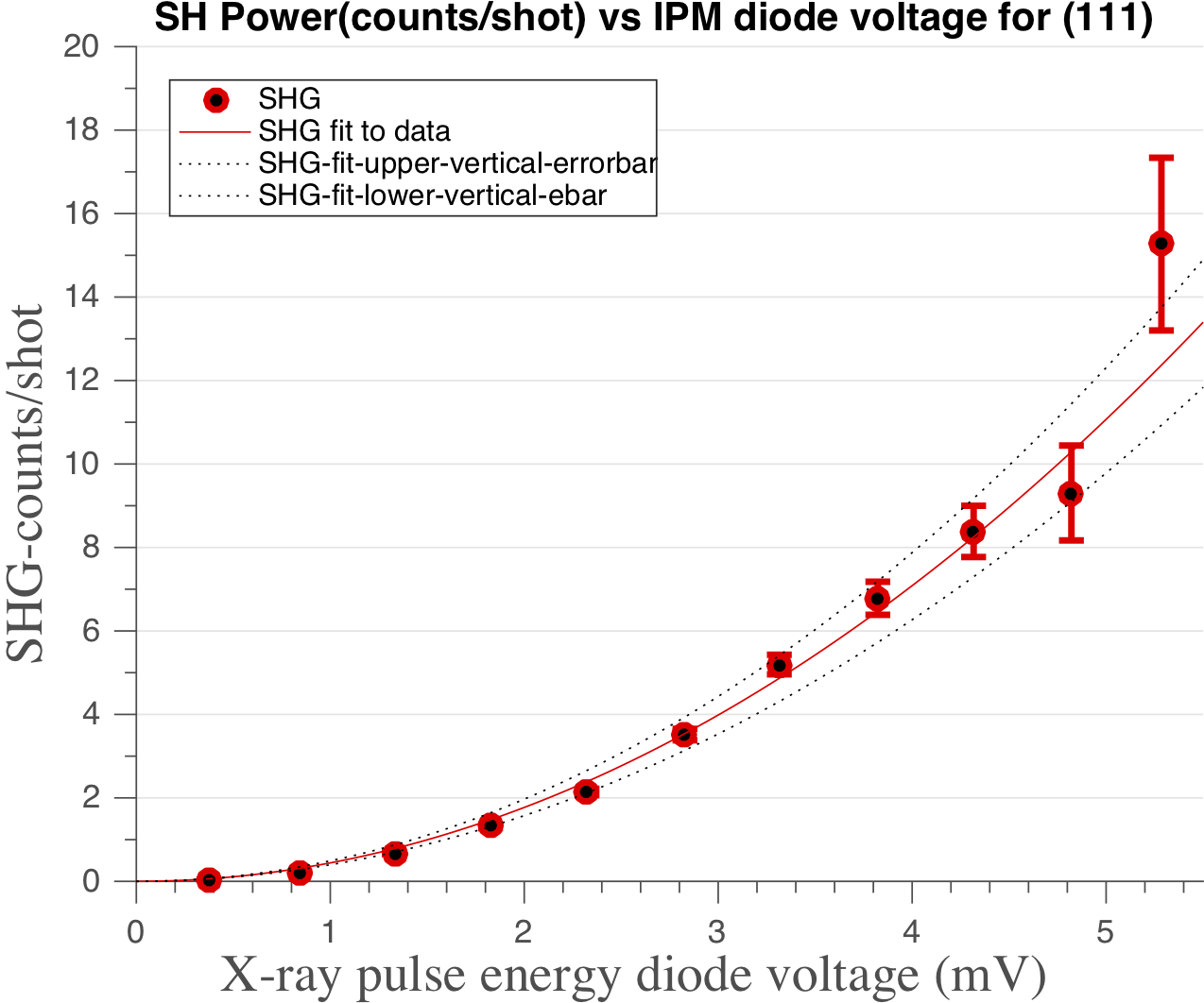 Figure 3
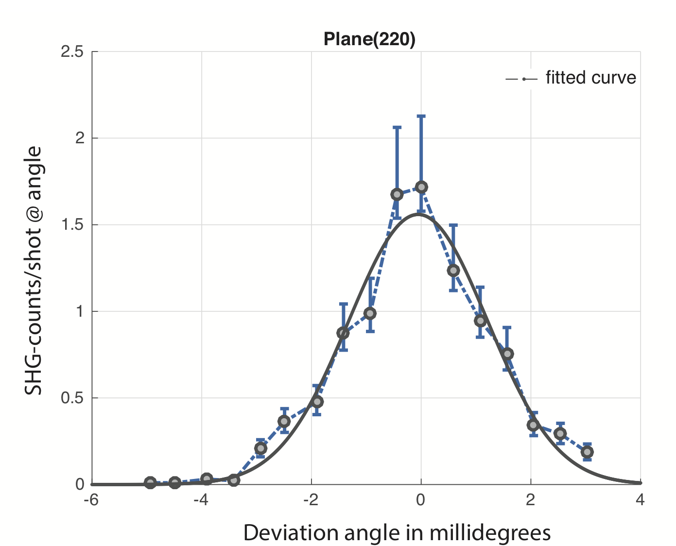 Figure 4
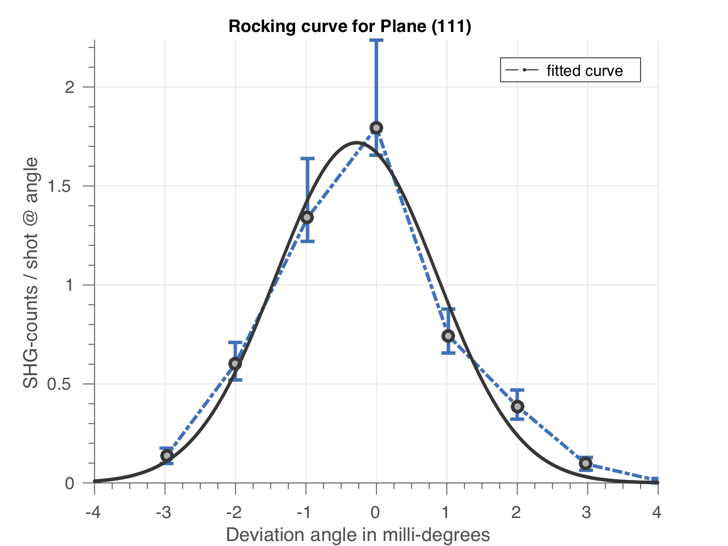 Figure 4
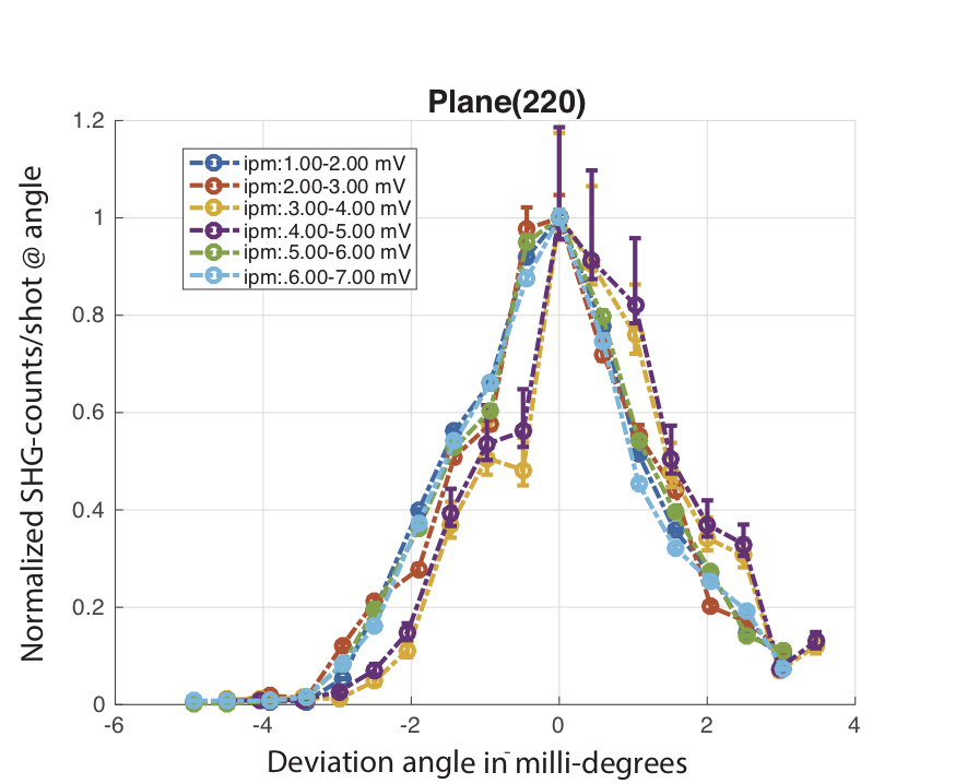 Figure 5
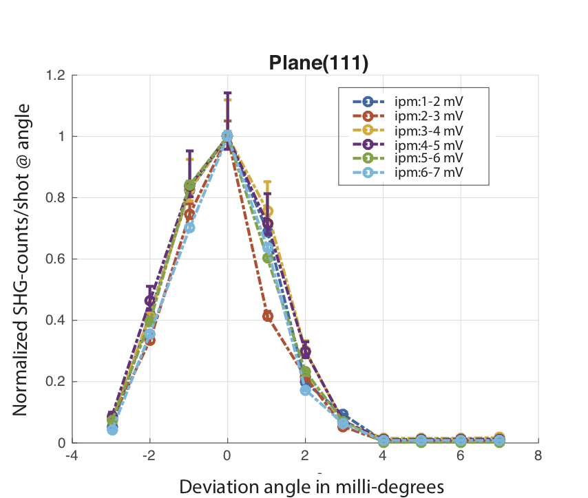 Figure 5
| Plane | Number of shots | ||||
| (111) | 60k | 35.26 | 44.07 | 17.63 | 8.81 |
| (11-1) | 126k | -35.26 | -26.44 | 17.64 | 8.82 |
| (220) | 60k | 0 | 14.5 | 28.99 | 14.5 |
| (113) | 83k | 64.76 | 81.82 | 34.14 | 17.06 |
| (00-4) | 122k | -90 | -69.27 | 41.47 | 20.73 |
| (331) | 120k | 13.26 | 35.94 | 45.39 | 22.68 |
| (660) | 77k | 0 | 48.67 | 97.4 | 48.67 |
| Plane | |||
|---|---|---|---|
| (111) | 0.5 | 7.3 | 1.42 |
| (11-1) | 0.3 | 7.1 | 1.4 |
| (220) | 0.4 | 6 | 1.1 |
| (113) | 0.07 | 3 | 0.66 |
| (00-4) | 0.2 | 2.7 | 0.52 |
| (331) | 0.05 | 3.2 | 0.61 |
| (660) | 0.02 | 0.5 | 0.11 |
| Plane | ||
|---|---|---|
| (111) | 0.45 | 1.8 |
| (11-1) | 0.45 | 1.72 |
| (220) | 0.7 | 2.34 |
| (113) | 0.5 | 1.6 |
| (00-4) | 0.73 | 2.19 |
| (331) | 0.6 | 1.4 |
| (660) | 0.63 | 0.33 |
| Experiment | ||
|---|---|---|
| IA | FWHM | |
| (111) | 2.8 | |
| (11-1) | 3 | |
| (220) | 3 | |
| (113) | 3.6 | |
| (00-4) | 4.2 | |
| (331) | 3.2 | |
| (660) | 3.3 | |
Peer Reviews
No public reviews on file for this paper yet. If you reviewed it on a platform where reviews are public (OpenReview, ICLR, NeurIPS, ICML), you can paste yours below so the community can read it here.
Videos
No videos yet. Explain this paper in a talk, walkthrough, or lecture? Add one.
Taxonomy
TopicsDiamond and Carbon-based Materials Research · High-pressure geophysics and materials · Laser-Plasma Interactions and Diagnostics
Multiple Fourier Component Analysis of X-ray Second Harmonic Generation in Diamond
P.Chakraborti B.Senfftleben B.Kettle S.W.Teitelbaum P.H.Bucksbaum S.Ghimire J.B.Hastings H.Liu S.Nelson T.Sato S. Shwartz Y.Sun C.Weninger D.Zhu D.A.Reis M.Fuchs1
1Department of Physics and Astronomy , University of Nebraska-Lincoln, Lincoln, Nebraska, USA
2Max Born Institute for Nonlinear Optics and Short Pulse Spectroscopy, Berlin, Germany
3Department of Physics, Imperial College, London, UK
4PULSE Institute, Stanford University, Palo Alto, California, USA
5LCLS, Stanford National Accelerator Lab, Menlo Park, California, USA
6Department of Physics, Barllan University, RamatGan, Israel
Abstract
The unprecedented brilliance of X-ray free-electron lasers (XFELs) Emma et al. (2004); Ishikawa et al. (2012) has enabled first studies of nonlinear interactions in the hard X-ray range. In particular, X-ray - optical mixing Glover et al. (2012), X-ray second harmonic generation (XSHG) Shwartz et al. (2014) and nonlinear Compton scattering (NLCS) Fuchs et al. (2015) have been recently observed for the first time using XFELs. The former two experiments as well as X-ray parametric downconversion (XPDC)Freund and Levine (1969); Eisenberger and McCall (1971a) are well explained by nonlinearities in the impulse approximationEisenberger and Platzman (1970), where electrons in a solid target are assumed to be quasi free for X-ray interactions far from atomic resonances. However, the energy of the photons generated in NLCS at intensities reaching up to exhibit an anomalous red-shift that is in violation with the free-electron model. Here we investigate the underlying physics of X-ray nonlinear interactions at intensities on order of . Specifically, we perform a systematic study of XSHG in diamond. While one phase-matching geometry has been measured in Shwartz et al.Shwartz et al. (2014), we extend these studies to multiple Fourier components and with significantly higher statistics, which allows us to determine the second order nonlinear structure factor. We measure the efficiency, angular dependence, and contributions from different source terms of the process. We find good agreement of our measurements with the quasi-free electron model.
pacs:
Valid PACS appear here
††preprint: APS/123-QED
I Introduction
At optical wavelengths, second harmonic generation is well understood since the experiment by Franken et al.Franken et al. (1961) in the 1960s. Classically, it can be explained by the nonlinear polarization induced by a strong oscillating electric field in an anharmonic atomic potential. In the dipole approximation it can be shown that second order interactions can only occur in non-centrosymmetric materials Boyd (1992). However, in the hard X-ray range, a different nonlinearity is dominant because of the unfavorable frequency scaling of the atomic nonlinearity, which for the second order polarization scales as far from resonances. At X-ray frequencies well above atomic resonances, the electrons in a solid can be treated as quasi free in the impulse approximation Eisenberger and Platzman (1970). In this case the nonlinear effects are based on the anharmonic motion of the quasi-free electrons under the influence of a strong electromagnetic wave, similar to a collisionless cold plasma Freund and Levine (1969); Eisenberger and McCall (1971b). Here, the second order polarization scales as . For a sufficiently large electric field, the electron can gain a relativistic momentum within half a light cycle. The Lorentz force due to the magnetic field becomes non-negligible and can be treated as a perturbationY.R.Shen (2002). Under such conditions, the electron performs a ’figure 8’ motion Vachaspati (1962), which in addition to an inhomogeneous charge density leads to the emission of radiation at harmonics of the incoming frequency. This nonlinearity is fundamentally different from that routinely used at optical wavelengths. For example the generation of second harmonic radiation is permitted even in centrosymmetric crystals. More importantly, it combines the potential of nonlinear optics with the atomic spatial resolution of X-ray wavelengths. Using nonlinear X-ray interactions, first experiments have been able to measure quantities otherwise not accessible, such as the induced local optical response with atomic resolution Tamasaku et al. (2011). In addition to potential applications, the investigation of this nonlinear response is of fundamental interest. Here we investigate X-ray second harmonic generation, the prototypical second order nonlinearity. We perform a systematic study of the nonlinearity by observing several Fourier components of the second order response and different boundary conditions, such as Laue and Bragg geometries. This allows us to measure the second order structure factor in diamond. We find that it is consistent with the quasi-free electron model, that has been successful in describing X-ray - matter nonlinearities such as parametric down conversion(PDC) Eisenberger and McCall (1971a), X-ray optical sum frequency generation(XOSFG) Glover et al. (2012) and non-resonant X-ray second harmonic generation experiment (XSHG) Shwartz et al. (2014). In the model we treat the Lorentz force due to the magnetic field as perturbation. This perturbation scales with the normalized vector potential , where is the Amplitude of the electric light field with frequency , the electron charge, the electron mass, and the speed of light. Although for X-rays this factor is comparably small because of their high frequency (in our case ), the plasma-like nonlinearity is significantly stronger than that due to the anharmonic atomic potential. We assume a plane wave of the form +c.c where , and are respectively the X-ray polarization vector, electric field amplitude, and wave vector of the incoming field. We expand the source current term in the free-electron approximation perturbatively, which at lowest (second) order leads to a current density oscillating at the second harmonic frequency () given by Nazarkin et al. (2003)
[TABLE]
Here, is the unperturbed electron charge density and the magnetic field. The first term (displacement term) is trivially zero for SHG in a plane wave approximation. The second current term is due to the nonlinear Lorentz force and oscillates in the propagation direction along . The third term, known as the Doppler term, only contributes in a non-uniform plasma. In particular, a perfect crystal has a periodically modulated charge density, which can be expanded in a Fourier series in terms of reciprocal lattice vectors as . As described below, this allows us to phase-match the process, which significantly increases the efficiency. To calculate the generated SH signal, we consider only the contributions from the Lorentz and Doppler terms. Owing to the low efficiency of SHG we assume our pump is undepleted and the outgoing field of the second harmonic, has a slowly varying envelope. Substituting the current term in the wave equation and making these additional assumptions, for a given reciprocal lattice vector the wave equation describing SHG in a crystal simplifies to the equation belowYudovich and Shwartz (2015)
[TABLE]
Here, is the angle of the SH signal with respect to the chosen crystal lattice plane (see figure 1), and are electric and magnetic constants, respectively. is the group velocity, the wave vector, the refractive index, and the polarization vector of the generated second harmonic field. We specify the coordinate along, and the coordinate transverse to the direction of the pump pulse propagation. describes the phase mismatch. The maximum signal can be achieved when the phase matching condition is satisfied, i.e. when (see figure 1). This condition leads to a small difference in angle from the linear Bragg diffraction of a photon due to slight differences in twice the refractive index for the fundamental wavelength and the index of the generated second harmonic. The two terms in square brackets on the right hand side of the equation are the Lorentz and Doppler term, respectively. Depending on the geometry, which is given by the choice of the reciprocal lattice vector used for phase matching, the relative strengths of the two terms can vary as shown in Table IV below. As can be seen from equation I, the generated SH field scales quadratically with the field of the pump pulse, as expected for a second order nonlinear process.
The highly non-colinear geometry between the pump and the SH pulse limits the length over which the two beams overlap during the interaction. In particular, the small focus size and the ultra-short duration of the pump pulse leads to spatial and temporal walk-off between the pump and signal over the crystal length. Within a walk-off length the SH signal grows quadratically with the interaction distance. However, for our geometry, where the effective crystal thickness is much smaller than the Rayleigh length but significantly longer than the walk-off length, the signal grows overall only linearly with crystal length. For a monochromatic 1D X-ray field with a frequency far from resonances and which exactly fulfills the phase-matching condition (), the SH intensity emitted into the phase-matching direction for the -th Fourier component is approximately given by
[TABLE]
Here is the normalized vector potential for the pump pulse, the walk-off length, and the effective crystal length, where is the crystal length.
For a quantitative comparison we numerically solve the wave equation (eq. I) for a temporally transform-limited Gaussian X-ray pulse with a finite bandwidth, and integrate over the length of the crystal. We then compare the theoretical efficiency of the SHG with the experimental value for different phase matching geometries.
II Experimental Setup
The experiment was performed at the XPP instrument at the LCLS free-electron laser using an X-ray energy of 9.8 keV (see figure 1). The X-ray beam was monochromatized to a bandwidth of 0.7 eV using a double crystal diamond (111) monochromator for scattering in the polarization plane. The X-ray pulse with an energy of 178 35 J and a duration of 30 10 fs (RMS) was focused to a spot size with waist radius of m, leading to an averaged intensity inside the focal spot of . The pulse energy on target can be varied by introducing Si attenuators of different thicknesses into the unfocused beam. The second FEL harmonic emission generated in the undulator was significantly reduced before reaching the target by the structurally forbidden (222) reflection of the diamond monochromator, two reflections from gracing incidence hard X-ray offset mirrors and additional harmonic rejection mirrors. In our measurements essentially no linear diffraction from the second FEL harmonic signal from the target crystal was observed. As a target for second harmonic generation, we used a (100)-cut diamond crystal with dimension of 4 4 0.8 mm3. We used various crystal lattice planes to investigate different geometries and multiple Fourier components of the charge density, both in Bragg reflection and in Laue transmission geometries. The crystal lattice planes and their respective scattering angles that were used as phase-matching geometries are listed in table 1.
As a detector, we used a pixelated 2D EPIX camera, which has an energy resolution of roughly 0.4 keV Zhu et al. (2014), which allows us to distinguish between fundamental and second harmonic photons. However, the detector cannot distinguish between a single second harmonic photon with energy and the pile-up of two or more lower-energy photons with a combined energy of detected in the same pixel. Similarly, a pixel count registered at an energy of could result from a single photon or from the pileup of two second harmonic photons detected in the same pixel. Due to the comparably small pixel size of 50 50 , there is significant charge sharing between the pixels. We correct for this by implementing a ’droplet algorithm’Nelson (1999) on the observed images for each shot. To significantly decrease the background and the probability of pileup from the FEL fundamental, a 1.5mm thick Al filter was placed directly in front of the EPIX detector, which for the fundamental has a transmission of only , while for the second harmonic .
The shot-to-shot pulse energy fluctuations of the XFEL beam are recorded using Intensity Position Monitor (IPM) diodes.These measurements allow us to more finely bin the intensity dependence of the measured SH signal as compared to only the Si attenuators. The on-target pulse energy was cross calibrated to the IPMs using an optical power meter.
III Experimental Results
The second harmonic signal is observed with a pixelated 2D array detector. Due to the comparably sparse signal and energy resolution of the detector, we can distinguish between photons with energies corresponding to the fundamental, second harmonic, and higher harmonics. A detector histogram for a run measuring the (111) Fourier component is shown in figure 2. The SH peak at 360 Analog-to-Digital Units (ADUs) is sufficiently strong to generate pile-up signal at (720 ADUs) and (1080 ADUs). From these histograms we can estimate the pile-up contributions from both the fundamental and the second harmonic photons. These estimates are included when calculating negative and positive contributions to the error bars of the experimental data.
For each Fourier component we study the intensity-dependence and the dependence of the XSHG signal on small deviations from the phase-matching condition. The X-ray pulse energy is coarsely varied by using Si attenuators of different thicknesses. The FEL pulse energy on target varies from shot to shot due to the intrinsic energy fluctuations of the SASE lasing process. In the monochromator the shot-to-shot fluctuations in the FEL spectrum are also transformed into additional pulse-energy fluctuations. By measuring the FEL pulse energy after the monochromator for each shot using Intensity-Position Monitors (IPMs), we can use the fluctuations to achieve a finer pulse-energy binning. The dependence of the SH signal on the FEL pulse energy can be seen in figure 3 for two representative phase-matching geometries. At the highest intensity, we observe comparably strong signals with several SH photons per shot after correction for the filter transmission and detector quantum efficiency. The number of generated photons show a quadratic dependence on the incoming X-ray pulse energy, as is expected for a second-order nonlinear process. To be more quantitative we fit the data with a second order polynomial function given by , where is the number of second harmonic counts per pulse and is the IPM diode voltage in milli-Volt (). and have the units of and respectively. The first term in the equation is the usual quadratic dependence of a second order nonlinear process while the second and third terms take into account parasitic linear scattering of the FEL second harmonic and any background. Table 2(b) shows the resulting fit parameters for each measured Fourier component. We only list the coefficient for the quadratic dependence since the fit coefficients and were negligible for all geometries. The fitted SH signal shows a clear quadratic dependence on the incoming pulse energy for all geometries. At the highest intensities for some of the geometries 10 photons are generated per pulse, leading to efficiencies on order of . Since the SH signal is emitted into a narrow beam, these photons are concentrated into only a few detector pixels. This leads to a significant pile-up of SH photons, generating peaks at even higher orders in the detector ADU histograms (see figure 2). Since the beamline transmission for photon energies higher than the FEL fundamental is extremely small and the higher-order peaks agree with Poisson statistics for pile-up, we also count these peaks as photons. We compare the measured efficiencies to theory in Table II.
The generated SH signal as a function of the crystal rocking curve is shown in figure 4 for the (220) and the (111) Fourier components. The observed signal has an angular offset from the expected linear elastic Bragg signal of the second FEL harmonic that agrees with the phase-matching condition described above. The SH signal is emitted into a narrow beam and is only well above background when the phase-matching condition is fulfilled. We calculate the width for each Fourier component in table 4).
IV Discussion of results
In order to explore the validity of the quasi-free electron model, we compare our measurements with theoretical results from the numerical solution of equation I. The different geometries also allow us to investigate the contributions of the different source terms described in equation 1(see TABLE III). We assume a temporally transform-limited Gaussian pulse and take into account the pump field depletion during the process from the long attenuation length in diamond (1.2 mm for 9.8 keV photons). For each geometry we integrate the wave equation along the effective propagation length of the pump pulse inside the crystal. For the integration we use an interval significantly smaller than the respective spatio-temporal walk-off length. From the obtained outgoing SHG field, , we calculate the theoretical efficiency according to
[TABLE]
where is the integrated second harmonic intensity at the exit of the crystal and the intensity of the incoming FEL fundamental. In table II(a) we compare the experimentally measured efficiencies to the numerical results for the highest FEL intensities. In case of the experimental values, the efficiency is given by the ratio of the number of observed SH photons corrected for filter transmission and detector quantum efficiency and the number of incoming FEL photons per pulse. We find that the highest experimental efficiencies, as well as the simulated efficiencies for our phase-matching condition, arise from the (111) and (11-1) planes in asymmetric Laue, and the (220) plane in symmetric Laue geometry(Table-II(a)). The efficiencies are of the order of for both the simulation and the experiment. However, the theoretical results are approximately a factor of 5-6 higher than the experimental efficiencies.
A simple calculation of the efficiency from the 1D analytical plane-wave solution (eqn. 2) significantly overestimates the efficiencies(), but it gives us a lot of qualitative insight into the SHG process. Due to the highly non-colinear geometry the SH signal gain is limited by walk-off. As the walk-off length is much smaller than both the Rayleigh range and the crystal thickness, the generated SH intensity scales bilinearly with and effective crystal length as . For our geometries we are mainly limited by spatial walk-off, given by , where is the focus diameter and the angle between the pump and the generated SH signal. The efficiency scales with the FEL pulse properties as , where is the pulse energy and the pulse duration. The relative efficiencies at similar intensities for the different Fourier components can be estimated by multiplying the source current terms with the walk-off lengths for each geometry. Specifically, in the plane wave case, the exactly phase-matched SH intensity is approximately given by , which is essentially equation 2. The magnitude of the two main source current density terms for each phase-matching configuration, namely the Lorentz and Doppler term in equation 1, are given in table 3. The listed current terms are the projections onto the SH polarization for each geometry. It can be seen that for most geometries the SH signal is mainly caused by the Doppler term. The high efficiencies of the (220) and (111) components can be explained by the comparably strong current terms and long walk-off lengths. For the (660) Fourier component the SH emission angle is close to 90∘ to the pump pulse propagation, along the FEL polarization direction. Although linear scattering in this direction using p-polarized light is highly suppressed, a substantial SH signal can be observed. For this component the Lorentz current term oscillating along the FEL propagation direction is larger than the Doppler term. We do not observe any unexpected behavior of the SHG for the geometry where the signal is mainly due to the Lorentz term compared to the ones where the signal is mainly due to the Doppler term.
The difference between experiment and simulation can be attributed to our assumptions of a monochromatic beam and perfect crystal quality. The error in the simulations is mainly due to uncertainties in the FEL parameters, specifically the FEL pulse energy, pulse duration, pulse structure, and focus size. The focus size and pulse duration not only affect the magnitude of the nonlinear source current term through the FEL intensity but also the walk-off lengths, over which the SH signal grows quadratically with distance. In order to account for this coupled error we have calculated the peak efficiencies for a set of FEL parameters within our uncertainties. From these numerical calculations we determine the upper and lower bounds for our error for each Fourier component. In particular we estimate the upper error bound using the highest X-ray pulse energy (216 J), smallest focus size (1.8 m), and shortest pulse duration (20 fs). The lower error bound is estimated at the lowest X-ray pulse energy (144 J), largest focus size (3.3 m), and longest pulse duration (40 fs). These results agree with the scalings obtained from the 1D model described above.
We also investigate the dependence of the generated SH signal for small deviations from the phase-matching condition. To this end we measure the SH signal as a function of the crystal rotation angle. In order to examine the shape of the crystal rocking curves we filter the data by FEL pulse energy. The curves with the highest statistics (IPM range of mV) for the (111) and (220) Fourier component are shown in figure 4. The shape of the rocking curves do not show a significant dependence on the FEL fundamental intensities, as can be seen in figure 5. Although the width of the observed rocking curves vary slightly for each Fourier component, the integrated reflectivities are nearly constant and within our uncertainties (see table 4). The measured rocking curves are significantly broader than what is expected from the linear X-ray diffraction crystal Darwin widths. The broadening is mainly due to the divergence of the incoming converging beam, which is considerably larger than the crystal Darwin widths for any of the reflections. For a beam divergence of ()(4 mdeg) we expect rocking curve widths of , which agrees reasonably well with the measured values and the integrated reflectivities.
V Conclusion
We have experimentally investigated phase-matched X-ray second harmonic generation in diamond at a photon energy of 9.8 keV. We observe clear nonlinear signals with efficiencies on order of at X-ray intensities of for multiple Fourier components of the second harmonic response. For each component we measure a quadratic dependence of the signal on the incoming FEL intensity, as expected for a second-order nonlinear process. Observable signals are only generated in well-defined geometries around the phase-matching condition. We have compared the measurements to a theoretical model based on the impulse approximation, which assumes the electrons in a solid to be quasi free during the interaction with X-rays. In particular, we have compared the measured efficiencies, phase-matching condition, and slight angular deviations from perfect phase-matching. The calculated efficiencies are roughly a factor of 5 higher than the experimental values, which is in reasonably good agreement within the parameters assumed for the simulation. The relative intensities of the different Fourier components also agree with theory. The intensity of the SH signal rapidly drops once outside the phase-matching condition. The widths of the crystal rocking curves is mostly due to the X-ray beam divergence. From these comparisons we can conclude that our measured second order structure factor is consistent with the quasi-free electron model within the uncertainties of the experimental values and the approximations of our calculations. This experiment gives valuable insights into a nonlinear response that is fundamentally different from that routinely used at optical wavelengths. The elementary understanding of these processes is of great importance as these nonlinear processes in the X-ray wavelength range allow the investigation of non-equilibrium dynamics with atomic time and length scales. Despite low conversion efficiencies, these nonlinear interactions have the potential to lead to novel diagnostics with capabilities beyond linear diffraction techniques, especially considering the availability of current and future high-repetition rate FEL facilities.
VI Acknowledgements
This material is based upon work supported by the U.S. Department of Energy, Office of Science, Basic Energy Sciences under Award Number DE-SC0016494 and by the AMOS program within the Chemical Sciences, Geosciences, and Biosciences Division of Basic Energy Sciences, U.S. Department of Energy under Award Number DE-AC02-76SF00515. Use of the Linac Coherent Light Source (LCLS), SLAC National Accelerator Laboratory, is supported by the U.S. Department of Energy, Office of Science, Office of Basic Energy Sciences under Contract No. DE-AC02-76SF00515.
The reference list from the paper itself. Each links out to its DOI / PubMed record.
- 1Emma et al. (2004) P. Emma, K. Bane, M. Cornacchia, Z. Huang, H. Schlarb, G. Stupakov, and D. Walz, Phys. Rev. Lett. 92 , 074801 (2004) . · doi ↗
- 2Ishikawa et al. (2012) T. Ishikawa, H. Aoyagi, T. Asaka, Y. Asano, N. Azumi, T. Bizen, H. Ego, K. Fukami, T. Fukui, Y. Furukawa, S. Goto, H. Hanaki, T. Hara, T. Hasegawa, T. Hatsui, A. Higashiya, T. Hirono, N. Hosoda, M. Ishii, T. Inagaki, Y. Inubushi, T. Itoga, Y. Joti, M. Kago, T. Kameshima, H. Kimura, Y. Kirihara, A. Kiyomichi, T. Kobayashi, C. Kondo, T. Kudo, H. Maesaka, X. M. Marechal, T. Masuda, S. Matsubara, T. Matsumoto, T. Matsushita, S. Matsui, M. Nagasono, N. Nariyama, H. Ohashi, · doi ↗
- 3Glover et al. (2012) T. E. Glover, D. M. Fritz, M. Cammarata, T. K. Allison, S. Coh, J. M. Feldkamp, H. Lemke, D. Zhu, Y. Feng, R. N. Coffee, M. Fuchs, S. Ghimire, J. Chen, S. Shwartz, D. A. Reis, S. E. Harris, and J. B. Hastings, Nature 488 , 603 (2012) . · doi ↗
- 4Shwartz et al. (2014) S. Shwartz, M. Fuchs, J. B. Hastings, Y. Inubushi, T. Ishikawa, T. Katayama, D. A. Reis, T. Sato, K. Tono, M. Yabashi, S. Yudovich, and S. E. Harris, Phys. Rev. Lett. 112 , 163901 (2014) . · doi ↗
- 5Fuchs et al. (2015) M. Fuchs, M. Trigo, J. Chen, S. Ghimire, S. Shwartz, M. Kozina, M. Jiang, T. Henighan, C. Bray, G. Ndabashimiye, P. H. Bucksbaum, Y. Feng, S. Herrmann, G. A. Carini, J. Pines, P. Hart, C. Kenney, S. Guillet, S. Boutet, G. J. Williams, M. Messerschmidt, M. M. Seibert, S. Moeller, J. B. Hastings, and D. A. Reis, Nat Phys 11 , 964 (2015) . · doi ↗
- 6Freund and Levine (1969) I. Freund and B. F. Levine, Phys. Rev. Lett. 23 , 854 (1969) . · doi ↗
- 7Eisenberger and Mc Call (1971 a) P. Eisenberger and S. L. Mc Call, Phys. Rev. Lett. 26 , 684 (1971 a) . · doi ↗
- 8Eisenberger and Platzman (1970) P. Eisenberger and P. M. Platzman, Phys. Rev. A 2 , 415 (1970) . · doi ↗