Soft labeling by Distilling Anatomical knowledge for Improved MS Lesion Segmentation
Eytan Kats, Jacob Goldberger, Hayit Greenspan

TL;DR
This paper introduces a soft labeling approach using anatomical knowledge and dilated masks to improve MS lesion segmentation accuracy with FCNNs, demonstrating enhanced performance on benchmark data.
Contribution
The study proposes a novel soft mask training method that incorporates neighboring pixel information to improve MS lesion segmentation accuracy.
Findings
Higher Dice similarity coefficient achieved
Improved precision-recall tradeoff demonstrated
Enhanced performance over independent expert annotations
Abstract
This paper explores the use of a soft ground-truth mask ("soft mask'') to train a Fully Convolutional Neural Network (FCNN) for segmentation of Multiple Sclerosis (MS) lesions. Detection and segmentation of MS lesions is a complex task largely due to the extreme unbalanced data, with very small number of lesion pixels that can be used for training. Utilizing the anatomical knowledge that the lesion surrounding pixels may also include some lesion level information, we suggest to increase the data set of the lesion class with neighboring pixel data - with a reduced confidence weight. A soft mask is constructed by morphological dilation of the binary segmentation mask provided by a given expert, where expert-marked voxels receive label 1 and voxels of the dilated region are assigned a soft label. In the methodology proposed, the FCNN is trained using the soft mask. On the ISBI 2015…
Click any figure to enlarge with its caption.
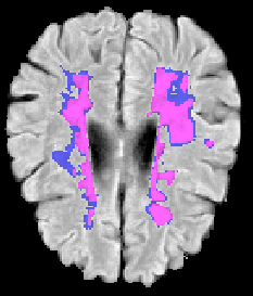 Figure 1
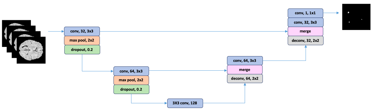 Figure 2
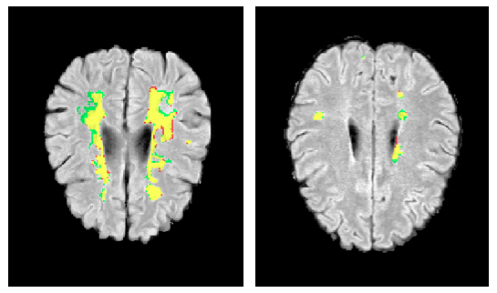 Figure 3
| Mask size | Dice | Precision | Recall | |||
| Rater 1 | Rater 2 | Rater 1 | Rater 2 | Rater 1 | Rater 2 | |
| 100% | 67.3 | 59.0 | 81.3 | 83.3 | 61.1 | 46.9 |
| 110% | 67.2 | 58.4 | 81.8 | 83.3 | 58.9 | 45.8 |
| 120% | 69.9 | 62.0 | 80.5 | 81.5 | 63.1 | 50.6 |
| 130% | 68.5 | 61.2 | 77.5 | 80.0 | 62.9 | 50.3 |
| 140% | 67.0 | 58.8 | 80.3 | 82.6 | 59.2 | 47.0 |
| Soft label | Dice | Precision | Recall | |||
| Rater 1 | Rater 2 | Rater 1 | Rater 2 | Rater 1 | Rater 2 | |
| 0.0 | 67.3 | 59.0 | 81.3 | 83.3 | 61.1 | 46.9 |
| 0.2 | 68.3 | 59.8 | 81.4 | 83.7 | 60.5 | 47.5 |
| 0.3 | 69.9 | 62.0 | 80.5 | 81.5 | 63.1 | 50.6 |
| 0.4 | 68.9 | 61.8 | 76.7 | 80.0 | 65.2 | 52.3 |
| Method | Dice | Precision | Recall |
|---|---|---|---|
| Binary mask | 56.0 | 82.9 | 44.6 |
| Soft labeled mask | 57.8 | 83.8 | 46.6 |
Peer Reviews
No public reviews on file for this paper yet. If you reviewed it on a platform where reviews are public (OpenReview, ICLR, NeurIPS, ICML), you can paste yours below so the community can read it here.
Videos
No videos yet. Explain this paper in a talk, walkthrough, or lecture? Add one.
Taxonomy
TopicsBrain Tumor Detection and Classification · Digital Imaging for Blood Diseases · AI in cancer detection
Soft labeling by Distilling Anatomical knowledge for Improved MS Lesion Segmentation
Abstract
This paper explores the use of a soft ground-truth mask (“soft mask”) to train a Fully Convolutional Neural Network (FCNN) for segmentation of Multiple Sclerosis (MS) lesions. Detection and segmentation of MS lesions is a complex task largely due to the extreme unbalanced data, with very small number of lesion pixels that can be used for training. Utilizing the anatomical knowledge that the lesion surrounding pixels may also include some lesion level information, we suggest to increase the data set of the lesion class with neighboring pixel data - with a reduced confidence weight. A soft mask is constructed by morphological dilation of the binary segmentation mask provided by a given expert, where expert-marked voxels receive label 1 and voxels of the dilated region are assigned a soft label. In the methodology proposed, the FCNN is trained using the soft mask. On the ISBI 2015 challenge dataset, this is shown to provide a better precision-recall tradeoff and to achieve a higher average Dice similarity coefficient. We also show that by using this soft mask scheme we can improve the network segmentation performance when compared to a second independent expert.
**Index Terms— ** Deep learning, multiple sclerosis, segmentation, soft labels
1 Introduction
Multiple Sclerosis (MS) is an autoimmune disease of the central nervous system that is characterized by the formation of delineated lesions visible in Magnetic Resonance Images (MRI). Accurate segmentation of MS lesions is essential for reliable disease onset detection, in tracking its progression and in evaluating treatment efficiency. In recent years, FCNNs have achieved promising results in lesion segmentation (see e.g. [1], [2], [3]). A major challenge in training FCNNs for MS lesion segmentation is the highly unbalanced data, as the number of lesion class voxels are often much lower than the number of non-lesion voxels. Models trained with unbalanced data yield segmentation that is biased towards the non-lesion class which is characterized by a greater amount of false negatives. There are various methods to address this data imbalance, including equal sampling [2], 2-phase training [4], and persistent loss functions [5] [6]. A second challenge facing delineation of MS lesions is the fact that the lesion contours are not well defined on the MRI images - leading to much ambiguity in the expert markings along the lesion contours. In Figure 1 we see two MRI scans overlaid with an expert ground-truth binary mask. In order to demonstrate the ambiguity, we show masks by 2 experts: One of the experts (rater 1) labels all the yellow and green voxels as lesion voxels (”1”). A second expert (rater 2) also labels all the yellow voxels as lesion voxels, but does not find that the green voxels belong to the lesion class. We note that the conflicting and more problematic voxels lie on the boundary of the lesion area.
A loss function used for FCNN training is expected to reflect the true delineations of the expert. As most of the inter-rater variability can be found along MS lesion contour voxels, we propose to modify the true delineations at those pixels by assigning soft class probabilities. Specifically, we present a loss function for training the FCNN that uses soft labeling within the framework of a Dice measure. This loss function provides additional information for the training process beyond the ground truth mask obtained by the expert. We evaluate the proposed training loss function on the ISBI 2015 MS lesion segmentation challenge dataset [7]. We show that utilizing the soft labeled masks for FCNN training leads to a better precision-recall result, with a dependence on the volume of the soft labeled region and the soft class probability value.
2 Extended Soft Labeling
Assume we train a FCNN for the task of lesion segmentation. A ground truth binary labeling for each voxel is provided by a manual annotation. As noted above the labeling is extremely unbalanced since most of the voxels are labeled as non-lesion. Training with imbalanced data is very problematic especially when the training evaluation measure is classification accuracy. A well-known alternative method for evaluating the performance of medical imaging systems is the Dice measure. However, Dice measure can also be skewed by imbalanced data.
Knowledge distillation is a popular method for transferring knowledge from a large model (a teacher) trained on a server to a smaller, compressed model (a student) simple enough to run on a device [8]. Here we applied a similar strategy to provide more information when training a lesion segmentation system. Voxels that are near contours of the segmented lesion are not well defined in MRI images, yet these voxels can carry additional anatomical information about the lesion structure. To effectively use this information during the training process we modified the manual delineations of the expert by soft labeling of the voxels in the proximity of lesion.
2.1 Soft Labeled Mask
To create the soft mask we expand the original binary mask by 3D morphological dilation. Using the clinical observation that lesions appear as hyper-intense regions in FLAIR images [9], we exclude from the dilated region those voxels with FLAIR intensity value lower than a defined threshold. Selected voxels from the dilated region are assigned a soft label which is interpreted as the probability of the voxel to be part of the lesion. In the experimental section we analyze the best value for . The label of the manually annotated voxels remains 1. Figure 2 shows an example of manual delineations of an expert and the soft labeling of pixels that are similar to the annotated voxels in both location and content.
2.2 Effective Loss Function
Dice is often serves as a loss function for training FCNN in medical imaging tasks. Dice is defined as follows:
[TABLE]
where TP - number of True Positive voxels, FP - number of False Positive voxels and FN - number of False Negative voxels.
By denoting ground truth mask as matrix T and predicted probabilistic mask as matrix P we can write the terms in the Dice definition as follows: , and where is a true value of the voxel , and is a predicted probability of the voxel . Using this notation the Dice loss function can be equivalently written as:
[TABLE]
By denoting the binary mask of the dilated region as matrix D and the soft label assigned to voxels of the dilated region as we can represent the soft labeled mask as . As a result, training FCNN with the soft labeled mask is equivalent to training with the following effective soft dice loss function:
[TABLE]
Below we compare between training FCNN with the Dice loss (2) and the soft Dice loss (3) and show that when using a Dice loss based on extended soft labels for training, we obtain better test time performance as measured by the Dice measure.
3 Experimental Results
In this section we evaluate the proposed loss function on a MS lesion dataset. We first find the optimal combination of dilation size and a soft label value on the training data using a cross-validation technique (Sections 3.3 and 3.4). After finding the optimal combination of parameter values, we use this to evaluate the performance of the method on the test portion of the data (Section 3.5) .
3.1 Dataset
The dataset of the ISBI 2015 MS lesion segmentation challenge [7] consists of 5 patients in the training set and 14 patients in the test set. Each case has 4 modalities: T1-weighted, T2-weighted, proton density-weighted and FLAIR. All images are manually delineated by two experts. The measured inter-rater Dice overlap of 0.634 indicates high inter-rater variability (see Figure 1).
To obtain the parameters that yield optimal results we evaluate the proposed method on the training set by applying a leave-one-out cross-validation approach. For each combination of parameters 5 identical models were trained on 4 patients and tested on the remaining patient in the training dataset. The final results of the 5 models were averaged to produce the final performance evaluation measures.
3.2 Network Architecture and Training Details
To demonstrate the results of the proposed approach we trained a U-net [10] based FCNN (see Figure 3). Similar to the U-net, the network architecture we used is divided into two pathways of corresponding layers which are connected to leverage both high- and low-level features. A contracting path alternates convolution layers and max-pooling layers with stride 2 for downsampling. The expansive path alternates convolution layers and transposed convolution layers as proposed by Brosch et al. [11]. All convolution layers, except for the last one, are followed by a rectified linear unit (ReLU) [12]. Activations of the last convolution layer are fed to a sigmoid function that produces a probabilistic segmentation map with values in the range of 0 and 1.
Manual delineations by the first rater were used as the original binary mask from which the soft labeled mask was constructed by the method described in Sec. 2.1. To slightly rectify the data imbalance we excluded slices that did not contain lesions according to the ground truth mask from the training data. The network was trained on 80% of the remaining slices, with 20% held out for validation. During training we applied random rotations on the images where rotation angles were drawn from a uniform distribution over a range of . To produce a binary mask from the probabilistic output of the model, we applied a fixed threshold that maximized the mean dice similarity coefficient over the training set. In the post-processing step, only connected components that contained more than voxels were selected to be lesions.
3.3 Dilation Size
First we evaluated our method for different dilation sizes whereas soft label value was fixed and equal to . The results (see Table 1) show that the model trained with a soft mask dilated to 120% of the manual segmentation size had the best precision-recall tradeoff and an improved overall Dice measure.
3.4 Soft Label
In this experiment the influence of soft label value on model performance was explored. The 120% dilation size of the manual segmentation was used for the evaluation. Based on the results shown in Table 2 the soft label value provided valuable information about near contour voxels during the training phase and achieved the highest Dice measure.
3.5 Test Results
Next, we trained the model on the whole training set using the optimal combination of the dilated size and soft label value equal to 120% and 0.3 respectively. To compare performance of suggested training strategy (3) the model was also trained on the whole training set while utilizing manual annotations of the first rater as the ground truth (2). The two versions were submitted to the challenge website 111https://smart-stats-tools.org/lesion-challenge. The test results are shown in Table 3. We can see that using the soft mask based loss function we gain a clear improvement in both Dice, precision and recall measures.
4 Conclusions
In this study we proposed a new loss function that is used for training FCNN for MS lesion segmentation task where the training data is highly unbalanced. We defined a soft labeled mask that is assigned to voxels which are similar to the ground truth in both location and intensity. These voxels are usually near contour regions and tend to be labeled differently by different experts. We showed that training the FCNN with the proposed modified Dice loss function leads to better model generalization. We demonstrate the propose method on MS lesion segmentation. This concept is general and can be harnessed to improve other medical image segmentation tasks.
The reference list from the paper itself. Each links out to its DOI / PubMed record.
- 1[1] K. Kamnitsas, C. Ledig, V. Newcombe, A. Kane J. Simpson, D. Menon, D. Rueckert, and B.Glocker, “Efficient multiscale 3d cnn with fully connected crf for accurate brain lesion segmentation,” Medical Image Analysis , vol. 36, pp. 61–78, February 2017.
- 2[2] S. Valverde, M. Cabezas, E. Roura, D. Pareto S. Gonzalez-Villa, J. Vilanova, L. Ramio-Torrenta, A. Rovira, A.Oliver, and X. Llado, “Improving automated multiple sclerosis lesion segmentation with a cascaded 3d convolutional neural network approach,” Neuro Image , vol. 155, pp. 159–158, July 2017.
- 3[3] S. Andermatt, S. Pezold, and P. Cattin, “Automated segmentation of multiple sclerosis lesions using multidimensional gated recurrent units,” In Brainlesion: Glioma, Multiple Sclerosis, Stroke and Traumatic Brain Injuries Workshop , 2017.
- 4[4] M. Havaei, A. Davy, D. Warde-Farley, A. Biard, A. Courville, Y. Bengio, C. Pal, P. Jodoina, and H. Larochellea, “Brain tumor segmentation with deep neural networks,” Medical Image Analysis , vol. 35, pp. 18–38, January 2017.
- 5[5] C. H. Sudre, W. Li, T. Vercauteren, S. Ourselin, and M. J. Cardoso, “Generalized dice overlap as a deep learning loss function for highly unbalanced segmentations,” In Deep Learning in Medical Image Analysis and Multimodal Learning for Clinical Decision Support Workshops , 2017.
- 6[6] S. Salehi, D. Erdogmus, and A. Gholipour, “Tversky loss function for image segmentation using 3d fully convolutional deep networks,” In International Workshop on Machine Learning in Medical Imaging , 2017.
- 7[7] A. Carass et al., “Longitudinal multiple sclerosis lesion segmentation: Resource & challenge,” Neuro Image , vol. 148, pp. 77–102, March 2017.
- 8[8] G. Hinton, O. Vinyals, and J. Dean, “Distilling the knowledge in a neural network,” ar Xiv , 2015.