Spatial separation of pyrrole and pyrrole-water clusters
Melby Johny, Jolijn Onvlee, Thomas Kierspel, Helen Bieker, Sebastian, Trippel, Jochen K\"upper

TL;DR
This paper demonstrates a method to spatially separate pyrrole and pyrrole-water clusters from other species in a supersonic beam, achieving high purity and low rotational temperatures supported by experimental and simulation data.
Contribution
The study introduces a technique for effectively separating pyrrole-water clusters with high purity from a molecular beam, supported by experimental validation and simulations.
Findings
Achieved ~100% purity of pyrrole-water clusters
Obtained rotational temperatures of 0.8 K and 0.4 K for different beam components
Validated experimental results with quantitative simulations
Abstract
We demonstrate the spatial separation of pyrrole and pyrrole(HO) clusters from the other atomic and molecular species in a supersonically-expanded beam of pyrrole and traces of water seeded in high-pressure helium gas. The experimental results are quantitatively supported by simulations. The obtained pyrrole(HO) cluster beam has a purity of ~100 %. The extracted rotational temperature of pyrrole and pyrrole(HO) from the original supersonic expansion is K, whereas the temperature of the deflected, pure-pyrrole(HO) part of the molecular beam corresponds to K.
Click any figure to enlarge with its caption.
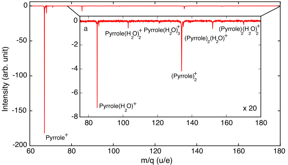 Figure 1
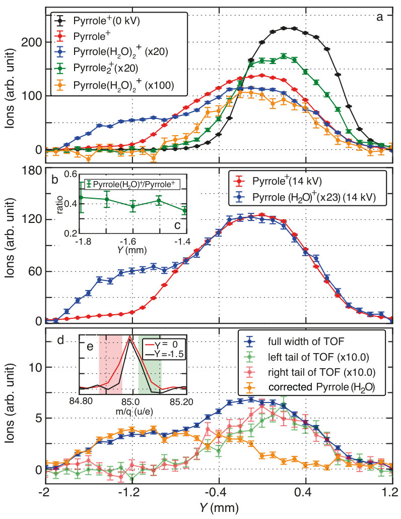 Figure 2
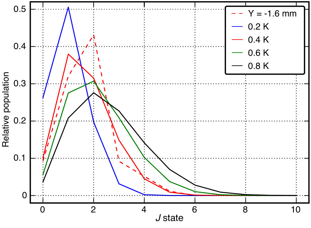 Figure 3
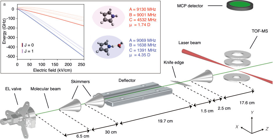 Figure 1
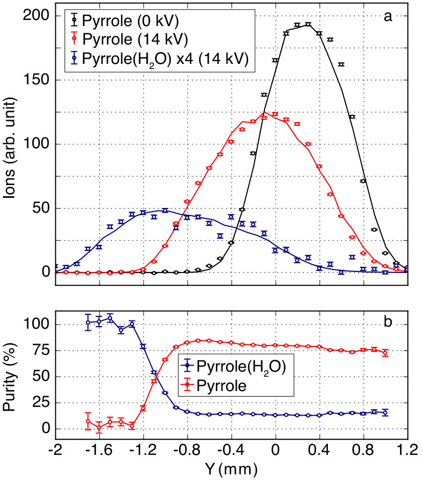 Figure 2
Peer Reviews
No public reviews on file for this paper yet. If you reviewed it on a platform where reviews are public (OpenReview, ICLR, NeurIPS, ICML), you can paste yours below so the community can read it here.
Videos
No videos yet. Explain this paper in a talk, walkthrough, or lecture? Add one.
Spatial separation of pyrrole and pyrrole-water clusters
Melby Johny
Jolijn Onvlee
Thomas Kierspel111Present address: Department of Chemistry, University of Basel, Klingelbergstrasse 80, Basel 4056, Switzerland
Helen Bieker
Sebastian Trippel
Jochen Küpper
Center for Free-Electron Laser Science, Deutsches Elektronen-Synchrotron DESY, Notkestrasse 85, 22607 Hamburg, Germany
Department of Physics, Universität Hamburg, Luruper Chaussee 149, 22761 Hamburg, Germany
The Hamburg Center for Ultrafast Imaging, Universität Hamburg, Luruper Chaussee 149, 22761 Hamburg, Germany
Abstract
We demonstrate the spatial separation of pyrrole and clusters from the other atomic and molecular species in a supersonically-expanded beam of pyrrole and traces of water seeded in high-pressure helium gas. The experimental results are quantitatively supported by simulations. The obtained cluster beam has a purity of %. The extracted rotational temperature of pyrrole and from the original supersonic expansion is K, whereas the temperature of the deflected, pure- part of the molecular beam corresponds to K.
keywords:
pyrrole, H_2O cluster, Stark effect, cold molecules, electric deflection, species separation
††journal: arXiv
url]https://www.controlled-molecule-imaging.org/
1 Introduction
Studies of solvation effects of biologically relevant aromatic molecules provide details on the influence of the molecule’s local environment on its function and on the nature of molecular interactions, as well as insights into the functions of complex biological systems [1]. Prototypical model chromophores such as imidazole and pyrrole were used to build synthetic polyamide ligands for the recognition of the Watson–Crick base pairs in the DNA minor groove [2]. Furthermore, pyrrole is a model of tryptophan’s indole chromophore, one of the strongest near-UV absorbers in proteins. Similarly, pyrrole is responsible for the photoconversion mechanism in the phytochrome enzyme [3] and it is a promising building block for organic dye-sensitized solar cells [4] as well as biological sensors [5].
A key reason for the intriguing photophysics of the above-mentioned chromophores is the excited state, which is repulsive along the N-H-stretching coordinate [6, 7, 8]. The photophysics and photochemistry of these molecules is fairly sensitive to the environment [9]. In a bottom-up approach, spectroscopic and theoretical investigations were performed for micro-solvated clusters to get fundamental insights into their photophysical and photochemical properties [10, 11, 7]. Recently performed experiments have provided evidence for ultrafast intermolecular relaxation processes in electronically-excited microsolvated tetrahydrofuran-water [12] and indole-water [13] clusters. This reflects one of the proposed efficient mechanisms for radiation damage processes of biomolecules via auto-ionization caused by secondary electrons [14]. Time-resolved experiments, such as photoion and photoelectron spectroscopy, aiming at the investigation of the photophysics and photochemistry of pyrrole, were performed to study the dynamics of H elimination from the N-H site of the molecule mediated by the excitation of the state [15, 16, 17, 7].
For clusters theoretical calculations predict that an electron is transferred across the hydrogen bond without photodissociation of the pyrrole moiety [8, 9]. Hence, detailed investigations of the photophysics of , and their comparison with the existing observations for [11, 7], promises to unravel these fundamental processes in the intermolecular interactions as well as the radiation damage of biological systems.
Advanced experiments aiming at unraveling these ultrafast dynamics often rely on pure samples of the individual species. Such controlled samples were previously exploited in investigations of the photophysics of indole [18] and clusters [13] following site-specific soft x-ray ionization and are even amenable to coherent-x-ray-diffraction studies [19]. These experiments relied on the spatial separation of individual species by the electrostatic deflector [20], which was previously demonstrated for the separation of from indole [21, 20, 22].
Here, we demonstrate the spatial separation of pyrrole, a sub unit of indole and tryptophan, and the microsolvated cluster. The structure of pyrrole and was studied using microwave spectroscopy [23, 24] and it was concluded that the singly-hydrogen-bonded cluster has a well defined structure, with the water attached to the N-H site of pyrrole [23, 8].
2 Experimental setup
A schematic representation of the experimental setup is shown in Figure 1 . An Even-Lavie valve [25] was used to generate a pulsed molecular beam by supersonic expansion of a few millibar of pyrrole (Sigma Aldrich, %) and traces of water seeded in bar of helium into vacuum. The valve was operated at a repetition rate of 250 Hz and was heated to . The expanded molecular beam was then skimmed twice using conical skimmers (Beam Dynamics, model 50.8, mm & model 40.5, mm), which were placed at distances of 6.5 cm and 30.2 cm downstream the nozzle, respectively. An inhomogeneous electric field created by the so-called -type deflector [26] was used to disperse the molecular beam according to the species’ effective-dipole-moment-to-mass ratio [20, 27, 28]. The molecular beam was cut by a vertically adjustable knife edge placed 1.5 cm downstream of the exit of the deflector, which allowed for an improved separation of all species present in the molecular beam [22]. The experiments were conducted by placing the knife edge at a height where it cut off the undeflected (0 kV applied on deflector electrodes) molecular beam at the center of the vertical column density profile. The molecular beam was further skimmed by a conical skimmer (Beam Dynamics, model 50.8 with mm) placed 4 cm downstream of the exit of the deflector. The transverse positions of the valve, skimmers, and the deflector were adjustable using motorized translation stages. A laser pulse with a duration of fs, a wavelength centered at 800 nm, focused to , and directed perpendicular to the molecular beam ionized molecules in the extraction region of a time-of-flight mass-spectrometer (TOF-MS) placed 17.6 cm downstream of the last skimmer. The peak intensity of the laser pulse was W/cm2. The ions generated due to the strong-field ionization were detected using a micro-channel plate (MCP), operated in single-shot readout.
3 Results and discussions
Figure 1 a shows the structure, rotational constants [23, 24], permanent dipole moment [23, 24], and the Stark energies of pyrrole (red curves) and (blue curves) for the rotational states. The Stark energies were calculated using CMIstark [29] within the rigid-rotor approximation, as justified by previous experimental and theoretical work on the cluster [21, 30]. Pyrrole has a % smaller Stark-energy shift than for the rotational state at an electric field strength of kV/cm. This enabled full spatial separation of from pyrrole in a cold molecular beam using the electrostatic deflector.
The molecular beam species underwent fragmentation following the strong-field-ionization process. Hence contributions of ionic fragments from larger clusters in specific mass gates were observed in our spectra. A % probability of fragmenting into pyrrole+ was experimentally obtained for our specific laser pulse properties. A detailed description of the fragmentation ratios and utilized mass gates for the specific ion signals is presented in the Supplementary Information. The fragmentation-corrected vertical column density profile for undeflected pyrrole is shown as black dots in Figure 2 a. The undeflected profiles of and other species in the TOF-MS spectra had similar shapes and are not shown. In addition, the fragmentation corrected deflection profiles of both, pyrrole (red dots) and (blue dots), are shown for voltages of kV applied across the deflector. All quantum states are strong-field seeking at the relevant electric field strengths experienced by the investigated molecules and clusters inside the deflector. Therefore, all species are deflected downward, in the negative direction [26]. The experimental deflection profile of shows the strongest deflection, down to mm, whereas pyrrole was only deflected down to mm. Significant deflection of is not expected due to its small effective dipole moment [31]. Larger cluster species, e. g., and , are also deflected less then pyrrole and ; these deflection profiles are shown and discussed in the Supplementary Information.
The solid lines in Figure 2 a are simulated profiles obtained from the results of Monte-Carlo trajectory calculations that take into account the geometrical constraints of the mechanical apertures in the experimental setup [27]. These simulated deflection profiles of pyrrole and matched the experimental data assuming an initial rotational temperature of the molecular beam entering the deflector of K. In the deflected part of the beam at mm the relative populations of the rotational states of were determined from the simulations. Although this rotational-state distribution is non-thermal, it approximately corresponds to a thermal distribution of K, see Supplementary Information. This indicates that an ensemble of very cold molecules is generated using the deflector [32, 33] and that ultracold ensembles, even of isolated well-defined molecular clusters, can be generated in the most deflected part of a dispersed molecular beam.
The purities of pyrrole and , as defined by the ratio of the specific signals to the sum of signals of all other species observed is shown in Figure 2 b, see Supplementary Information for details of the analysis. This demonstrates that a molecular beam of with a purity of % was produced at vertical positions . The column density profile of helium is not shown here, but is expected to be only slightly broader than the undeflected (0 kV) profile of the molecular species, owing to its lighter mass. Furthermore, helium is expected to be only marginally deflected by a few µm due to its small polarizability [26]. Hence, the extracted pure molecular beam of should also be free from helium gas at mm.
4 Conclusion
We demonstrated the spatial separation of the cluster in a molecular beam, i. e., from pyrrole, water, larger clusters, and the seed gas. A purity of % of was obtained in the most deflected part of the molecular beam. Simulated deflection profiles were in excellent agreement with the experiment. They yielded a rotational temperature of K in the initial molecular beam for both, pyrrole and , and of K in the deflected pure beam.
These pure beams of provide a crucial ingredient for photophysics studies aiming at time-resolved hydrogen bond formation/dissociation dynamics, e. g., in ultrafast laser pump and x-ray probe experiments. Further interesting aspects will be the control of the orientation in the laboratory frame by laser aligning or mixed-field orienting clusters [32, 34, 30]. The separated pure species are also ideally suited for experiments to image the structure and dynamics of the complex in the molecular frame, e. g., through molecular-frame photoelectron angular distributions (MFPADs), gas-phase x-ray diffraction, or laser-induced electron diffraction (LIED) experiments [35, 36, 19, 37, 38].
Acknowledgments
This work has been supported by the European Union’s Horizon 2020 research and innovation program under the Marie Skłodowska-Curie Grant Agreement 641789 “Molecular Electron Dynamics investigated by Intense Fields and Attosecond Pulses” (MEDEA), by the excellence cluster “The Hamburg Center for Ultrafast Imaging – Structure, Dynamics and Control of Matter at the Atomic Scale” of the Deutsche Forschungsgemeinschaft (CUI, DFG-EXC1074), by the European Research Council under the European Union’s Seventh Framework Program (FP7/2007-2013) through the Consolidator Grant COMOTION (ERC-Küpper-614507), and by the Helmholtz Association through the Virtual Institute 419 “Dynamic Pathways in Multidimensional Landscapes” and the “Initiative and Networking Fund”. J.O. gratefully acknowledges a fellowship by the Alexander von Humboldt Foundation.
References
- [1]
M. F. Colombo, D. C. Rau, V. A. Parsegian, Protein Solvation in Allosteric regulation: A Water Effect on Hemoglobin, Science 256 (5057) (1992) 655–659.
URL http://science.sciencemag.org/content/256/5057/655
- [2]
S. White, J. W. Szewczyk, J. M. Turner, E. E. Baird, P. B. Dervan, Recognition of the four WatsonCrick base pairs in the DNA minor groove by synthetic ligands, Nature 391 (1998) 468–471.
URL http://dx.doi.org/10.1038/35106
- [3]
A. T. Ulijasz, G. Cornilescu, C. C. Cornilescu, J. Zhang, M. Rivera, J. L. Markley, R. D. Vierstra, Structural basis for the photoconversion of a phytochrome to the activated Pfr form, Nature 463 (2010) 250–254.
URL https://www.nature.com/articles/nature08671
- [4]
Y.-S. Yen, Y.-C. Hsu, J. T. Lin, C.-W. Chang, C.-P. Hsu, D.-J. Yin, Pyrrole-based organic dyes for dye-sensitized solar cells, J. Phys. Chem. C 112 (32) (2008) 12557–12567.
URL https://doi.org/10.1021/jp801036s
- [5]
F. Tan, L. Cong, X. Li, Q. Zhao, H. Zhao, X. Quan, J. Chen, An electrochemical sensor based on molecularly imprinted polypyrrole/graphene quantum dots composite for detection of bisphenol A in water samples, Sens. Actuator B Chem. 233 (2016) 599–606.
doi:10.1016/j.snb.2016.04.146.
URL http://linkinghub.elsevier.com/retrieve/pii/S0925400516306281
- [6]
A. L. Sobolewski, W. Domcke, C. Dedonder-Lardeux, C. Jouvet, Excited-state hydrogen detachment and hydrogen transfer driven by repulsive states: A new paradigm for nonradiative decay in aromatic biomolecules, Phys. Chem. Chem. Phys. 4 (2002) 1093–1100.
URL http://dx.doi.org/10.1039/B110941N
- [7]
H. Lippert, H.-H. Ritze, I. V. Hertel, W. W Radloff, Femtosecond time–resolved hydrogen–atom elimination from photoexcited pyrrole molecules, Chem. Phys. Lett. 5 (2004) 1423–1427.
URL https://doi.org/10.1002/cphc.200400079
- [8]
I. Frank, K. Damianos, Excited state dynamics in pyrrole-water clusters: First principles simulation, Chem. Phys. 343 (2008) 347–352.
doi:10.1016/j.chemphys.2007.08.029.
URL https://doi.org/10.1016/j.chemphys.2007.08.029
- [9]
A. L. Sobolewski, W. Domcke, Photoejection of electrons from pyrrole into an aqueous environment: ab initio results on pyrrole-water clusters, Chem. Phys. Lett. 321 (5-6) (2000) 479–484.
doi:10.1016/S0009-2614(00)00404-8.
URL http://linkinghub.elsevier.com/retrieve/pii/S0009261400004048
- [10]
H. Lippert, V. Stert, L. Hesse, C. P. Schulz, I. V. Hertel, W. Radloff, Ultrafast photoinduced processes in indole–water clusters, Chem. Phys. Lett. 376 (1-2) (2003) 40–48.
doi:10.1016/S0009-2614(03)00921-7.
URL http://linkinghub.elsevier.com/retrieve/pii/S0009261403009217
- [11]
A. L. Sobolewski, W. Domcke, Photoinduced charge separation in indole–water clusters, Chem. Phys. Lett. 329 (1-2) (2000) 130–137.
doi:10.1016/S0009-2614(00)00983-0.
URL http://linkinghub.elsevier.com/retrieve/pii/S0009261400009830
- [12]
X. Ren, E. Wang, A. D. Skitnevskaya, A. B. Trofimov, G. Kirill, A. Dorn, Experimental evidence for ultrafast intermolecular relaxation processes in hydrated biomolecules, Nat. Phys. 79 (2018) 1745.
doi:10.1038/s41567-018-0214-9.
URL https://doi.org/10.1038/s41567-018-0214-9
- [13]
T. Kierspel, Imaging structure and dynamics using controlled molecules, Dissertation, Universität Hamburg, Hamburg, Germany (2016).
- [14]
E. Alizadeh, T. M. Orlando, L. Sanche, Biomolecular damage induced by ionizing radiation: The direct and indirect effects of low-energy electrons on DNA, Annu. Rev. Phys. Chem. 66 (2015) 379–398.
doi:10.1146/annurev-physchem-040513-103605.
URL https://www.annualreviews.org/doi/pdf/10.1146/annurev-physchem-040513-103605
- [15]
M. Ashfold, B. Cronin, A. L. Devine, R. Dixon, M. G. D. Nix, The role of \pi$$\sigma^{*} excited states in the photodissociation of heteroaromatic molecules, Science 312 (2006) 1637–1640.
URL http://science.sciencemag.org/content/312/5780/1637/tab-pdf
- [16]
G. M. Roberts, C. A. Williams, H. Yu, A. S. Chatterley, J. D. Young, S. Ullrich, V. G. Stavros, Probing ultrafast dynamics in photoexcited pyrrole: timescales for 1\pi$$\sigma^{*} mediated H-atom elimination, Faraday Disc. 163 (2013) 95–115.
URL http://pubs.rsc.org/en/content/articlepdf/2013/fd/c2fd20140b
- [17]
O. M. Kirkby, M. A. Parkes, S. P. Neville, G. A. Worth, H. H. Fielding, Non-radiative relaxation dynamics of pyrrole following excitation in the range 249.5–200 nm, Chem. Phys. Lett. 683 (2017) 179–185.
doi:10.1016/j.cplett.2017.04.035.
URL https://doi.org/10.1016/j.cplett.2017.04.035
- [18]
T. Kierspel, C. Bomme, M. Di Fraia, J. Wiese, D. Anielski, S. Bari, R. Boll, B. Erk, J. S. Kienitz, N. L. M. Müller, D. Rolles, J. Viefhaus, S. Trippel, J. Küpper, Photophysics of indole upon x-ray absorption, Phys. Chem. Chem. Phys. 20 (2018) 20205.
arXiv:1802.02964, doi:10.1039/C8CP00936H.
URL https://dx.doi.org/10.1039/C8CP00936H
- [19]
J. Küpper, S. Stern, L. Holmegaard, F. Filsinger, A. Rouzée, A. Rudenko, P. Johnsson, A. V. Martin, M. Adolph, A. Aquila, S. Bajt, A. Barty, C. Bostedt, J. Bozek, C. Caleman, R. Coffee, N. Coppola, T. Delmas, S. Epp, B. Erk, L. Foucar, T. Gorkhover, L. Gumprecht, A. Hartmann, R. Hartmann, G. Hauser, P. Holl, A. Hömke, N. Kimmel, F. Krasniqi, K.-U. Kühnel, J. Maurer, M. Messerschmidt, R. Moshammer, C. Reich, B. Rudek, R. Santra, I. Schlichting, C. Schmidt, S. Schorb, J. Schulz, H. Soltau, J. C. H. Spence, D. Starodub, L. Strüder, J. Thøgersen, M. J. J. Vrakking, G. Weidenspointner, T. A. White, C. Wunderer, G. Meijer, J. Ullrich, H. Stapelfeldt, D. Rolles, H. N. Chapman, X-ray diffraction from isolated and strongly aligned gas-phase molecules with a free-electron laser, Phys. Rev. Lett. 112 (2014) 083002.
arXiv:1307.4577, doi:10.1103/PhysRevLett.112.083002.
URL https://dx.doi.org/10.1103/PhysRevLett.112.083002
- [20]
Y.-P. Chang, D. A. Horke, S. Trippel, J. Küpper, Spatially-controlled complex molecules and their applications, Int. Rev. Phys. Chem. 34 (2015) 557–590.
arXiv:1505.05632, doi:10.1080/0144235X.2015.1077838.
URL http://dx.doi.org/10.1080/0144235X.2015.1077838
- [21]
S. Trippel, Y.-P. Chang, S. Stern, T. Mullins, L. Holmegaard, J. Küpper, Spatial separation of state- and size-selected neutral clusters, Phys. Rev. A 86 (2012) 033202.
arXiv:1208.4935, doi:10.1103/PhysRevA.86.033202.
URL http://pra.aps.org/abstract/PRA/v86/i3/e033202
- [22]
S. Trippel, M. Johny, T. Kierspel, J. Onvlee, H. Bieker, H. Ye, T. Mullins, L. Gumprecht, K. Długołęcki, J. Küpper, Knife edge skimming for improved separation of molecular species by the deflector, Rev. Sci. Instrum. 89 (2018) 096110.
arXiv:1802.04053, doi:10.1063/1.5026145.
URL https://doi.org/10.1063/1.5026145
- [23]
M. J. Tubergen, A. M. Andrews, R. L. Kuczkowski, Microwave spectrum and structure of a hydrogen-bonded pyrrole-water complex, J. Phys. Chem. 97 (29) (1993) 7451–7457.
URL https://doi.org/10.1021/j100131a011
- [24]
U. Nygaard, J. Nielsen, J. Kirchheiner, G. Maltesen, C. S, J. Rastrup-Andersen, G. Sørensen, Microwave spectra of isotopic pyrroles: Molecular structure, dipole moment and quadrupole coupling constants of pyrrole, J. Mol. Struct. 3 (6) (1969) 491–506.
doi:10.1016/0022-2860(69)80031-1.
URL https://doi.org/10.1016/0022-2860(69)80031-1
- [25]
U. Even, J. Jortner, D. Noy, N. Lavie, N. Cossart-Magos, Cooling of large molecules below 1 K and He clusters formation, J. Chem. Phys. 112 (2000) 8068–8071.
URL http://dx.doi.org/10.1063/1.481405
- [26]
J. S. Kienitz, K. Długołęcki, S. Trippel, J. Küpper, Improved spatial separation of neutral molecules, J. Chem. Phys. 147 (2) (2017) 024304.
arXiv:1704.08912, doi:10.1063/1.4991479.
URL https://doi.org/10.1063/1.4991479
- [27]
F. Filsinger, J. Küpper, G. Meijer, L. Holmegaard, J. H. Nielsen, I. Nevo, J. L. Hansen, H. Stapelfeldt, Quantum-state selection, alignment, and orientation of large molecules using static electric and laser fields, J. Chem. Phys. 131 (2009) 064309.
arXiv:0903.5413, doi:10.1063/1.3194287.
URL http://scitation.aip.org/content/aip/journal/jcp/131/6/10.1063/1.3194287
- [28]
F. Filsinger, U. Erlekam, G. von Helden, J. Küpper, G. Meijer, Selector for structural isomers of neutral molecules, Phys. Rev. Lett. 100 (2008) 133003.
arXiv:0802.2795, doi:10.1103/PhysRevLett.100.133003.
URL http://dx.doi.org/10.1103/PhysRevLett.100.133003
- [29]
Y.-P. Chang, F. Filsinger, B. Sartakov, J. Küpper, CMIstark: Python package for the stark-effect calculation and symmetry classification of linear, symmetric and asymmetric top wavefunctions in dc electric fields, Comp. Phys. Comm. 185 (2014) 339–349.
arXiv:1308.4076, doi:10.1016/j.cpc.2013.09.001.
URL http://www.sciencedirect.com/science/article/pii/S0010465513003019
- [30]
L. V. Thesing, A. Yachmenev, R. González-Férez, J. Küpper, Analyzing laser-induced alignment of weakly-bound molecular aggregates, Phys. Rev. A 98 (2018) 053412.
arXiv:1808.01206, doi:10.1103/PhysRevA.98.053412.
URL https://journals.aps.org/pra/abstract/10.1103/PhysRevA.98.053412
- [31]
D. A. Horke, Y.-P. Chang, K. Długołęcki, J. Küpper, Separating para and ortho water, Angew. Chem. Int. Ed. 53 (2014) 11965–11968.
arXiv:1407.2056, doi:10.1002/anie.201405986.
URL http://onlinelibrary.wiley.com/doi/10.1002/anie.201405986/abstract
- [32]
L. Holmegaard, J. H. Nielsen, I. Nevo, H. Stapelfeldt, F. Filsinger, J. Küpper, G. Meijer, Laser-induced alignment and orientation of quantum-state-selected large molecules, Phys. Rev. Lett. 102 (2009) 023001.
arXiv:0810.2307, doi:10.1103/PhysRevLett.102.023001.
URL http://dx.doi.org/10.1103/PhysRevLett.102.023001
- [33]
J. H. Nielsen, P. Simesen, C. Z. Bisgaard, H. Stapelfeldt, F. Filsinger, B. Friedrich, G. Meijer, J. Küpper, Stark-selected beam of ground-state OCS molecules characterized by revivals of impulsive alignment, Phys. Chem. Chem. Phys. 13 (2011) 18971–18975.
arXiv:1105.2413, doi:10.1039/c1cp21143a.
- [34]
S. Trippel, J. Wiese, T. Mullins, J. Küpper, Communication: Strong laser alignment of solvent-solute aggregates in the gas-phase, J. Chem. Phys. 148 (10) (2018) 101103.
arXiv:1801.08789, doi:10.1063/1.5023645.
URL http://aip.scitation.org/doi/10.1063/1.5023645
- [35]
L. Holmegaard, J. L. Hansen, L. Kalhøj, S. L. Kragh, H. Stapelfeldt, F. Filsinger, J. Küpper, G. Meijer, D. Dimitrovski, M. Abu-samha, C. P. J. Martiny, L. B. Madsen, Photoelectron angular distributions from strong-field ionization of oriented molecules, Nat. Phys. 6 (2010) 428.
arXiv:1003.4634, doi:10.1038/NPHYS1666.
URL https://www.nature.com/articles/nphys1666
- [36]
C. I. Blaga, J. Xu, A. D. DiChiara, E. Sistrunk, K. Zhang, P. Agostini, T. A. Miller, L. F. DiMauro, C. D. Lin, Imaging ultrafast molecular dynamics with laser-induced electron diffraction, Nature 483 (7388) (2012) 194–197.
URL https://www.nature.com/articles/nature10820
- [37]
J. Yang, M. Guehr, X. Shen, R. Li, T. Vecchione, R. Coffee, J. Corbett, A. Fry, N. Hartmann, C. Hast, K. Hegazy, K. Jobe, I. Makasyuk, J. Robinson, M. S. Robinson, S. Vetter, S. Weathersby, C. Yoneda, X. Wang, M. Centurion, Diffractive imaging of coherent nuclear motion in isolated molecules, Phys. Rev. Lett. 117 (2016) 153002.
doi:10.1103/PhysRevLett.117.153002.
URL https://link.aps.org/doi/10.1103/PhysRevLett.117.153002
- [38]
A. Trabattoni, S. Trippel, U. D. Giovannini, J. F. Olivieri, J. Wiese, T. Mullins, J. Onvlee, S.-K. Son, A. R. Biagio Frusteri, J. Küpper, Setting the clock of photoelectron emission through molecular alignmentarXiv:1802.06622.
The reference list from the paper itself. Each links out to its DOI / PubMed record.
- 1[1] M. F. Colombo, D. C. Rau, V. A. Parsegian, Protein Solvation in Allosteric regulation: A Water Effect on Hemoglobin , Science 256 (5057) (1992) 655–659. doi:10.1126/science.1585178 . URL http://science.sciencemag.org/content/256/5057/655 · doi ↗
- 2[2] S. White, J. W. Szewczyk, J. M. Turner, E. E. Baird, P. B. Dervan, Recognition of the four Watson − - Crick base pairs in the DNA minor groove by synthetic ligands , Nature 391 (1998) 468–471. doi:10.1038/35106 . URL http://dx.doi.org/10.1038/35106 · doi ↗
- 3[3] A. T. Ulijasz, G. Cornilescu, C. C. Cornilescu, J. Zhang, M. Rivera, J. L. Markley, R. D. Vierstra, Structural basis for the photoconversion of a phytochrome to the activated Pfr form , Nature 463 (2010) 250–254. doi:10.1038/nature 08671 . URL https://www.nature.com/articles/nature 08671 · doi ↗
- 4[4] Y.-S. Yen, Y.-C. Hsu, J. T. Lin, C.-W. Chang, C.-P. Hsu, D.-J. Yin, Pyrrole-based organic dyes for dye-sensitized solar cells , J. Phys. Chem. C 112 (32) (2008) 12557–12567. doi:10.1021/jp 801036 s . URL https://doi.org/10.1021/jp 801036 s · doi ↗
- 5[5] F. Tan, L. Cong, X. Li, Q. Zhao, H. Zhao, X. Quan, J. Chen, An electrochemical sensor based on molecularly imprinted polypyrrole/graphene quantum dots composite for detection of bisphenol A in water samples , Sens. Actuator B Chem. 233 (2016) 599–606. doi:10.1016/j.snb.2016.04.146 . URL http://linkinghub.elsevier.com/retrieve/pii/S 0925400516306281 · doi ↗
- 6[6] A. L. Sobolewski, W. Domcke, C. Dedonder-Lardeux, C. Jouvet, Excited-state hydrogen detachment and hydrogen transfer driven by repulsive π 1 σ ∗ superscript 𝜋 1 superscript 𝜎 {}^{1}\pi\sigma^{*} states: A new paradigm for nonradiative decay in aromatic biomolecules , Phys. Chem. Chem. Phys. 4 (2002) 1093–1100. doi:10.1039/B 110941 N . URL http://dx.doi.org/10.1039/B 110941 N · doi ↗
- 7[7] H. Lippert, H.-H. Ritze, I. V. Hertel, W. W Radloff, Femtosecond time–resolved hydrogen–atom elimination from photoexcited pyrrole molecules , Chem. Phys. Lett. 5 (2004) 1423–1427. doi:10.1002/cphc.200400079 . URL https://doi.org/10.1002/cphc.200400079 · doi ↗
- 8[8] I. Frank, K. Damianos, Excited state dynamics in pyrrole-water clusters: First principles simulation , Chem. Phys. 343 (2008) 347–352. doi:10.1016/j.chemphys.2007.08.029 . URL https://doi.org/10.1016/j.chemphys.2007.08.029 · doi ↗