Spatio-angular fluorescence microscopy II. Paraxial 4$f$ imaging
Talon Chandler, Hari Shroff, Rudolf Oldenbourg, and Patrick La, Rivi\`ere

TL;DR
This paper analyzes the spatio-angular properties of a paraxial 4f fluorescence microscope, revealing its ability to record out-of-plane fluorophore orientations and highlighting biases caused by monopole approximations.
Contribution
It demonstrates the spatio-angular band limit of the system and assesses the impact of monopole approximation on fluorophore concentration estimates.
Findings
Out-of-plane fluorophore orientations are recorded by the system.
Monopole approximation biases are small with many randomly oriented fluorophores.
Efficient simulations are enabled by exploiting the band limit.
Abstract
We investigate the properties of a single-view fluorescence microscope in a 4 geometry when imaging fluorescent dipoles without using the monopole or scalar approximations. We show that this imaging system has a spatio-angular band limit, and we exploit the band limit to perform efficient simulations. Notably, we show that information about the out-of-plane orientation of ensembles of in-focus fluorophores is recorded by paraxial fluorescence microscopes. Additionally, we show that the monopole approximation may cause biased estimates of fluorophore concentrations, but these biases are small when the sample contains either many randomly oriented fluorophores in each resolvable volume or unconstrained rotating fluorophores.
Click any figure to enlarge with its caption.
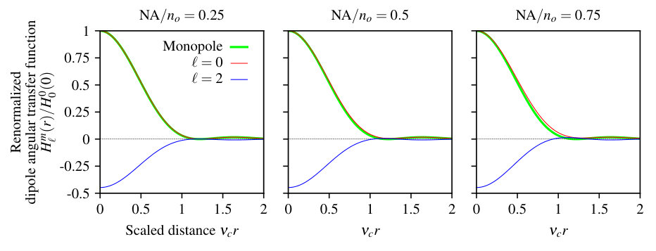 Figure 1
 Figure 2
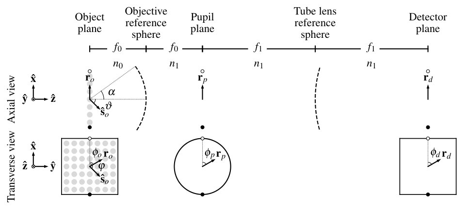 Figure 3
 Figure 4
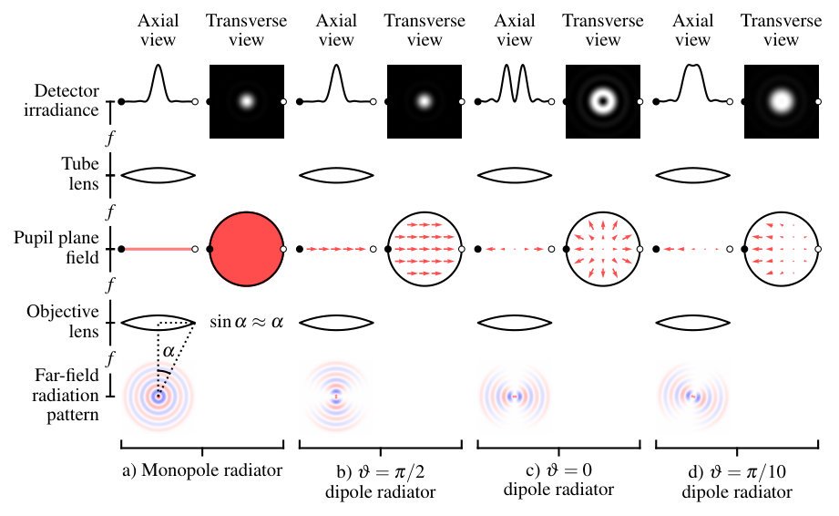 Figure 5
 Figure 6
 Figure 7
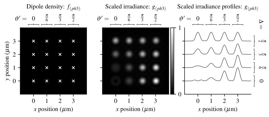 Figure 8
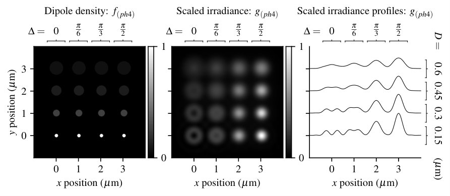 Figure 9
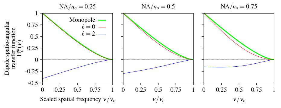 Figure 10
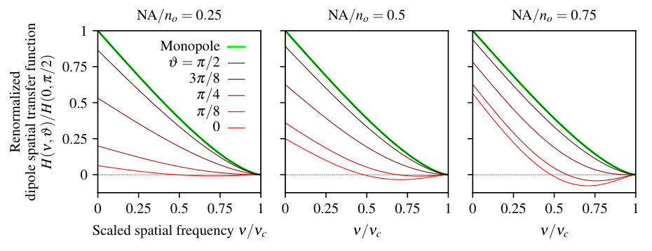 Figure 11
 Figure 12
| Quantity | Symbol | Relationships |
|---|---|---|
| Monopole density | — | |
| Monopole spectrum | ||
| Monopole coherent spread function | — | |
| Monopole coherent transfer function | ||
| Monopole point spread function | ||
| Monopole transfer function | ||
| Scaled irradiance | ||
| Scaled irradiance spectrum |
| Quantity | Symbol | Relationships |
|---|---|---|
| Dipole density | — | |
| Dipole spatial spectrum | ||
| Dipole angular spectrum | ||
| Dipole spatio-angular spectrum | ||
| Dipole coherent spread function | — | |
| Dipole coherent transfer function | ||
| Dipole point spread function | ||
| Dipole spatial transfer function | ||
| Dipole angular transfer function | ||
| Dipole spatio-angular transfer function | ||
| Scaled irradiance | ||
| Scaled irradiance spectrum | ||
Peer Reviews
No public reviews on file for this paper yet. If you reviewed it on a platform where reviews are public (OpenReview, ICLR, NeurIPS, ICML), you can paste yours below so the community can read it here.
Videos
No videos yet. Explain this paper in a talk, walkthrough, or lecture? Add one.
Spatio-angular fluorescence microscopy
II. Paraxial 4f imaging
Talon Chandler
\authormark1,* Hari Shroff
\authormark2,3 Rudolf Oldenbourg
\authormark3 and Patrick La Rivière\authormark1,3
\authormark1University of Chicago, Department of Radiology, Chicago, Illinois 60637, USA
\authormark2Section on High Resolution Optical Imaging, National Institute of Biomedical Imaging and Bioengineering, National Institutes of Health, Bethesda, Maryland 20892, USA
\authormark3Marine Biological Laboratory, Bell Center, Woods Hole, Massachusetts 02543, USA
††journal: osajournal††articletype: Research Article
{abstract*}
We investigate the properties of a single-view fluorescence microscope in a geometry when imaging fluorescent dipoles without using the monopole or scalar approximations. We show that this imaging system has a spatio-angular band limit, and we exploit the band limit to perform efficient simulations. Notably, we show that information about the out-of-plane orientation of ensembles of in-focus fluorophores is recorded by paraxial fluorescence microscopes. Additionally, we show that the monopole approximation may cause biased estimates of fluorophore concentrations, but these biases are small when the sample contains either many randomly oriented fluorophores in each resolvable volume or unconstrained rotating fluorophores.
1 Introduction
In the first paper of this series we developed a new set of transfer functions that can be used to analyze spatio-angular fluorescence microscopes [1]. In this work we will demonstrate these transfer functions by analyzing a single-view fluorescence microscope in a geometry.
A central goal of this work is to examine the validity of the monopole approximation in fluorescence microscopy. Although many works implicitly apply the monopole approximation, we have encountered two explicit justifications: (1) the sample contains many randomly oriented fluorophores within a resolvable volume or (2) the sample contains unconstrained rotating fluorophores. While both of these situations yield monopole-like emitters, neither yields emitters that are perfectly described by the monopole model. We investigate the dipole model of fluorophores in detail and find the conditions under which the monopole approximation is justified.
We begin in section 2 by specifying the imaging geometry and defining pupil functions for imaging systems with and without the monopole approximation. We explicitly relate the pupil functions to the coherent transfer functions to establish a connection between physical calculations and the transfer functions. Next, in section 3 we calculate the monopole and dipole transfer functions in closed form, and we use these transfer functions to perform efficient simulations with four numerical phantoms. Finally, in section 4 we discuss the results and expand on how the pupil functions can be used to develop improved models for spatio-angular microscopes.
2 Theory
During our initial modeling [1] we considered an aplanatic optical system imaging a sample of in-focus fluorophores—either a monopole density, , or a dipole density, —by recording the scaled irradiance on a two-dimensional detector, . A central result was that we could express the relationship between the object and the data as a linear Hilbert-space operator, and we showed that these operators took the form of an integral transform in a delta function basis. For monopoles the integral transform takes the form
[TABLE]
where is the monopole point spread function. For dipoles the integral transform takes the form
[TABLE]
where is the dipole point spread function. Note that we have written Eqs. (1) and (2) in their demagnified forms. We will use primes to denote the unscaled detector coordinate, , and unscaled point spread functions, .
After expressing the operators in a delta function basis we explored the form of the operators with several other choices of basis functions. Tables 1 and 2 summarize our results.
Our task is to calculate the form of the monopole and dipole transfer functions for a specific imaging geometry. In this work we will consider an aplanatic optical system in a * configuration* with an arbitrary first lens (the objective lens) and a paraxial second lens (the tube lens) as shown in Fig. 2. A lens can be considered paraxial if the angle between the optical axis of the lens and the marginal ray is small enough that . As a rule of thumb, non-paraxial effects only become significant when the numerical aperture of a lens exceeds 0.7 [2, ch. 6], but this is only a rough guideline. Commercial microscopes with infinity-corrected objectives can almost always can be modeled by considering the tube lens as paraxial.
The reference list from the paper itself. Each links out to its DOI / PubMed record.
- 1[1] T. Chandler, H. Shroff, R. Oldenbourg, and P. J. La Rivière, “Spatio-angular fluorescence microscopy I. basic theory,” \Journal Title https://arxiv.org/abs/1812.07093 (2018).
- 2[2] M. Gu, Advanced Optical Imaging Theory , Springer Series in Optical Sciences (Springer, 2000).
- 3[3] H. Barrett and K. Myers, Foundations of Image Science (Wiley-Interscience, 2004).
- 4[4] J. Goodman, Introduction to Fourier Optics (Mc Graw-Hill, 1996).
- 5[5] D. Axelrod, “Fluorescence excitation and imaging of single molecules near dielectric-coated and bare surfaces: a theoretical study.” \Journal Title Journal of Microscopy 247 2 , 147–60 (2012).
- 6[6] A. S. Backer and W. E. Moerner, “Extending single-molecule microscopy using optical Fourier processing,” \Journal Title J. Phys. Chem. B 118 , 8313–8329 (2014).
- 7[7] C. J. R. Sheppard, M. Gu, Y. Kawata, and S. Kawata, “Three-dimensional transfer functions for high-aperture systems,” \Journal Title J. Opt. Soc. Am. A 11 , 593–598 (1994).
- 8[8] M. R. Arnison and C. J. Sheppard, “A 3D vectorial optical transfer function suitable for arbitrary pupil functions,” \Journal Title Optics Communications 211 , 53–63 (2002).