Goldilocks Zone for Enhanced Ionization in Strong Laser Fields
M. M\"oller, A. M. Sayler, P. Wustelt, L. Yue, S. Gr\"afe, and G. G., Paulus

TL;DR
This study identifies a specific 'Goldilocks Zone' of laser parameters where enhanced ionization of H₂⁺ occurs, confirmed through experiments and simulations, clarifying why this phenomenon is rare and difficult to observe.
Contribution
The paper experimentally and theoretically characterizes the narrow parameter space for enhanced ionization in H₂⁺, providing a model that explains its elusive nature.
Findings
Enhanced ionization occurs only within a small laser parameter space.
Experimental measurements match simulation results for electron spectra.
A model explains the limited conditions for observing enhanced ionization.
Abstract
Utilizing a benchmark measurement of laser-induced ionization of an H molecular ion beam target at infrared wavelength around 2 m, we show that the characteristic two-peak structure predicted for laser-induced enhanced ionization of H and diatomic molecules in general, is a phenomenon which is confined to a small laser parameter space --- a Goldilocks Zone. Further, we control the effect experimentally and measure its imprint on the electron momentum. We replicate the behavior with simulations, which reproduce the measured kinetic-energy release as well as the correlated-electron spectra. Based on this, a model, which both maps out the Goldilocks Zone and illustrates why enhanced ionization has proven so elusive in H, is derived.
Click any figure to enlarge with its caption.
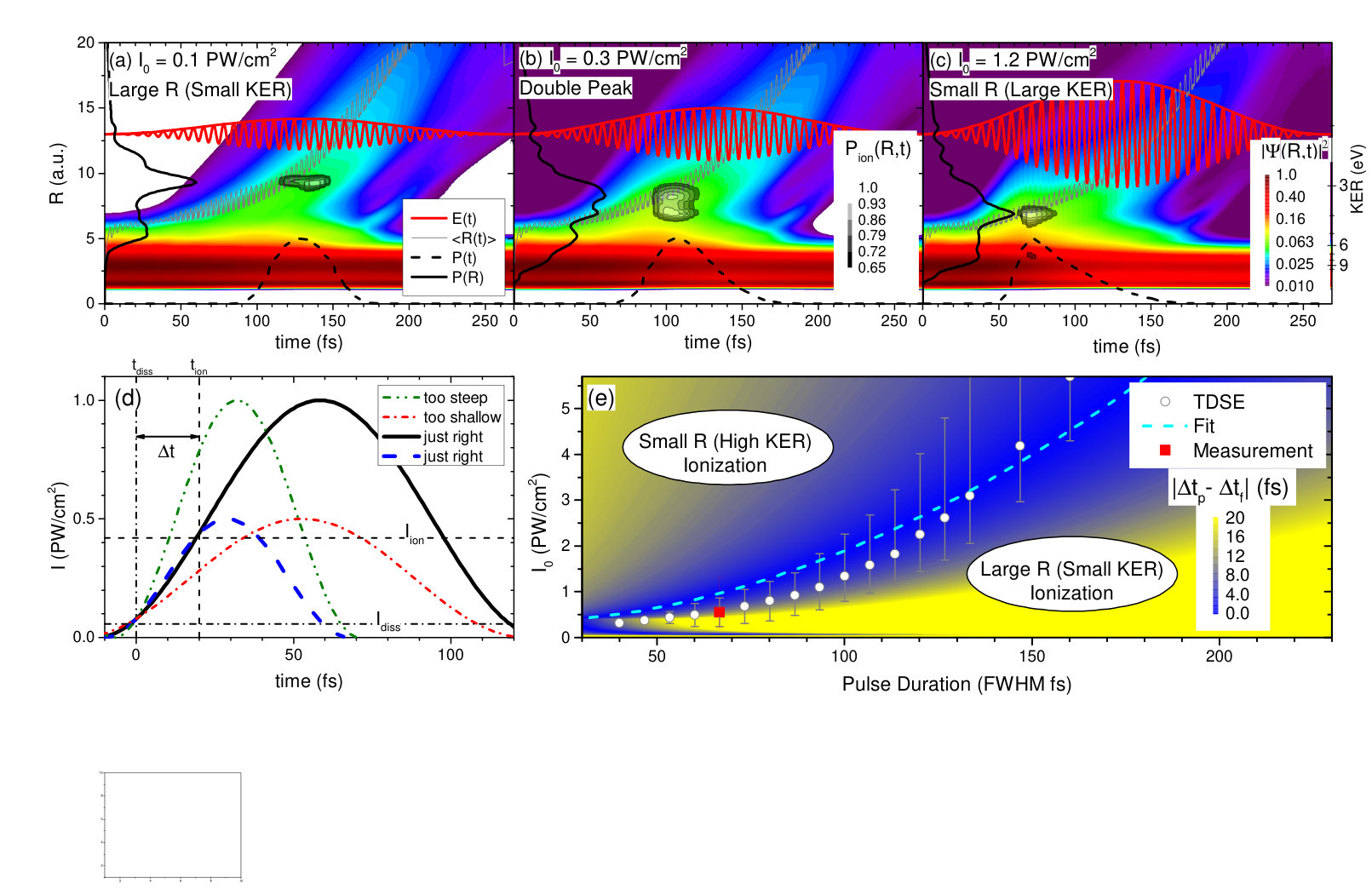 Figure 1
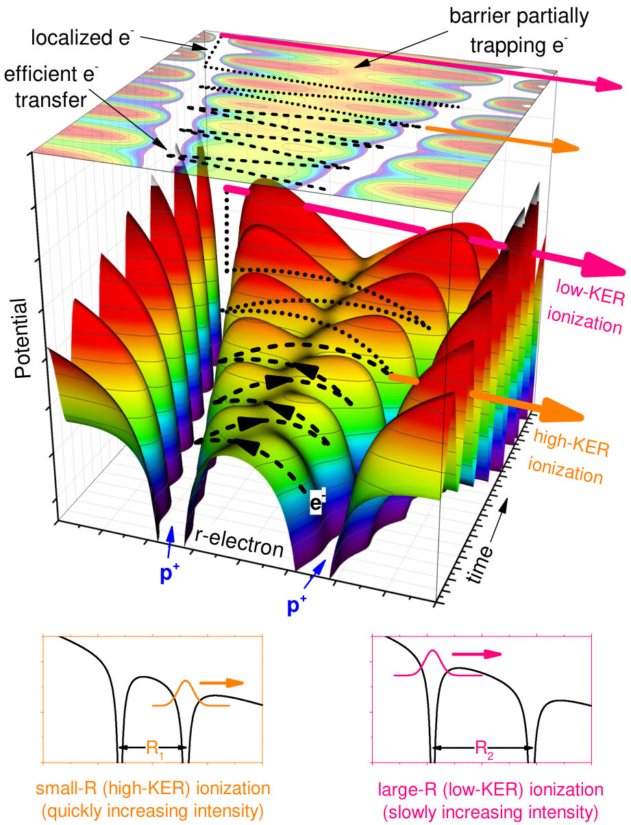 Figure 2
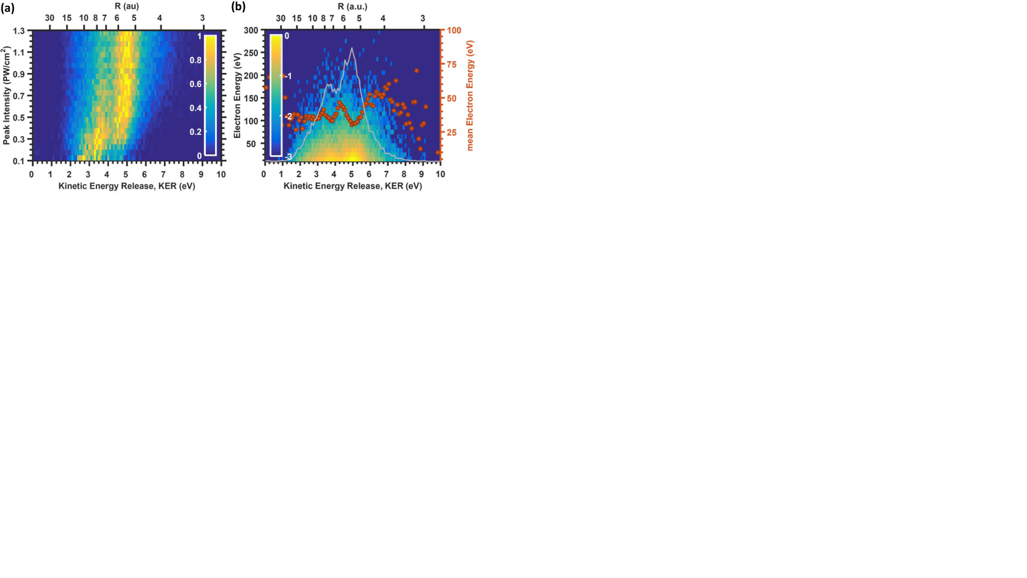 Figure 2
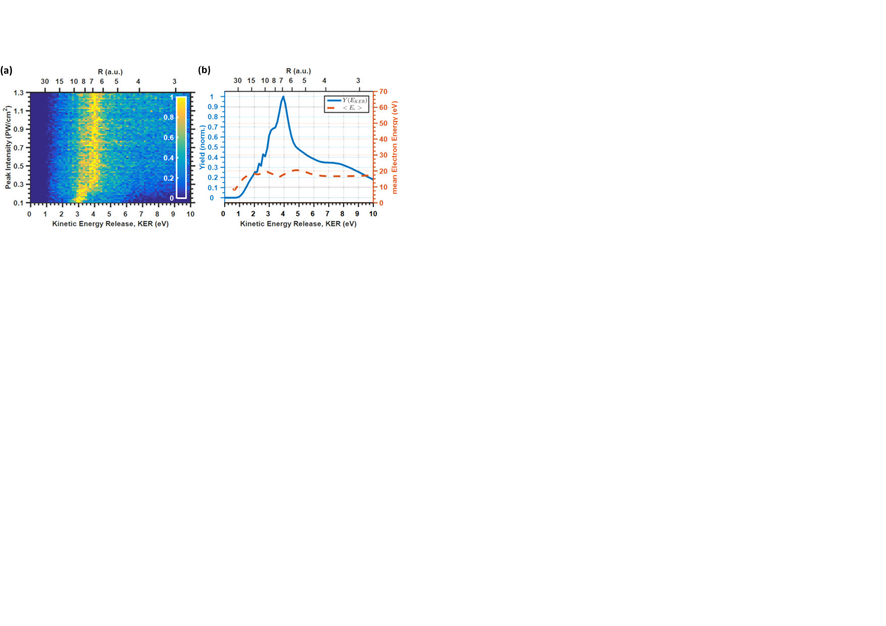 Figure 3
Peer Reviews
No public reviews on file for this paper yet. If you reviewed it on a platform where reviews are public (OpenReview, ICLR, NeurIPS, ICML), you can paste yours below so the community can read it here.
Videos
No videos yet. Explain this paper in a talk, walkthrough, or lecture? Add one.
Taxonomy
TopicsLaser-Matter Interactions and Applications · Mass Spectrometry Techniques and Applications · Laser-induced spectroscopy and plasma
Goldilocks Zone for Enhanced Ionization in Strong Laser Fields
M. Möller
Institute of Optics and Quantum Electronics, Abbe Center of Photonics, Friedrich Schiller University Jena, Max-Wien-Platz 1, 07743 Jena, Germany
Helmholtz Institut Jena, Fröbelstieg 3, 07743 Jena, Germany
A. M. Sayler
Institute of Optics and Quantum Electronics, Abbe Center of Photonics, Friedrich Schiller University Jena, Max-Wien-Platz 1, 07743 Jena, Germany
Helmholtz Institut Jena, Fröbelstieg 3, 07743 Jena, Germany
P. Wustelt
Institute of Optics and Quantum Electronics, Abbe Center of Photonics, Friedrich Schiller University Jena, Max-Wien-Platz 1, 07743 Jena, Germany
Helmholtz Institut Jena, Fröbelstieg 3, 07743 Jena, Germany
L. Yue
Institute of Physical Chemistry and Abbe Center of Photonics, Friedrich Schiller University Jena, Helmholtzweg 4, 07743 Jena, Germany
S. Gräfe
Institute of Physical Chemistry and Abbe Center of Photonics, Friedrich Schiller University Jena, Helmholtzweg 4, 07743 Jena, Germany
G. G. Paulus
Institute of Optics and Quantum Electronics, Abbe Center of Photonics, Friedrich Schiller University Jena, Max-Wien-Platz 1, 07743 Jena, Germany
Helmholtz Institut Jena, Fröbelstieg 3, 07743 Jena, Germany
Abstract
Utilizing a benchmark measurement of laser-induced ionization of an H molecular ion beam target at infrared wavelength around 2 m, we show that the characteristic two-peak structure predicted for laser-induced enhanced ionization of H and diatomic molecules in general, is a phenomenon which is confined to a small laser parameter space — a Goldilocks Zone. Further, we control the effect experimentally and measure its imprint on the electron momentum. We replicate the behavior with simulations, which reproduce the measured kinetic-energy release as well as the correlated-electron spectra. Based on this, a model, which both maps out the Goldilocks Zone and illustrates why enhanced ionization has proven so elusive in H, is derived.
pacs:
32.80.-t, 42.65.-k, 31.15.A-
Since its first complete quantum description in the 1920’s Burrau (1927), the H bond has served as the prototype for all molecular systems Ibrahim et al. (2018). This is particularly true for the attosecond dynamics of molecular bonding in strong fields, where the insights gained from H have served as a foundation for the understanding of more complex bonds. For example, the concepts of bond hardening, bond softening, above-threshold dissociation and above-threshold ionization were first determined from H and are now ubiquitous in the descriptions of laser-induced molecular dynamics Ibrahim et al. (2018).
However, another foundational process — enhanced ionization (EI) — continues to be evasive and contentious Ben-Itzhak et al. (2008); Xu et al. (2015), partially due to the difficulty of direct measurements of H. The EI process was predicted by early time-dependent Schrödinger equation (TDSE) calculations for H with fixed internuclear distances and showed that ionization, i.e. H, is enhanced for specific internuclear distances Zuo and Bandrauk (1995). Since, EI has frequently been used as one of the processes invoked to explain and predict laser-induced molecular dynamics for small and more complex molecules alike Normand and Schmidt (1996); Hishikawa et al. (1999); Liekhus-Schmaltz et al. (2015); Jiang et al. (2010); Xie et al. (2014); Cornaggia (2016); Liu and Barth (2017).
In a common picture of EI in H, depicted in Fig. 1, there are two distinct mechanisms leading to ionization. If the nuclear separation, , is relatively small and the laser field is relatively large, then the electronic wavefunction will follow the laser, moving to the downhill side of the potential with each optical half cycle and tunnel out with sufficient laser intensity around the peaks of the field. Alternatively, if the laser field ramps up slowly, the molecule stretches and the potential barrier between the two protons grows. This can effectively trap a portion of the electron wavepacket on the uphill proton and allow ionization from the upper potential over the deformed inner barrier Liu and Barth (2017). Thus, ionization is enhanced near -values corresponding to the aforementioned cases and a characteristic double peak structure should emerge in the - or KER-dependent electron yield Zuo and Bandrauk (1995).
Although this is a logical and straightforward explanation and the double-peak structure is obvious in the calculated static-field ionization rate of fixed-nuclei H molecules Plummer and McCann (1996, 1997), many studies of H over the past two decades could not clearly identify EI, which has fueled the debate over the relevance of the concept Takemoto and Becker (2010); Odenweller et al. (2011); Silva et al. (2013); Xu et al. (2015); Yue and Madsen (2016); Xu et al. (2017). Moreover, experimental effects; such as the initial vibrational state distribution of a H target, the imprint of the prerequisite ionization of a H2 target, depletion, intensity-volume effects, conversion from the measured KER to the inferred -values and the coupled electron-nuclear dynamics; make the process and interpretation much more complex Ben-Itzhak et al. (2008); Xu et al. (2015). Measurements, which use neutral H2 as a target, often leave uncertainty about which observed effects are, at least partially, due to prerequisite ionization. For example, indications of EI have only been clearly seen when creating a traveling nuclear wavepacket from a ionization from a neutral target, , and probing the resulting dissociative state with time-delayed few-cycle pulses Xu et al. (2015). However, this leaves many question about the prevalence and importance of the phenomenon for typical laser fields.
To settle these long-standing questions and clearly demonstrate the EI effect in H, here, we implement, the first to our knowledge, intensity-dependent measurement of the short-wave infrared (SWIR) laser-induced ionization of a H molecular-ion-beam target, which captures both the momenta of the nuclear fragments and the correlated electron momentum. In this largely unexplored territory of wavelength and intensity, we conclusively observe the two-peak structure characteristic of EI and are able to control it with the intensity envelope of the laser pulse. Next, using specifically developed nuclear-wavepacket propagation calculations that include ionization, which are further validated by the correlated KER and electron momenta spectra, we determine the Goldilocks Zone, i.e. the limited laser parameter space, where EI is visible. Finally, this data is used to formulate a clear-cut model with great explanatory and predictive power.
The experimental challenges arise from the dilute ion-beam target and conditions needed to measure the momenta of both protons and the electron Ben-Itzhak et al. (2005); Rathje et al. (2013); Wustelt et al. (2015), see supplemental material for details 111See Supplemental Material for a PDF document further detailing the experimental methods and numerical simulations.. The electron momentum, , is determined using the sum momentum of the proton, i.e. without directly detecting the electron. This requires an extremely high experimental precision and well-collimated ion beam. Further, despite the significantly more complex laser setup needed, we chose to use SWIR pulses to increase the electron momentum, . Although our measurement of the electron momentum is blurred with the momentum distribution that arises from the temperature of the H molecular ion beam, compared to a direct measurement of the electron momentum Odenweller et al. (2011, 2014), our approach reduces the experimental cost and complexity significantly and is, to the best of our knowledge, the first application of this technique to a molecular ion beam laser interaction.
The measured intensity- and KER-dependent laser-induced ionization yield for H with 65 fs, 2 m pulses is shown in Fig. 2(a). Here one sees that at low intensity ( 0.5 PW/cm2) the yield is peaked near 3.5 eV ( a.u.). For these intensities, the laser field ramps up slowly and the molecule stretches, trapping a portion of the electron wavepacket in the upper potential well, which is then ionized. At high intensity ( 0.3 PW/cm2) the yield is peaked near 5 eV ( a.u.) and slowly increases with intensity. Here, the steep increase in intensity facilitates ionization at smaller s, where the electron wavepacket is effectively transferred to the lower potential well each half cycle. This depletes the dissociative nuclear wavepacket before it reaches thereby reducing the peak at lower KER. The characteristic double-peak structure we are in search of only occurs in the narrow overlapping transition intensity range ( 0.40.2 PW/cm2), where both processes can occur.
Although this interpretation, based on the measurement of the nuclear fragments, tells us a great deal about the underlying dynamics, it is only half the picture. To gain full access to the dynamics at play, we simultaneously measure the nuclear fragment momenta to produce the joint electron-nuclear energy distribution (JED) shown in Fig. 2(b). Here we see that reductions in the width of the electron spectrum are correlated with increases in the yield. Although calculated JEDs, where the electron and the nuclear dynamics are treated in equal footing Silva et al. (2013); Yue and Madsen (2013, 2016), show hints of this behavior, that work typically focuses on diagonal energy-conserving lines, which have also been measured, e.g. by Wu et al. Wu et al. (2013). In contrast, here we are focused on the large-scale behavior.
Unlike some of the more complex models of H ionization, which look at details of the -dependent timing of the liberated electron wave-packet Takemoto and Becker (2010, 2011), the measurement behavior here has a relatively straightforward qualitative explanation. Namely, in addition to increasing the yield, enhancing the ionization rate at certain s lowers the average intensity required for ionization. This, in turn, reduces the photoelectron energy, which scales with the intensity Becker et al. (2002). Thus, peaks in the KER-dependent ionization yield should overlap with minima in the width of the correlated electron spectrum as observed.
To further understand the experimental results and extend the control of EI to other laser parameters, e.g. wavelength and pulse duration, we implement a two-surface time-dependent Schrödinger calculations Schwendner et al. (1997) and augmented them for ionization Staudte et al. (2007) including the correlated electron energy, see supplemental material. Using the measured laser parameters, this results in the spectra shown in Fig. 3. Here we see that the enhanced two-surface model is a good qualitative match to the measured data and accurately predicts the double-peak structure for roughly the same narrow intensity range. Moreover, the corresponding KER-dependent electron momentum also follows the measured trend, which confirms that the model is capturing the relevant underlying dynamics. The minor differences between measurement and theory, i.e. slightly different peak positions, are likely due to several imperfections of the model detailed in the supplemental material.
To identify the dynamics at play we examine the calculated nuclear dynamics for the three characteristic situations noted above in Fig. 4(a)–(c). In Fig. 4(a), when the intensity is too small, the leading edge of the laser pulse begins the dissociation process, i.e. stretching in of the nuclear wavepacket, and the intensity only becomes sufficient to ionize after the molecule has stretched to a.u. (KER 3 eV). In Fig. 4(c), when the intensity is too big, the intensity ramps up quickly enough that ionization depletes the nuclear wavepacket at a.u. (KER 4.5 eV) before much stretching can occur. In Fig. 4(b), when the intensity is just right, the intensity is high enough to ionize near , but low enough to allow part of the wavepacket to survive and stretch to before ionization, which results in the double peak shown.
This leads to an intuitive model for predicting when the characteristic double-peak structure of EI will be visible. Assuming that stretching of the molecule is initiated by the laser field at relatively low intensities, , and small internuclear distances, , then ionization will occur at a later time, , after the molecule has stretched to and the intensity has increased sufficiently to ionize the molecule, . Further, if there are two preferred internuclear distances for ionization, and , then the double-peaked structure will be lost, if the laser intensity does not ramp up in the very particular way described above. This places constraints on the intensity envelope of the laser pulse. For example, if one wishes to maintain the same timing while increasing the intensity, the pulse length must be increased, see the thick dashed and solid lines in Fig. 4(d).
To determine these parameters, we first map out this region with our measurements and wavepacket-propagation calculations. Specifically, to parameterize the double peak structure, we fit a double Gaussian to the calculated data over a large range of laser parameters. Then we find the ratio of the peaks, , where is the amplitude of the lower of the two peaks and is the the amplitude of the higher of the two peaks. This allows one to determine the Goldilocks Zone where the double peak occurs and to what extent it is visible. The intensity where the maximum value of this ratio, , occurs is plotted in Fig. 4(e) (circles) as a function of the FWHM pulse duration for the calculations at m and is marked by the bars.
This data can then be used to test the aforementioned model, see supplemental material, and extract the fit parameters from the intensity averaged calculations: , and , see line in Fig. 4(e). Here the positions of the calculated maximum ratio between the two peaks fit nicely to the simple model and the fitted values are consistent with existing measurements and calculations. Therefore, this remarkably simple model can serve as a guide to control EI by balancing the pulse length and intensity of the laser. Additionally, the time for the pulse to ramp up from to relative to is plotted in false color to illustrate why EI is so elusive, particularly for the short pulses typically used in strong-field physics.
In conclusion, we have measured the intensity- and KER-dependent ionization yield, along with the electron momentum, for the benchmark molecular ion, H, starting directly from a molecular ion beam in the relatively unexplored short-wave infrared (SWIR) regime. We demonstrate that the characteristic double-peak feature of enhanced ionization (EI) can only be observed in a very limited laser parameter space — the Goldilocks Zone — where pulse duration and laser intensity are carefully balanced and the interplay between nuclear stretching dynamics and ionization allows for ionization from a broad nuclear wave packet. This directly address a long-standing debate, explains the elusive nature of enhanced ionization, and serves as a guide for how to manipulate laser parameters to coherently control the phenomenon. Moreover, as the behavior of H serves as the prototype for all molecular systems and EI is generally believed to play a decisive role in more complex systems, this has broad ramifications for strong-field physics in general.
Acknowledgements.
We acknowledge helpful discussions with F. Grossmann. This work was supported by grants PA730/5 and GR4482/2 of the German Research Foundation (DFG) as well as by laserlab europe.
The reference list from the paper itself. Each links out to its DOI / PubMed record.
- 1Burrau (1927) Ø. Burrau, The Science of Nature (Naturwissenschaften) 15 , 16 (1927) . · doi ↗
- 2Ibrahim et al. (2018) H. Ibrahim, C. Lefebvre, A. D. Bandrauk, A. Staudte, and F. Légaré, J. Phys. B: At. Mol. Opt. Phys. 51 , 042002 (2018) .
- 3Ben-Itzhak et al. (2008) I. Ben-Itzhak, P. Q. Wang, A. M. Sayler, K. D. Carnes, M. Leonard, B. D. Esry, A. S. Alnaser, B. Ulrich, X. M. Tong, I. V. Litvinyuk, C. M. Maharjan, P. Ranitovic, T. Osipov, S. Ghimire, Z. Chang, and C. L. Cocke, Phys. Rev. A 78 , 063419 (2008) . · doi ↗
- 4Xu et al. (2015) H. Xu, F. He, D. Kielpinski, R. T. Sang, and I. V. Litvinyuk, Sci. Rep. 5 , 1 (2015) . · doi ↗
- 5Zuo and Bandrauk (1995) T. Zuo and A. D. Bandrauk, Phys. Rev. A 52 , 2511 (1995).
- 6Normand and Schmidt (1996) D. Normand and M. Schmidt, Phys. Rev. A 53 , R 1958 (1996) . · doi ↗
- 7Hishikawa et al. (1999) A. Hishikawa, A. Iwamae, and K. Yamanouchi, Phys. Rev. Lett. 83 , 1127 (1999) . · doi ↗
- 8Liekhus-Schmaltz et al. (2015) C. E. Liekhus-Schmaltz, I. Tenney, T. Osipov, A. Sanchez-Gonzalez, N. Berrah, R. Boll, C. Bomme, C. Bostedt, J. D. Bozek, S. Carron, R. Coffee, J. Devin, B. Erk, K. R. Ferguson, R. W. Field, L. Foucar, L. J. Frasinski, J. M. Glownia, M. Gühr, A. Kamalov, J. Krzywinski, H. Li, J. P. Marangos, T. J. Martinez, B. K. Mc Farland, S. Miyabe, B. Murphy, A. Natan, D. Rolles, A. Rudenko, M. Siano, E. R. Simpson, L. Spector, M. Swiggers, D. Walke, S. Wang, T. Weber, P. H. · doi ↗