Test of SensL SiPM coated with NOL-1 wavelength shifter in liquid xenon
D. Yu. Akimov, V. A. Belov, O. V. Borshchev, A. A. Burenkov, Yu. L., Grishkin, A. K. Karelin, A. V. Kuchenkov, A. N. Martemiyanov, S. A., Ponomarenko, G. E. Simakov, V. N. Stekhanov, N. M. Surin, V. S. Timoshin, O., Ya. Zeldovich

TL;DR
This study compares the performance of a SensL SiPM coated with NOL-1 to a Hamamatsu VUV3 MPPC in liquid xenon, measuring their efficiency in detecting 175-nm scintillation light.
Contribution
It provides the first direct comparison of SensL SiPM and Hamamatsu MPPC performance in liquid xenon for VUV light detection.
Findings
SensL SiPM achieved 13.1% detection efficiency.
Hamamatsu MPPC achieved 6.0% detection efficiency.
SensL SiPM shows higher efficiency in liquid xenon detection.
Abstract
A SensL MicroFC-SMT-60035 6x6 mm silicon photo-multiplier coated with a NOL-1 wavelength shifter have been tested in the liquid xenon to detect the 175-nm scintillation light. For comparison, a Hamamatsu vacuum ultraviolet sensitive MPPC VUV3 3x3 mm was tested under the same conditions. The photodetection efficiency of % and %, correspondingly, is obtained.
Click any figure to enlarge with its caption.
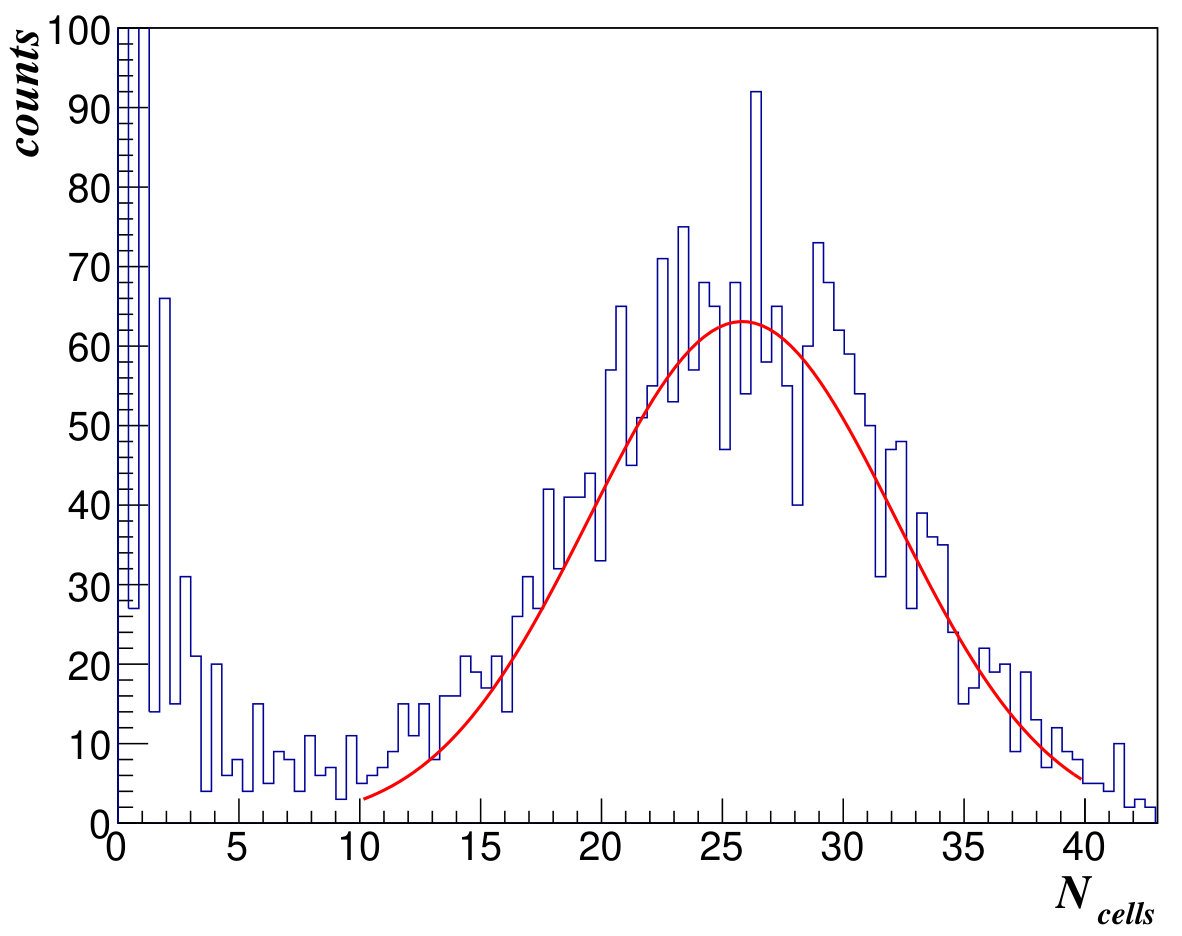 Figure 1
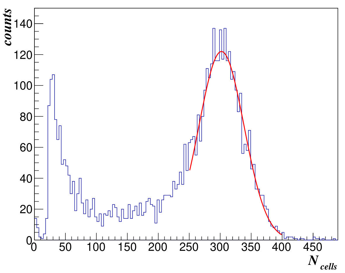 Figure 2
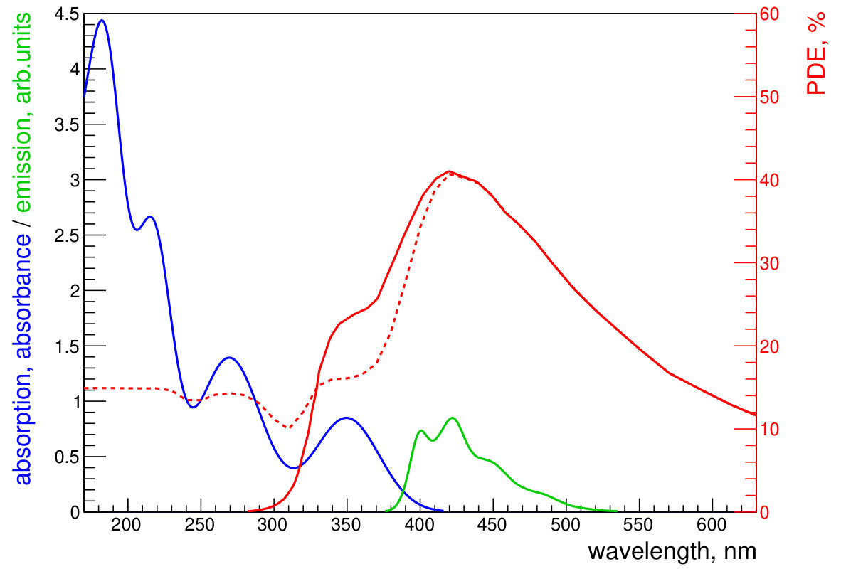 Figure 3
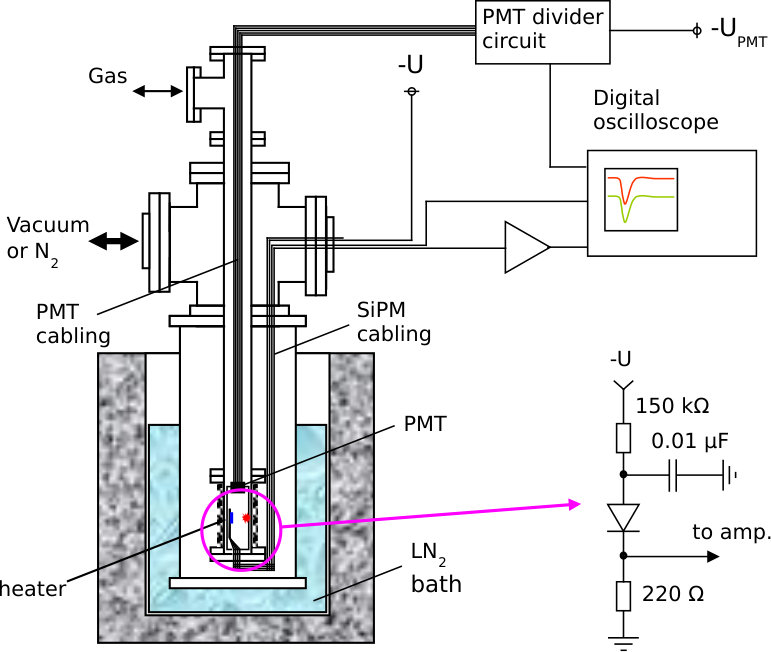 Figure 4
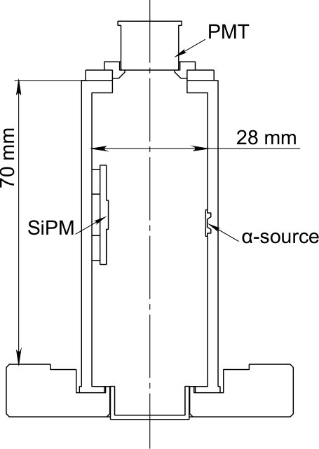 Figure 5
Peer Reviews
No public reviews on file for this paper yet. If you reviewed it on a platform where reviews are public (OpenReview, ICLR, NeurIPS, ICML), you can paste yours below so the community can read it here.
Videos
No videos yet. Explain this paper in a talk, walkthrough, or lecture? Add one.
\affiliation
[a]Institute for Theoretical and Experimental Physics named by A. I. Alikhanov of National Research Center “Kurchatov Institute”, 25 Bolshaya Cheremushkinskaya st., 117218 Moscow, Russian Federation \affiliation[b]National Nuclear Research University “MEPhI”, 31 Kashirskoe sh., 115409 Moscow, Russian Federation \affiliation[c]Enikolopov Institute of Synthetic Polymer Materials, Russian Academy of Science, 70 Profsoyuznaya st., 117393, Moscow, Russian Federation \affiliation[d]Moscow Institute of Physics and Technology, 9 Institutskij per., 141700, Moscow reg. Russian Federation \affiliation[e]Azimuth Photonics, 11 Khavskaya st., 115162, Moscow, Russian Federation \[email protected]
Test of SensL SiPM coated with NOL-1 wavelength shifter in liquid xenon
D. Yu. Akimov
V. A. Belov,\noteCorresponding author
O. V. Borshchev
A. A. Burenkov
Yu. L. Grishkin
A. K. Karelin
A. V. Kuchenkov
A. N. Martemiyanov
S. A. Ponomarenko
G. E. Simakov
V. N. Stekhanov
N. M. Surin
V. S. Timoshin
O. Ya. Zeldovich
Abstract
A SensL MicroFC-SMT-60035 66 mm2 silicon photo-multiplier coated with a NOL-1 wavelength shifter have been tested in the liquid xenon to detect the 175-nm scintillation light. For comparison, a Hamamatsu vacuum ultraviolet sensitive MPPC VUV3 33 mm2 was tested under the same conditions. The photodetection efficiency of % and %, correspondingly, is obtained.
\keywords
Photon detectors for UV, visible and IR photons (solid-state) (PIN diodes, APDs, Si-PMTs, G-APDs, CCDs, EBCCDs, EMCCDs etc); Cryogenic detectors; Noble-liquid detectors (scintillation, ionization, two-phase)
1 Introduction
A silicon photomultiplier (SiPM, MPPC, etc.) technology is very attractive for the use in low-background experiments in replacement for photomultiplier tubes (PMTs) due to the potentially very low level of radioactivity of semiconductor materials. Silicon photomultipliers (here and after, we refer to them as SiPM) have a gain of about , comparable to that of PMT, have the low operating voltage and power consumption. The devices have linear response to the light intensity when the number of detected photons is not very high in compare to the number of SiPM cells. SiPM is not sensitive to electric or magnetic fields. Manufacturing is based on the same technology as for many other devices in semiconductor industry, that opens the possibility for inexpensive mass production. SiPMs have already replaced regular PMTs in a variety of cases, including LHCb SciFi upgrade [1], CMS HCal upgrade [2] and MEG II [3]. Several collaborations like GERDA, NEXT, nEXO are planning to use VUV sensitive SiPMs in their detectors.
The first test of SiPM in liquid xenon was performed in 2005 [4]. Unfortunately, SiPMs don’t suit well for noble gas based detectors because the emitted light of noble gases has wavelengths in a VUV region (below 200 nm). The SiPM microstructure is usually not optimized for such wavelengths; moreover, protection layers are usually not transparent in this region. Several manufacturers have embarked in an extensive development program in order to achieve the reasonable sensitivity to the VUV light. Specially dedicated devices (e.g. MPPC) have been developed and they are already on practical stage for the use in a liquid xenon (emission wavelength is 175 nm) environment in MEG experiment. The alternative way to solve this problem is to use a wavelength shifter (WLS).
The aim of this work is to demonstrate experimentally the performance of SensL SiPM with a WLS to detect the liquid xenon scintillation light and to compare its characteristics with those of the VUV-sensitive Hamamatsu Photonics VUV3 MPPC device. It was obtained in our previous studies that the photodetection efficiency (PDE) of WLS + SiPM system can reach up to 50% of the SiPM PDE in the blue region [5]. An important feature of our study is that the photodetectors are tested directly in the liquid xenon environment.
2 Wavelength shifter
A nanostructured organosilicon luminophore (NOL) is a new type of WLS which combines an absorber and an emitter in one molecule. It has the very high energy transfer efficiency of the electronic excitation from the outer fragments of a molecule absorbing the light at the short wavelengths, to the inner fragment having the high photoluminescence quantum yield (QY) at longer wavelengths [6, 7]. This type of luminophores are of interest for various technical applications [5, 8]. The NOL-1 type was chosen for shifting of the VUV light emission of liquid xenon (175 nm) to the region of maximal PDE of SiPM (420 nm). The WLS can be deposited as a thin film layer directly on the front surface of SiPM over the protective optically transparent compound. Spectral properties of NOL-1 are suited very well for this task [9]. Absorption spectrum of the transparent 200-nm NOL-1 film is plotted in figure 1 by blue curve in terms of absorbance , where is a wavelength of light. It consists of four absorption bands with maxima at the following wavelengths: 182 nm, 216 nm, 270 nm and 350 nm. The first three of them correspond to excitation of the outer fragments. The absorption band with a maximum at 350 nm corresponds to excitation of the central fragment. The emission spectrum of NOL-1 is shown in figure 1 by the green curve. It corresponds to de-excitation of the central fragment and doesn’t depend on the excitation wavelength. This spectrum has two maxima: at 400 and 422 nm. The effective photodetection efficiency , defined as an overall photodetection efficiency of the WLS + SiPM system, is express according to the formula:
[TABLE]
where is photodetection efficiency of the SensL SiPM (shown in figure 1 by red), and are the boundaries of the interval where . The factor 0.57–0.61 describes the amount of emitted light that reach SiPM face. It is bigger than a naive assumption value of 0.5 because we also take into account reflection of the emitted light from surface of WLS layer that comes from the difference in the refractive indices of materials. The value is approximately equal to 73% at nm that corresponds to absorption of light by the outer fragments of molecule and equal to 80% for around 350 nm that corresponds to absorption of light by the inner fragment of molecule. The effective photodetection efficiency calculated with the use of formula above for the 200-nm NOL-1 layer deposited on the SensL SiPM is shown in figure 1 by the red dashed curve.
The choice of the 200-nm thickness of NOL-1 layer is based on our previous studies [5]. The WLS was deposited directly on the front surface of SiPM over the protective compound in the form of solution in toluene. Then it was dried up forming a solid amorphous film. Such a film has higher adhesion to the surface of the protective compound than that of a polycrystalline film formed by other frequently used WLSs p-terphenyl and tetraphenyl butadiene. This, along with a 7 times larger molar mass, makes NOL-1 coating much more steady in liquid xenon, resulting in significant reduce of solubility in liquid xenon, compared to p-terphenyl. That could make it safe from the point of view of production of a volume distributed effects on scintillation light, even when it is in direct contact with liquid xenon. The absorbance at 175 nm for the WLS layer of such a thickness is higher than 4. This means that the layer absorbs 99.99% of the liquid xenon emission light. The absorbance in the region of overlapping of the absorption (blue) and the emission (green) spectra is less than . This results in reabsorption losses of below 6%. One can see that the is almost zero at the wavelengths shorter than 300 nm. However, depositing of the 200-nm layer of NOL-1 on SiPM provides equal to 13–15% at these wavelengths. The expected value of for 175 nm is %.
3 Experimental setup
The experimental setup used for measurements is shown schematically in figure 2. The chamber is assembled from standard CF vacuum pieces. The test cell which contains SiPM and PMT is made of CF nipple (35 mm inner diameter) with a feedthrough flange on the bottom. The cell in installed in a bigger CF nipple which can be evacuated or filled with gaseous N2 for thermo conductivity. A liquid N2 bath is used for cooling the chamber. A resistive wire heater wound around the CF nipple servers for maintaining the required temperature inside the test cell. The design of the inner part of the test cell is shown in figure 3.
Inside the cell, there is a construction made of a stainless steel (ss) and standing on four ss legs. This construction can hold several SiPMs on the legs and a small PMT on the top. An 241Am alpha-source, which produces scintillation light in the liquid xenon, is placed on the leg opposite to the SiPM. The SiPM and its electrical circuit are mounted on a ceramic plate. A Hamamatsu R7400-06 PMT having a synthetic silica window with bialkali photocathode and QE 15% for the xenon light was used for triggering. The test chamber was pumped out by a zeolite and then by a titanium discharge pump down to torr without being heated. Heating of the cell was not allowed because of the photodetector installed inside. Xenon gas had undergone purification procedure with a Mykrolis Megaline purifier before filling the chamber.
Voltage power supply as well as signal readout was provided with the use of coaxial cables and vacuum feedthroughs. The SiPM signals were additionally amplified with a gain of 5 by custom made fast amplifiers built on the basis of OPA656N OpAmp. For SensL SiPM, the amplifier circuit was located on the back side of the ceramic plate; for Hamamtsu SiPM, outside of the cryostat. Signals from the SiPMs and PMT were recorded with the Tektronix TDS5034 digital oscilloscope at a digitization frequency of 125 MHz. The scope was triggered by SiPM in anticoincidence with PMT for noise runs and in coincidence with PMT for alpha runs. The threshold in SiPM channel was set low enough to register single cell signals. Signal waveforms of a total length of 10 s, with trigger located in a middle, were recorded for later analysis.
Two photodetectors were consequently tested: the non-VUV-sensitive SensL MicroFC-60035-SMT SiPM coated with the 200-nm NOL-1 WLS layer and the VUV-sensitive Hamamatsu Photonics VUV3 MPPC for comparison. Characteristics of these photodetectors can be found in table 1. The overvoltage for Hamamatsu device was set according to the manufacturer recommendations. For SensL SiPM, the overvoltage was selected as a balance between gain and noise characteristics to optimize the alpha-peak resolution. For each SiPM, three sets of measurements were performed. These include a noise run in vacuum plus noise and source runs the liquid xenon environment. All these measurements were performed at the temperature of .
4 Data analysis
Recorded waveforms of registered events were processed with a dedicated software on the event-by-event basis. Signal pulses above the software threshold were selected. The software threshold was set depending on noise level and gain for each SiPM. For each pulse, a sequence of approximately 100 samples having the amplitude above the threshold defined the pulse boundaries and was used to calculate pulse amplitude, area and width. It was then baseline-corrected by subtracting the average of the previous 100 samples of waveform.
Calibration of SiPMs was made by dedicated noise runs in anti-coincidence with PMT signal. On the resulting area spectrum, the distance between the first and the second peak corresponds to a single cell signal area, and the ratio between the second and the first peaks served as a crosstalk estimation. We didn’t carry out a deep study of afterpulses, but within our time window we did not observe any.
The main datasets with alpha-peak were collected with the use of coincidence with PMT. For production of VUV light an 241Am radioactive source was used. It emits alpha-particles with a compact set of energies, that results in an average energy MeV. The source was made by shallow ion implantation to a substrate i.e. with an open surface. Thus, the source emitted the alpha-particles almost without energy losses in source material. In liquid xenon, the alpha-particles of such energy have the stopping range of approximately 40 m. Therefore, the radioactive source served as a point-like scintillation light source. We used the average energy for scintillation photon production by alpha-particles eV [10] to convert the deposited energy to the number of emitted photons. The test chamber and the assembly were made of a stainless steel. The chamber walls were unpolished. The reflection coefficient for the VUV xenon light (wavelength equals 175 nm) for such a material is known to be below 10%. Thus, we consider a non-direct portion of the light to be small enough in our geometry. Consequently, we may estimate the number of photons that reach the SiPM surface exclusively by the use of the solid angle spanned from the point of the source location to the SiPM photosensitive surface: . The resulting SiPM efficiency was calculated as , where is the alpha-peak position in terms of number of fired cells, and is estimated cross-talk.
The energy spectra measured for alpha-particles in the liquid xenon are shown in figure 4. From the alpha-peak positions, the PDE values were calculated to be % and % for Hamamatsu and SensL SiPM, correspondingly. The estimated relative error of these PDE values (19%) is comprised of the uncertainty of light collection efficiency (16%; includes both the error of solid angle and unaccounted reflections), error on the crosstalk value (9%), the alpha-particle energy and energy per photon uncertainty (5%).
We couldn’t find publications of measurements of Hamamatsu VUV3 in liquid xenon. Recently, several measurements were performed in liquid xenon, but using different device of similar type (Hamamatsu 10943-3186(X) Type A), and showed various results. MEG II experiment presented PDE value to be over 15% for selected samples [3]. In another test with this device for DARWIN experiment [11] author didn’t calculate PDE value explicitely, but used the same method of measurements. For a 1212 mm2 sample he obtained 300 for alpha-peak with 241Am source installed in 20 mm apart from the SiPM. Given 15 times bigger SiPM surface area, one can see that a factor of 12 difference is reasonably compatible with our result. There is a separate result for the VUV3 measurements in liquid argon, where a PDE value of 8% [12] for 128 nm light was demonstrated. Using wavelength dependance presented by the manufacturer [13], one can scale this to the wavelength of xenon light (175 nm) and can obtain the value 6.8%. This is also in agreement with out result.
5 Summary
We studied the applicability of silicon photomultipliers (SiPM) for the VUV light detection in liquid xenon experiments. We have demonstrated that the SensL MicroFC-SMT-60035 SiPM coated with the NOL-1 WLS shows PDE % for 175 nm light in liquid xenon. The VUV sensitive Hamamatsu Photonics VUV3 MPPC device demonstarated PDE % under the same conditions. The former result indicates that regular commercial SiPMs coated with the NOL-1 WLS can be used in liquid xenon detectors as a replacement of PMTs. Such SiPMs are much better understood and have a fine-tuned manufacturing process. We developed an easy way of NOL-1 thin film deposition on a SiPM surface directly over the protective cover. The experimentally measured PDE value for the SensL SiPM coated with the NOL-1 is in a good agreement with the expected one.
\acknowledgments
We are very grateful to “YE International” (“Hamamatsu Photonics” distributor) and to “Azimuth Photonics” (“SensL” distributor) for supplying us with the samples of photodetectors. This study was supported by RFBR, projects no. 14-02-00675-a, 14-22-03028-ofi_m and 15-02-06874-a.
The reference list from the paper itself. Each links out to its DOI / PubMed record.
- 1[1] T. Kirn (for the LH Cb collaboration), Sci Fi – A large scintillating fibre tracker for LH Cb , NIM A 845 (2017) 481–485.
- 2[2] N. Strobbe (for the CMS collaboration), The upgrade of the CMS hadron calorimeter with silicon photomultipliers , JINST 12 (2017) C 01080.
- 3[3] Sh. Ogawa (for the MEG II collaboration), Liquid xenon calorimeter for MEG II experiment with VUV-sensitive MPP Cs , NIM A 845 (2017) 528–532.
- 4[4] E. Aprile, P. Cushman, K. Ni and P. Shagin, Detection of liquid xenon scintillation light with a silicon photomultiplier , NIM A 556 (2006) 215–218.
- 5[5] D.Yu. Akimov et al., Development of VUV wavelength shifter for the use with a visible light photodetector in noble gas filled detectors , NIM A 695 (2012) 403–406.
- 6[6] S.A. Ponomarenko et al., Nanostructured organosilicon luminophores and their application in highly efficient plastic scintillators , Sci. Rep. 4 (2014) 6549.
- 7[7] S.A. Ponomarenko et al., Nanostructured organosilicon luminophores as a new concept of nanomaterials for highly efficient down-conversion of light , Proc. SPIE 9545 (2015) 954509.
- 8[8] T. Uekert et al., Nanostructured organosilicon luminophores in highly efficient luminescent down-shifting layers for thin film photovoltaics , Solar Energy Materials and Solar Cells 155 (2016) 1–8.