Capturing ultrafast magnetic dynamics by time-resolved soft x-ray magnetic circular dichroism
Kou Takubo, Kohei Yamamoto, Yasuyuki Hirata, Yuichi Yokoyama, Yuya, Kubota, Shingo Yamamoto, Susumu Yamamoto, Iwao Matsuda, Shik Shin, Takeshi, Seki, Koki Takanashi, and Hiroki Wadati

TL;DR
This paper demonstrates the use of time-resolved soft x-ray magnetic circular dichroism with sub-50 ps resolution to observe ultrafast demagnetization in FePt thin films, highlighting the technique's potential for studying rapid magnetic dynamics.
Contribution
It introduces a novel experimental setup combining Tr-XMCD with high time resolution and compares PEY and TFY modes for analyzing ultrafast magnetic phenomena.
Findings
Ultrafast demagnetization observed within 50 ps in FePt films
PEY mode provides less-distorted spectra suitable for sum rule analysis
Demagnetization threshold depends on photon density
Abstract
Experiments of time-resolved x-ray magnetic circular dichroism (Tr-XMCD) and resonant x-ray scattering at a beamline BL07LSU in SPring-8 with a time-resolution of under 50 ps are presented. A micro-channel plate is utilized for the Tr-XMCD measurements at nearly normal incidence both in the partial electron and total fluorescence yield (PEY and TFY) modes at the L2,3 absorption edges of the 3d transition-metals in the soft x-ray region. The ultrafast photo-induced demagnetization within 50 ps is observed on the dynamics of a magnetic material of FePt thin film, having a distinct threshold of the photon density. The spectrum in the PEY mode is less-distorted both at the L2,3 edges compared with that in the TFY mode and has the potential to apply the sum rule analysis for XMCD spectra in pump-probed experiments.
Click any figure to enlarge with its caption.
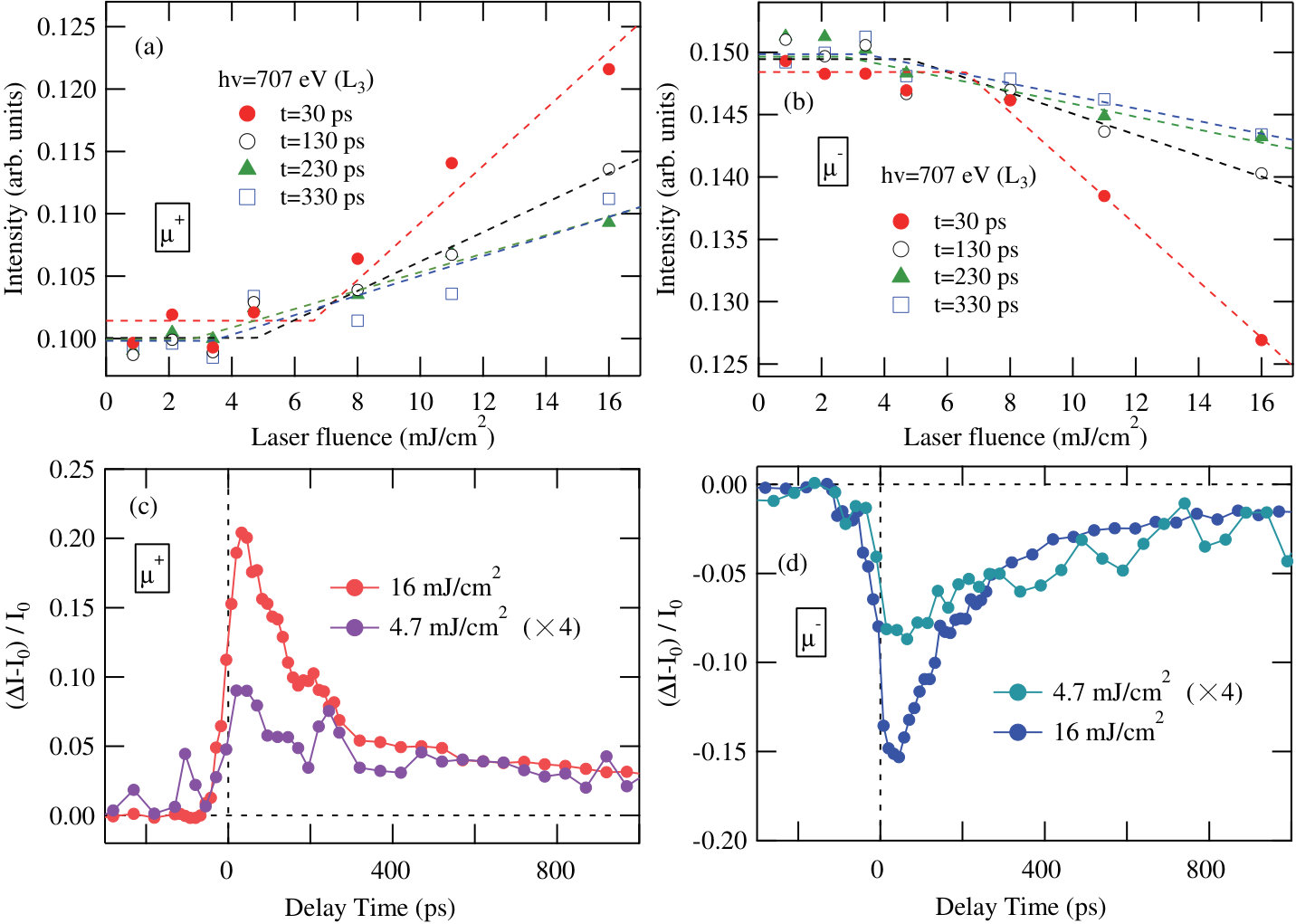 Figure 1
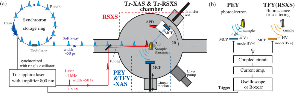 Figure 1
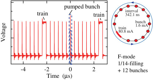 Figure 2
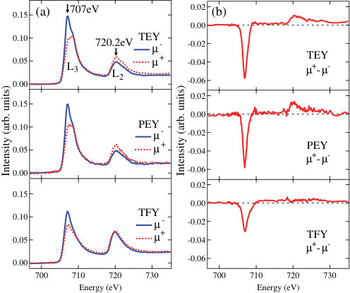 Figure 3
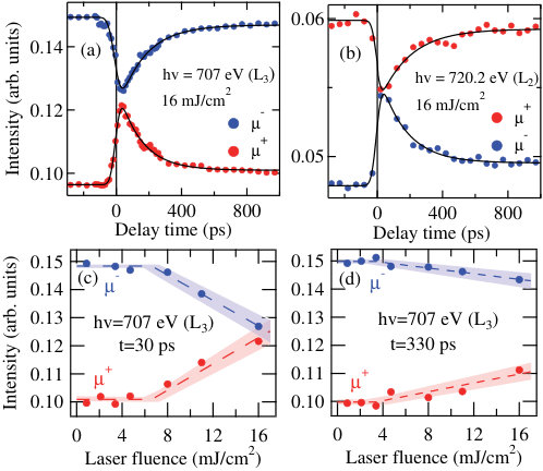 Figure 4
Peer Reviews
No public reviews on file for this paper yet. If you reviewed it on a platform where reviews are public (OpenReview, ICLR, NeurIPS, ICML), you can paste yours below so the community can read it here.
Videos
No videos yet. Explain this paper in a talk, walkthrough, or lecture? Add one.
Capturing ultrafast magnetic dynamics by time-resolved soft x-ray magnetic circular dichroism
Kou Takubo
Institute for Solid State Physics, University of Tokyo, Kashiwa 277-8581, Japan
Kohei Yamamoto
Institute for Solid State Physics, University of Tokyo, Kashiwa 277-8581, Japan
Yasuyuki Hirata
Institute for Solid State Physics, University of Tokyo, Kashiwa 277-8581, Japan
Yuichi Yokoyama
Institute for Solid State Physics, University of Tokyo, Kashiwa 277-8581, Japan
Yuya Kubota
Institute for Solid State Physics, University of Tokyo, Kashiwa 277-8581, Japan
Shingo Yamamoto
Institute for Solid State Physics, University of Tokyo, Kashiwa 277-8581, Japan
Susumu Yamamoto
Institute for Solid State Physics, University of Tokyo, Kashiwa 277-8581, Japan
Iwao Matsuda
Institute for Solid State Physics, University of Tokyo, Kashiwa 277-8581, Japan
Shik Shin
Institute for Solid State Physics, University of Tokyo, Kashiwa 277-8581, Japan
Takeshi Seki
Institute for Materials Research, Tohoku University, Sendai 980-8577, Japan
Koki Takanashi
Institute for Materials Research, Tohoku University, Sendai 980-8577, Japan
Hiroki Wadati
Institute for Solid State Physics, University of Tokyo, Kashiwa 277-8581, Japan
Abstract
Experiments of time-resolved x-ray magnetic circular dichroism (Tr-XMCD) and resonant x-ray scattering at a beamline BL07LSU in SPring-8 with a time-resolution of under 50 ps are presented. A micro-channel plate is utilized for the Tr-XMCD measurements at nearly normal incidence both in the partial electron and total fluorescence yield (PEY and TFY) modes at the absorption edges of the 3 transition-metals in the soft x-ray region. The ultrafast photo-induced demagnetization within 50 ps is observed on the dynamics of a magnetic material of FePt thin film, having a distinct threshold of the photon density. The spectrum in the PEY mode is less-distorted both at the edges compared with that in the TFY mode and has the potential to apply the sum rule analysis for XMCD spectra in pump-probed experiments.
Control of electron, magnetic, and lattice states by optical excitations in magnetically ordered materials has attracted considerable attention due to their potential applications in electronic and magnetic recording media functioning on an ultrafast time scale below nanosecond (ns, 10*-9* second, GHz range), since the observation of the ultrafast demagnetization in Ni within 1 picosecond (ps).Beaurepaire96 The ultrafast photo-induced changes of magnetic states are non-equilibrium phenomena and several mechanisms have been proposed to understand them.Nasu ; Kirilyuk10 ; Koopmans These phenomena generally involve fast structural changes near surface region Thomsen ; Jal and therefore require cooperative effects, inevitably having a threshold of the photon density.
To capture their non-equilibrium dynamics, ultrafast time-resolved experiments have been carried out using ultra-short laser pulses.Kirilyuk10 Recently, the development of a bunched synchrotron light source,Saes03 ; Cavalleli05 ; Stamm07 ; Boeglin10 ; Holldack14 and x-ray free electron lasersYamamoto15 ; Higley16 ; Bostedt16 have enabled investigation of the dynamic phenomena with element selectivity by tuning the photon energy of the x-ray to the absorption edges of the constituent elements. Especially, time-resolved soft x-ray spectroscopy has many unique characteristics, such as element specificity, chemical specificity and surface sensitivity, which make them versatile for application in a wide range of scientific fields including spintronics and environmental science. Owing to its importance, various time-resolved soft x-ray spectroscopy studies have been carried out.Holldack10 ; Wietstruk11 ; Radu11 ; Eschenlohr13 ; Tsuyama16 ; Ogawa12 ; Yamamoto13 The time-resolved x-ray magnetic circular dichroism (Tr-XMCD) and resonant soft x-ray scattering (Tr-RSXS) measurements for magnetic and electronic materials have been conducted in LCLS,Higley16 ; Bostedt16 ALS,Stamm07 and BESSY II slicing facilities.Boeglin10 ; Holldack10 ; Radu11 ; Wietstruk11 ; Holldack14 ; Eschenlohr13 ; Tsuyama16 The time-resolved x-ray magneto optical Kerr effect (XMOKE) measurements have also been conducted in FERMI.Yamamoto15 Tr-XMCD measurements at the absorption below 100 eV have also been performed by using high harmonics generation from tabletop lasers in recent years.Kfir
XMCD at the absorption of 3 transition-metals in the soft x-ray region ( 400 eV) is a powerful tool to evaluate the magnetic moments at the specific sites.Chen90 ; Thole92 Sum rules for XMCD at the edges in x-ray absorption spectroscopy (XAS) allow us to determine the orbital and spin contributions to the magnetic moments. The static soft XAS and XMCD in the total electron yield (TEY) mode are the most functional and simple methods, in which the photo-current induced by x-ray absorption is measured. In the pump-probe experiments, however, the photo-current induced by the pump laser exceeds that by the probe x ray. Thus, previous pump-probe XAS and XMCD experiments have been performed in the transmission setting for thin films on transmissive substrates or foils like Si3N4, Al, and so forth.Cavalleli05 ; Stamm07 ; Boeglin10 ; Radu11 ; Eschenlohr13 However, the good qualities of magnetic materials such as a perpendicular magnetic FePt thin film are unable to be achieved on the transparent Si3N4. The x-ray reflectivity and XMOKE measurements have also been performed for non-transparent thin films or bulk samples but with rather grazed settings for their magnetized axis.Holldack14 ; Yamamoto15 ; Tsuyama16
Here, we presents a setting for Tr-XAS and Tr-XMCD in the partial electron yield (PEY) and total fluorescence yield (TFY) modes to measure non-transmissive as-grown samples at nearly normal incidence. The PEY mode, in which the emitted photoelectrons are measured, is rather surface sensitive but these spectra are known to usually be similar to those obtained in the TEY.Lau02 On the other hand, the TFY mode, in which the fluorescence of an x-ray is measured, is rather bulk sensitive compared to EY. Despite its bulk-sensitivity, the relationship between FY and the absorption coefficient is non-trivial and saturation effects become important in the TFY.Troger92 ; Achkar11 The saturation effects in the TFY often change the ratio of XMCD at the and edges and preclude applying the sum rule analysis to them.
FePt thin films have drawn intense research interest owing to their potential for high density recording applications by using their magnetism.Platt02 ; Shima02 ; Seki11 ; Bedanta15 ; Iwama16 FePt has a high uniaxial magnetic anisotropy when the L10 ordered structure is formed, and exhibits perpendicular magnetization in L10-FePt (001) thin films, which makes it a candidate for heat-assisted magnetic recording (HAMR),Weller00 which is an already-working technology that incorporates laser heating into the recording process. Recently, it has been shown that proper heat-sink layers optimize the recording-time window down to 0.2 ns in HAMR of FePt.Weller15 The challenge of achieving shorter recording-time has led to an intensive research of the ultrafast magnetization dynamics. Very recently, Lambert et al. have showed that circularly polarized laser pulses can induce a small helicity-dependent magnetization in FePtAgC granular film.Lambert14 This finding suggests the possibility of all-optical switching of the magnetism in FePt having very short recording-time windows on the ps time scale. However, the debate on the origin of this effect is on-going, and more experiments are necessary to clarify if FePt could be switched on the ultrafast time scale.Nieves16
In this letter, the results of Tr-XMCD experiments for as-grown FePt films with non-transmissive substrates are reported. By virtue of the pump-probe technique, a photo-induced ultrafast demagnetization at the Fe sites of the FePt thin film within the experimental time-resolution of 50 ps for Tr-XMCD at SPring-8.
Figure 1 shows an overview of the experimental setup for Tr-XAS, Tr-XMCD, and Tr-RSXS measurements in the soft x-ray region at BL07LSU of SPring-8.Yamamoto14 Tr-RSXS is performed at the side of the experimental chamber. is the angle between the x-ray and sample-surface as the standard definition of x-ray diffraction. The scattering is detected by the micro-channel plate (MCP) or avalanche photodiode (APD) installed on the 2 motion of the diffractometer.future On the other hand, XAS and XMCD in the PEY or TFY modes are measured at the side of the chamber. Emitted photoelectrons or x-ray fluorescence are caught by another MCP topped on the linear motion, which could be positioned as close as 2 cm from the sample surface. The femtosecond Ti:sapphire laser with a wavelength of 800 nm housed at the laser station of BL07LSUOgawa12 ; Yamamoto13 ; Yamamoto14 is introduced into the XAS and RSXS chamber. The laser irradiates samples 10∘ below the x-ray and photo-induced dynamics of the electronic and structural evolutions are examined by means of a pump-probed technique. The laser pulses with 1 kHz repetition rate are synchronized with selected bunches of the synchrotron and delayed electronically. The pulse width of the Ti:sapphire laser is 50 fs. On the other hand, the single bunch width of the x-rays in the H-mode and F-mode at SPring-8SPring8 is 50 ps, which limits the experimental time-resolution. Figure 2 shows an example of the time-profile of the signals in the F-mode. The signals of the MCP or APD are amplified and gated on the oscilloscope or Boxcar integrator which is triggered by the laser pulses [Fig.1 (b)].
A chevron-type (dual-plate) MCP Wiza79 is leveraged for XAS and XMCD measurements in the PEY and TFY modes. Each surface channel of the MCP detects both charged particles (photoelectrons) and energetic photons (x-ray) and amplifies them as the electric currents. When a positive voltage is applied to MCP-in terminal of the surface plate, both the photoelectrons and x-ray fluorescence are detected, as illustrated in Fig. 1(b). However, since the current induced by the photoelectron is much larger than that by x-ray, the spectrum can be regarded to arise from the PEY. Alternatively, when a negative high voltage (HV) is applied to the MCP-in, the photoelectrons are bounded and only the fluorescence reaches to MCP. In this case, the TFY spectrum is obtained.
A FePt (001) thin film with the thickness of 50 nm was epitaxially grown at 500∘C on an MgO (100) single crystal substrate in an ultrahigh vacuum magnetron sputtering system.Seki11 The high temperature deposition process promoted the L10 ordering of FePt, resulting in the perpendicular magnetization. Right and left circularly polarized x-rays ( and ) for the XMCD measurement were obtained by combinations of horizontal and vertical figure-8 undulators in BL07LSU.Yamamoto14 The x-ray beams were focused using a mirror and its size was 100 m 200 m on the sample. The magnetic field was applied by a 260mT permanent magnet placed behind the substrate. XMCD was measured at 10 degrees off normal incidence ( = 100∘). The laser was linearly polarized and focused to a size 1mm2 on the sample.
Figure 3 shows the static XMCD spectra at the Fe edges for the FePt thin film in the TEY, PEY, and TFY modes. Intense XMCD was observed both at the edges, -40 at 707 eV and 6 at 720.2eV for the original XAS in TEY and PEY. A spectral difference between the TEY and PEY mode is barely observable. On the other hand, XMCD at the edge in the TFY mode is scarcely observed owing to the distortion caused by the saturation effect, although TFY has a bulk-sensitivity. Using the sum rules, the magnetic moments at the Fe site are estimated to be = 2.63 and = 0.10 for TEY, and = 2.74 and = 0.15 for PEY, respectively. Here, the number of the 3 electrons was assumed to be =6.6. These values are basically consistent with previous studies,XMCD1 ; XMCD2 ; XMCD6 while it has already been argued that there were some systematic discrepancies arising from the errors in the background subtraction procedures.XMCD6 On the other hand, the magnetic moments are estimated to be = 1.53 and = 0.08 for the TFY spectra, which exhibited a large discrepancy arising from their strong distortion.
The photo-induced dynamics of the FePt thin film were examined in the less-distorted PEY mode. The time-evolutions of the FePt thin film with 16 mJ/cm2 laser irradiation are given in Fig. 4 (a) at =707.0 eV ( edge) and (b) at 720.2 eV ( edge), respectively. As can be seen, almost similar time-evolutions are observed for the Fe edges. XMCD, namely the difference of the intensities at and , decreases immediately after the pumped pulse irradiation both at the Fe edges. XMCD exhibits a reduction in its intensity by 90 of the original value at both the edges 30 ps after the pump pulse. Then the XMCD exhibits a slow recovery of the magnetization. The time-evolutions are fitted by the following function
[TABLE]
convoluted with the Gaussian response function of the time-resolution (=50 ps). The 4 time-evolutions can be fitted with similar time constants of ps and ps [solid lines in Fig. 4(a) and (b)]. On analysis, the photo-induced demagnetization occurs within the experimental time resolution 50 ps after the pump pulse, and is relaxed with a time constant of 150 ps. It should be noted that the time constant of 150 ps for the demagnetization recovery in the FePt thin film is much slower than that observed in the elementary metals Fe, Ni, and so forth of 10 ps, Kirilyuk10 and similar to the slow recovery of several hundred ps time scale observed for the intermetallic GdFeCoKirilyuk10 ; Radu11 and Co/Pt multi layers,Kanzantseva08 which also show magnetization reversals induced by the laser pulses. In addition, the magnetization is not recovered to the original value even at =1500 ps, which is completed before the next bunch arrives after 342 ns. The time constant of 150 ps will correspond to the time for spin and electron temperature transferring into the lattice, as discussed below.
Figure 4 (c)(d) gives the Tr-XMCD intensities ( and ) at (c) =30 ps and (d) =330 ps as a function of the laser fluence. Threshold-like behaviour is observed at 6.5 mJ/cm2 for =30 ps, which is estimated by the linear fittings for the fluence dependences. The threshold seems to decrease as the time increases (See supplementary for more information). These results indicate that the photo-induced effect will not be a simple thermal effect.Nasu ; Tsuyama16 The threshold-like behaviour, namely the abrupt increase of the change ratio at around a few tens ps scale with high fluence seems to be a consequence of the critical magnetization fluctuation near the Curie temperature, which was suggested in previous Tr-MOKE studies.Mendil ; Kimling The demagnetization dynamics for general metallic magnets have been described by a so-called three-temperature modelKirilyuk10 ; Kimling ; Koopmans in the Tr-MOKE studies, while Tr-MOKE detects macroscopic magnetizations and does not have an element selectivity. Sub-ps demagnetization and following ps remagnetization are observed as the first step of the dynamics, that may be hindered by the present temporal resolution. Then sub-ps de- or remagnetization is followed by a slower step (extend to several hundred ps) as the lattice temperature increases, that is clearly seen in the present dynamics. One may consider that all of demagnetization processes can be described by temperature dependences of the electron and spin specific heats and does not relate to a cooperative effect including the ’lattice’. However, the photo irradiation also causes a sudden volume expansion near the surface just after the phonon temperature becomes to raise, which is usually lasting for several hundred ps.Thomsen In addition, the slow relaxation of FePt with time constant of 150 ps indicates that the ’lattice’ temperature is also important especially on this time scale (See supplementary again). Furthermore, a recent time-resolved x-ray resonant magnetic reflectivity measurement revealed that demagnetization dynamics in a Ni film involves a structural change near the surface region.Jal Therefore, it will be reasonable that the photo-induced demagnetization of FePt involves a kind of lattice cooperative effect and is not a simple thermal effect. Further time-resolved structural study including x-ray diffraction and reflectivity measurements will be very suggestive in order to clarify this point, which can be performed in same chamber in situ.
Here, we report the advantages of the present Tr-XAS and XMCD measurements, which are (1) the precise sum rule analysis in the PEY mode at nearly normal incidence, and (2) the depth-dependent measurements utilizing the difference of probing depths between PEY and TFY. When some negative retarding voltages are applied to the MCP-in terminal in the PEY mode, the surface sensitivity can be tuned between about 1 nm and 10 nm. On the other hand, the TFY mode can detect the bulk region of 100 nm. The depth-resolved dynamical measurements will be archived by using this technique. The difference of photo-induced dynamics between surface and bulk region on several materials including surface and/or interface magnetisms will be clarified. The less-distorted PEY method will provide an opportunity for applying a sum rule analysis to XMCD spectra in the ultrafast pump-probed spectroscopy for various magnetic materials, which would be very useful to explore the all-optical switching in spintronics devices such as FePt, Co/Pt multi-layers and so forth. On the other hand, the bulk sensitive TFY method will allow us to measure the dynamics of bulk samples showing various quantum phenomena including high- superconductors, heavy fermion metals and so forth. In conclusion, we have studied the photo-induced magnetic dynamics of the FePt thin films at the Fe site using the Tr-XMCD technique at nearly normal incidence. The ultrafast photo-induced demagnetization within 50 ps and its slow relaxation taking a few hundred ps are clarified with a distinct threshold-like behaviour. MCP has been used for Tr-XMCD both in the electron and fluorescence yield modes at the edges of the 3 transition-metals in the soft-x-ray region. The spectrum in the PEY mode is less distorted and basically similar to that obtained in the TEY mode. No difference of the change ratio is observed between the Fe and edges in the time-resolved measurement, taking advantage of the PEY method.
See supplementary material for additional data of laser fluence dependence at various pump-probe delays.
We acknowledge kind support by Dr. Y. Tanaka, Dr. T. Ohkochi and Prof. T. Kinoshita. Tr-XMCD measurements were performed with the approval of Synchrotron Radiation Research Organization, the University of Tokyo (No. 2016A7504, 2016A7403, 2016B7403, 2016B7518). This research was supported by the Japan Society the Promotion of Science (JSPS) of Grant-in-Aid for Young Scientists (B) (Nos. 16K20997 and 16K17722) and for Scientific Research (C) (No. 26400328). This work was also supported by the Ministry of Education, Culture, Sports, Science and Technology of Japan (X-ray Free Electron Laser Priority Strategy Program).
References
[S1] J. Mendil, P. Nieves, O. Chubykalo-Fesenko, J. Walowski, T. Santos, S. Pisana, and M. Münzenberga, Sci.
Rep. 4, 3980 (2014).
[S2] J. Kimling, J. Kimling, R. B. Wilson, B. Hebler, M. Albrecht, and D. G. Cahill, Phys. Rev. B 90, 224408
(2014).
[S3] C. Thomsen, H. T. Grahn, H. J. Maris, and J. Tauc, Phys. Rev. B 34, 4129 (1986).
[S4] E. Jal, V. López-Flores, N. Pontius, C. Schüßler-Langeheine, T. Ferté, N. Bergeard, C. Boeglin, B. Vodungbo,
J. Lüning, and N. Jaouen, arXiv:1701.01375v1.
The reference list from the paper itself. Each links out to its DOI / PubMed record.
- 1(1) E. Beaurepaire, J.-C. Merle, A. Daunois, and J.-Y. Bigot, Phys. Rev. Lett. 76 , 4250 (1996).
- 2(2) Photoinduced Phase Transitions , edited by K. Nasu, (World Scientific, Singapore, 2004).
- 3(3) A. Kirilyuk, A. V. Kimel, and T. Rasing, Rev. Mod. Phys. 82 , 2731 (2010) and reference there in.
- 4(4) B. Koopmans, G. Malinowski, F. Dalla Longa, D. Steiauf, M. Fähnle, T. Roth, M. Cinchetti, and M. Aeschlimann, Nat. Mater. 9 , 259-265 (2010).
- 5(5) C. Thomsen, H. T. Grahn, H. J. Maris, and J. Tauc, Phys. Rev. B 34 , 4129 (1986).
- 6(6) E. Jal, V. López-Flores, N. Pontius, C. Schüßler-Langeheine, T. Ferté, N. Bergeard, C. Boeglin, B. Vodungbo, J. Lüning, and N. Jaouen, ar Xiv:1701.01375 v 1.
- 7(7) M. Saes, C. Bressler, R. Abela, D. Grolimund, S. L. Johnson, P. A. Heimann, and M. Chergui, Phys. Rev. Lett. 90 , 047403(2003).
- 8(8) A. Cavalleri, M. Rini, H. H. W. Chong, S. Fourmaux, T. E. Glover, P. A. Heimann, J. C. Kieffer, and R. W. Schoenlein, Phys. Rev. Lett. 95 , 067405 (2005).