Understanding properties of the master effector of phage shock operon in Mycobacterium tuberculosis via bioinformatics approach
Zarrin Basharat, Azra Yasmin

TL;DR
This paper explores the role of the PspA protein in the phage shock operon of Mycobacterium tuberculosis using bioinformatics methods.
Contribution
The study models and simulates PspA's conformational changes in M. tuberculosis, revealing structural transitions.
Findings
PspA undergoes significant conformational transitions at the far end in Mycobacterium tuberculosis.
The structural behavior of PspA influences the overall function of the Psp system.
The study is investigating the impact of phosphorylation and structural fluctuations during PspA complex formation.
Abstract
The phage-shock-protein (psp) is a part of the psp operon, which assists in safeguarding the survival of bacterium in stress and shields the cell against proton motif force challenge. It is strongly induced by bacterium allied phages, improperly localized mutant porins and various other stresses. Master effector of the operon, PspA has been modeled and simulated, illustrating how it undergoes significant conformational transition at the far end in Mycobacterium tuberculosis. Association of this key protein of the operon influences action of Psp system on the whole. We are further working on the impact of phosphorylation perturbation and the impact of structural fluctuations during complex formation of PspA with other moieties of interest.
Genes, proteins, chemicals, diseases, species, mutations and cell lines named across the full text — each resolved to its canonical identifier and authoritative record.
Click any figure to enlarge with its caption.
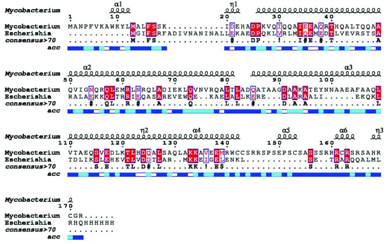 Figure 1
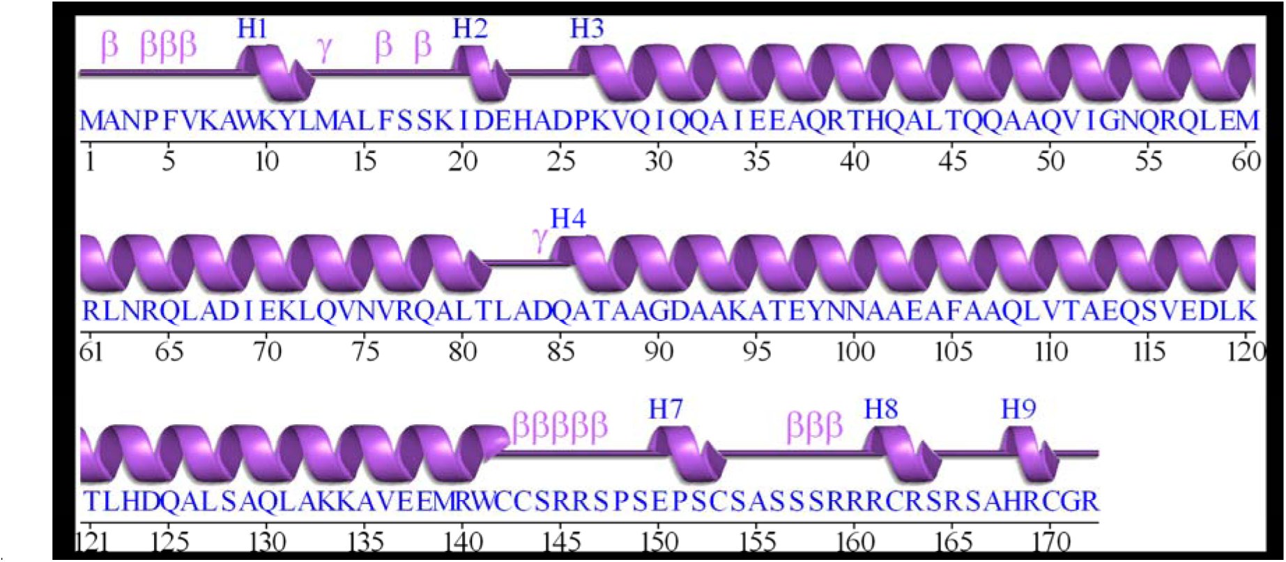 Figure 2A
 Figure 2B
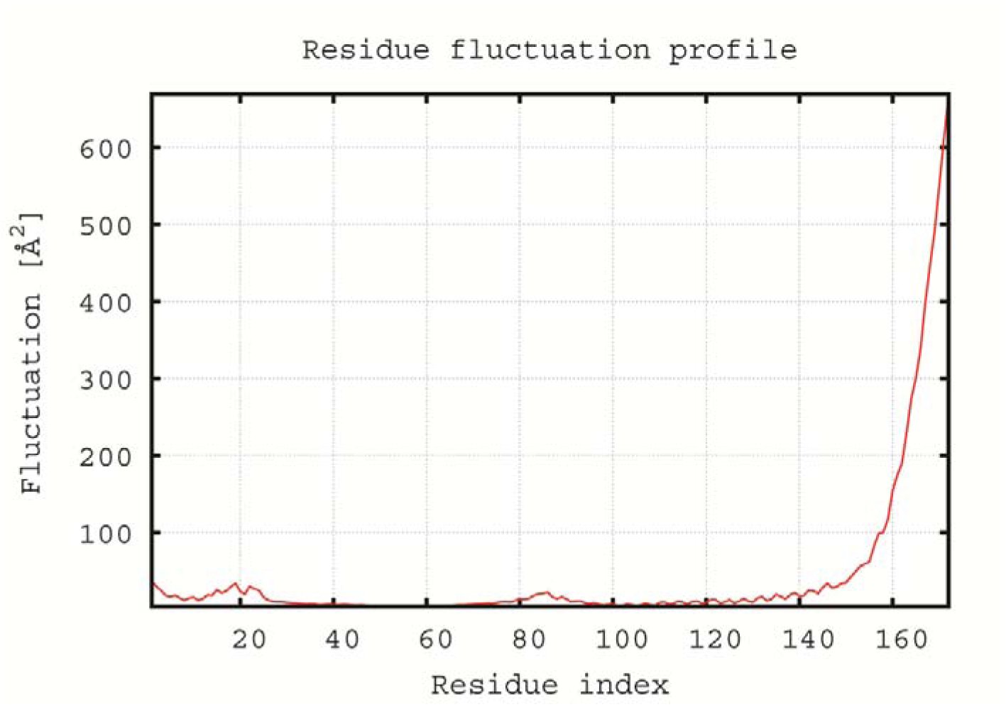 Figure 3A
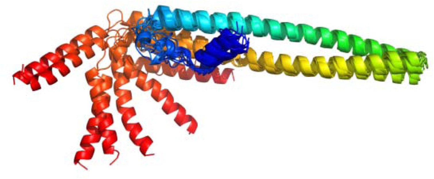 Figure 3B
Peer Reviews
No public reviews on file for this paper yet. If you reviewed it on a platform where reviews are public (OpenReview, ICLR, NeurIPS, ICML), you can paste yours below so the community can read it here.
Videos
No videos yet. Explain this paper in a talk, walkthrough, or lecture? Add one.
Taxonomy
TopicsBacteriophages and microbial interactions · RNA and protein synthesis mechanisms · Bacterial Genetics and Biotechnology
We hereby report ab initio structure model of the Mycobacterium tuberculosis phage shock protein A (PspA). PspA is the central constituent of bacterial stress response machinery, encoded by phage shock operon (Huvet et al, 2010). PspA, regulates not only it’s own transcription but that of the whole operon as well (Elderkin et al, 2005; Male et al, 2014). PspA is inferred to be a dual-function protein (Guegwen et al, 2009; Jovanovic et al, 2014) and localized amid cytoplasmic and inner membrane interface of the bacterium (Engl et al, 2009). It is responsible for maintaining the cell membrane integrity along with restoration of the proton motive force (Male et al, 2014; Engl et al, 2009; Wan et al, 2015).
Mycobacterium tuberculosis is a pathogenic bacterium responsible for causing disease in humans and veterinary species (Sakamoto, 2012). A lot of work has been carried out on Mycobacterium phages for therapeutic purpose. However, to the best of our knowledge, no report of the phage shock protein analysis for this pathogen exists at the moment. We retrieved PspA protein sequence of Mycobacterium tuberculosis from the Uniprot database with Accession number: R4M912 and analyzed the sequence and the structure using computational tools. Although, PspA belongs to the highy conserved PspA/IM30 family but Mycobacterium tuberculosis PspA shared a very low sequence homology with Escherichia coli PspA, revealed using Clustal Omega (Fig. 1) with seeded guide trees and HMM profile-profile technique for alignment generation at the backend (Sievers et al., 2011). The secondary structure analyzed by PROMOTIF tool (Hutchison and Thornton, 1996) revealed that the protein consisted of 9 helices, 6 helix-helix interacs, 14 β–turns and 3 Ↄ–turns (Fig. 2A).
Multiple sequence alignment of the Escherichia coli and Mycobacterium tuberculosis PspA. Conserved residues are shown in red color. Helices are denoted by squiggles at the top of the alignment. Solvent accessibility is depicted by a bar below the sequence (blue = accessible, cyan = intermediate, white = buried).
(A) Secondary structure of Mycobacterium tuberculosis PspA (helices labelled H1, H2…H9). β depicts beta turn and γ is for gamma turn.
Due to low homology with experimentally determined structures available in the RCSB Protein databank, 3D structure (Fig. 2B) was modeled by I–TASSER (Roy et al, 2010; yang et al, 2015) using Escherichia coli PspA as a template (PDB ID: 4WHE). The C–score based on the significance of template alignment threading and simulations of the structure assembly convergence parameters of the model chosen for analysis was -1.29 (lies between ideal range of -5 to 2), indicating good quality model. Estimated root mean square deviation of the predicted model from the Escherichia coli model was 7.8±4.4Å. The constructed structure resembled a helical bundle with important binding site residues predicted to occur at position 66, 69, 70, 73, 92, 96, 99, 100, 102 and 103. Despite low sequence conservation, the structure is however, well conserved due to the underlying fact that the protein folds remain conserved in similar function proteins. PspA structure in Escherichia coli is known to self–assemble into ring (Standar et al., 2008) or striated and indented rod–shaped complexes (Male et al., 2014) based on electron microscopy and helical rods based on X–ray crystallography analysis (Osadnik et al., 2015). PspA homologue LiaH in Bacillus subtilis (Wolf et al., 2008) and holins of bacteriophage lambda (Savva et al., 2010) have also been reported to self-assemble to rod-like structures from ring shaped protein complexes. The Mycobacterium tuberculosis PspA is also rod shaped and it is implied that these rod–like structures could form a support framework and aid in the maintenance of membrane integrity during phage shock response (Male et al., 2014).
(B) 3D structure of Mycobacterium tuberculosis PspA.
CABS–flex procedure based on the well–established coarse–grained CABS protein model (Fraga et al., 2014) was employed for the fast simulation of near–native dynamics of PspA. CABS is a computationally efficient alternative to classical all–atom molecular dynamics (Jamroz et al., 2013). The 3D modeled structure was input and used as a starting point for the all-atom, explicit water, 10-nanosecond dynamic simulation. Analysis was carried out at the backend and automatically analyzed trajectory (Fig. 3) was obtained to study the dynamic behaviour of protein. A set of eight (all-atom) protein model sets were obtained with global distance test score ranging from 0.6–0.7. Most dominant structural fluctuations appeared at the last beta turn region including 8^th^ and 9^th^ helix. Relative propensity of protein residues to deviate from an average dynamics structure increased substantially at the ending helices with fluctuation increasing from 100 Å at residue 160 to to >600 Å at residue 172. Understanding of flexibility of PspA can be of aid in research areas as molecular evolution (Boehr et al., 2009).
(A) Structural flexibility profile of simulated Mycobacterium tuberculosis PspA with fluctuations for individual protein residues shown via red line. The output is based on all-atom model via trajectory clustering.
(B) Refinement of the model and superpositioning (Provided 3D model as base) is centred on maximum likelihood superposition method of THESEUS (Theobald and Wuttke, 2006).
Phage infection has also been demonstrated to induce substantial fluctuations in host protein phosphorylation (Rieul et al., 1987; Russel and Model, 2006) and this was suggestive of PspA potential for phosphorylation as well. NetPhos Bac 1.0 (Miller et al., 2009) was used for prediction of possible phosphorylation residues. Seven serine residues (S144, S149, S156, S157, S158, S164, S166) were predicted to have phosphorylation potential based on neural network approach. However, none of these exhibited a knack to occur on predicted binding residues and hence, their exact role in PspA function of Mycobacterium tuberculosis yet remains to be elucidated.
Our findings pave way for further experimental studies and are of aid in understanding the Mycobacterium tuberculosis PspA response to the extracytoplasmic stresses that may damage the cytoplasmic membrane. We have used computational approach for the prediction of 3D structure of this protein but to furthur understand the function of the rod-like structure of Mycobacterium tuberculosis PspA, additional studies are required which can confirm and enhance the reported information along with elucidation of in depth biological function and interactions of Mycobacterium tuberculosis PspA.
Competing interests
The authors declare that no competing interests exist.
The reference list from the paper itself. Each links out to its DOI / PubMed record.
- 1Huvet, M., Toni, T., Sheng, X., Thorne, T., Jovanovic, G., Engl, C., Buck, M., Pinney, J.W. and Stumpf, M.P.H. (2010). The evolution of the Phage shock protein response system: Interplay between protein function, Genomic organization, and system function. Mol. Biol. Evol. 28, 1141–1155.
- 2Elderkin, S., Bordes, P., Jones, S., Rappas, M. and Buck, M. (2005). Molecular determinants for Psp A-mediated repression of the AAA transcriptional activator Psp F. J. Bacteriol. 187, 3238–3248.
- 3Male, A.L., Oyston, P.C.F. and Tavassoli, A. (2014). Self-Assembly of Escherichia coli Phage Shock Protein A. Adv. Microbiol. 4, 353–359.
- 4Guegwen, E., Savitzky, D.C., Darwin, A.J. (2009). Analysis of the Yersinia enterocolitica Psp BC proteins defines functional domains, essential amino acids and new roles within the phage-shock-protein response. Mol. Microbiol. 74, 619–633.
- 5Jovanovic, G., Mehta, P. and Buck, M. (2014). Anionic lipids and the cytoskeletal proteins Mre B and Rod Z define the spatio–temporal distribution and function of membrane stress controller Psp A in Escherichia coli. Microbiology. 160, 2374–86.
- 6Engl, C., Jovanovic, G., Lloyd, L.J., Murray, H., Spitaler, M., Ying, L., Errington, J. and Buck, M. (2009). In vivo localizations of membrane stress controllers Psp A and Psp G in Escherichia coli. Mol. Microbiol. 73, 382–396.
- 7Wan, F., Mao, Y., Dong, Y., Ju, L., Wu, G. and Gao, H. (2015). Impaired cell envelope resulting from arc A mutation largely accounts for enhanced sensitivity to hydrogen peroxide in Shewanella oneidensis. Sci. Rep. 5, 10228.
- 8Sakamoto, K. (2012).The Pathology of Mycobacterium tuberculosis Infection. Vet. Pathol. 49, 423–439.