Lateral Genetic Transfers Between Eukaryotes and Bacteriophages
Sarah R Bordenstein, Seth R Bordenstein

TL;DR
The study finds that eukaryotic genes can transfer to bacteriophages, revealing new insights into lateral genetic transfer and viral evolution.
Contribution
The discovery of eukaryotic protein domains in bacteriophage WO genomes suggests lateral transfer from animals to phages.
Findings
Eukaryotic protein domains like latrotoxin-CTD are found in intact bacteriophage genomes.
These domains are enriched with eukaryotic protease cleavage sites and form large phage genes.
The findings imply lateral transfer from animal to bacteriophage genomes.
Abstract
Viruses are trifurcated into eukaryotic, archaeal and bacterial categories. This domain-specific ecology underscores why eukaryotic genes are typically co-opted by eukaryotic viruses and bacterial genes are commonly found in bacteriophages. However, the presence of bacteriophages in symbiotic bacteria that obligately reside in eukaryotes may promote eukayotic DNA transfers to bacteriophages. By sequencing full genomes from purified bacteriophage WO particles of Wolbachia, we discover a novel eukaryotic association module with various animal proteins domains, such as the black widow latrotoxin-CTD, that are uninterrupted in intact bacteriophage genomes, enriched with eukaryotic protease cleavage sites, and combined with additional domains to forge some of the largest bacteriophage genes (up to 14,256 bp). These various protein domain families are central to eukaryotic functions and have…
Genes, proteins, chemicals, diseases, species, mutations and cell lines named across the full text — each resolved to its canonical identifier and authoritative record.
Click any figure to enlarge with its caption.
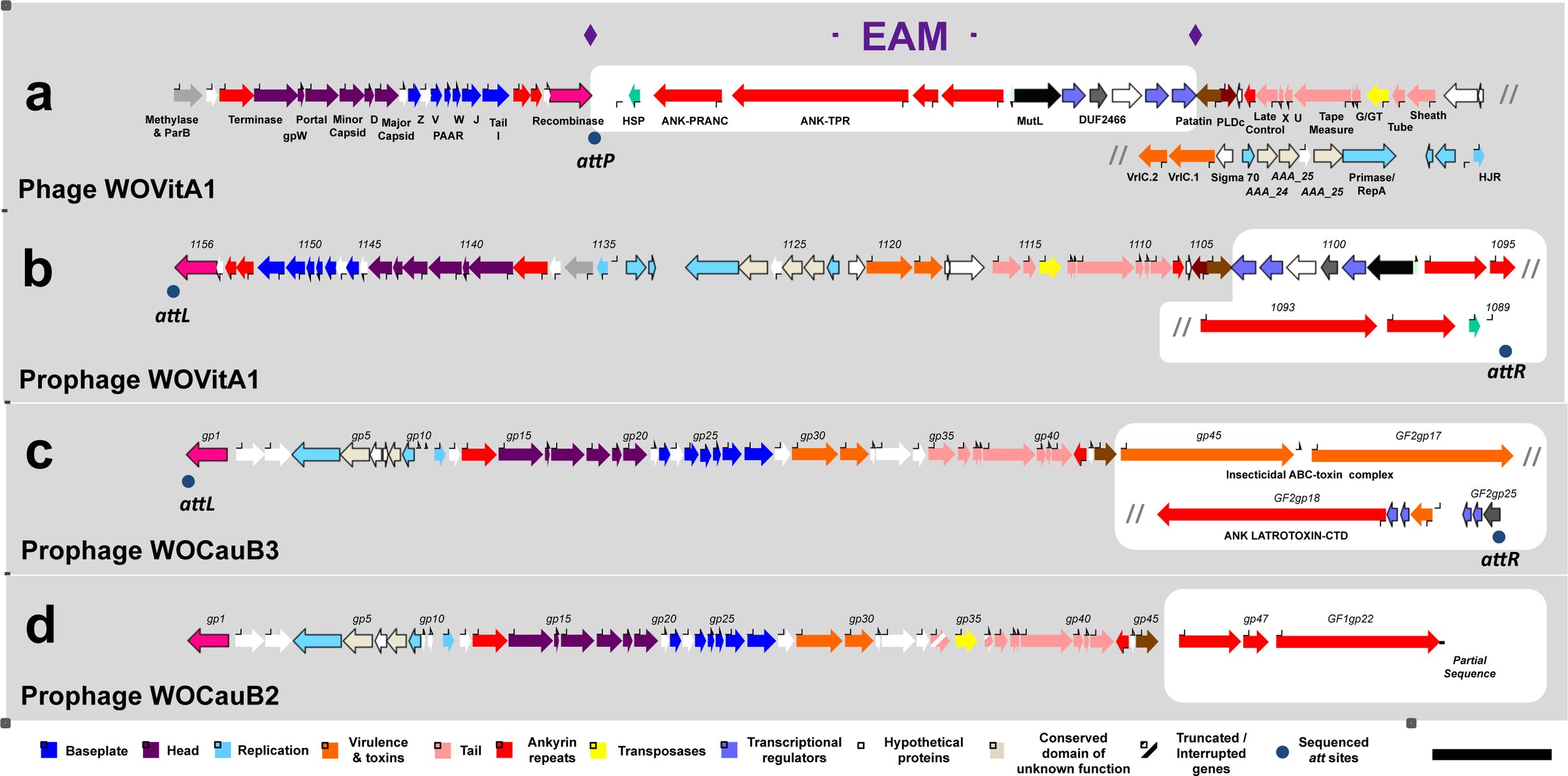 Figure 1
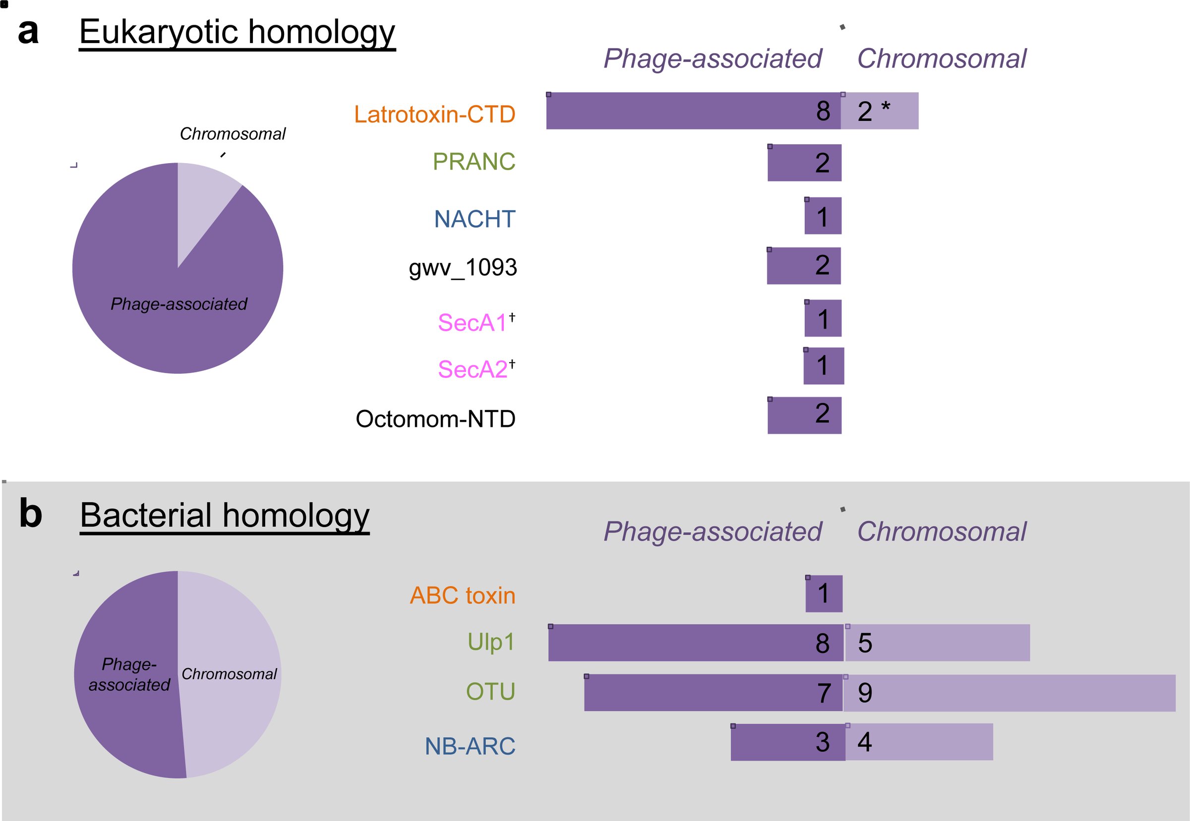 Figure 2
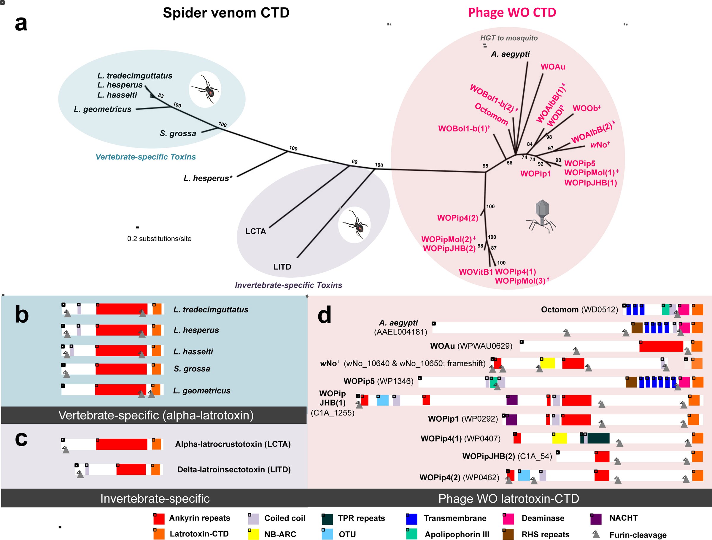 Figure 3
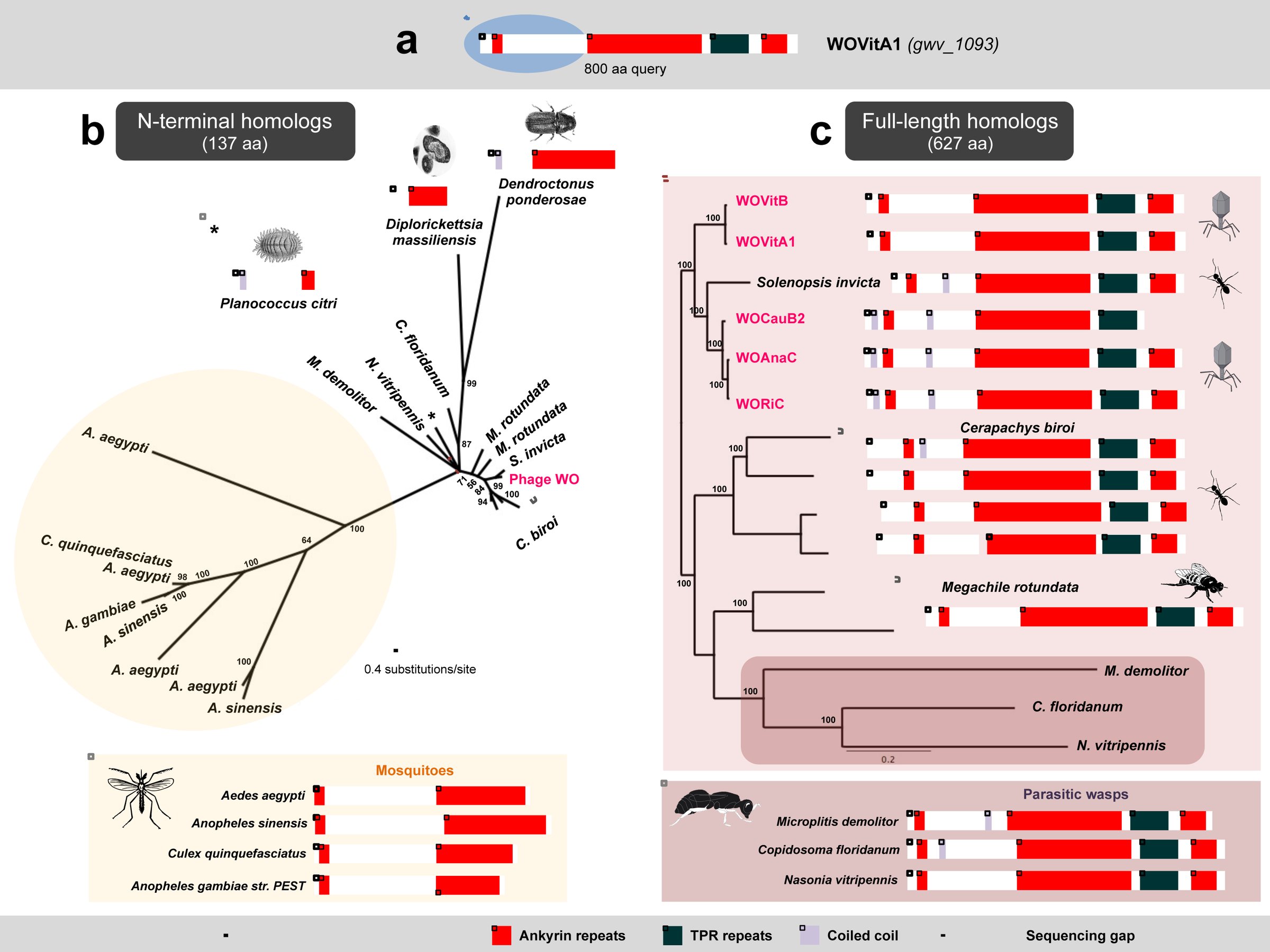 Figure 4
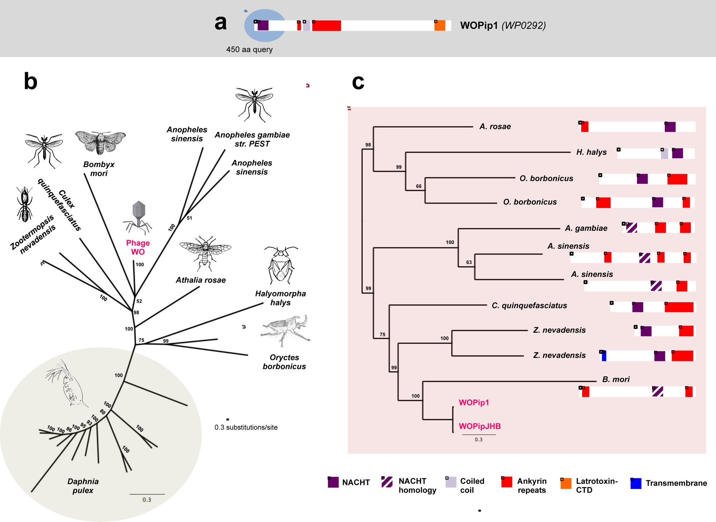 Figure 5
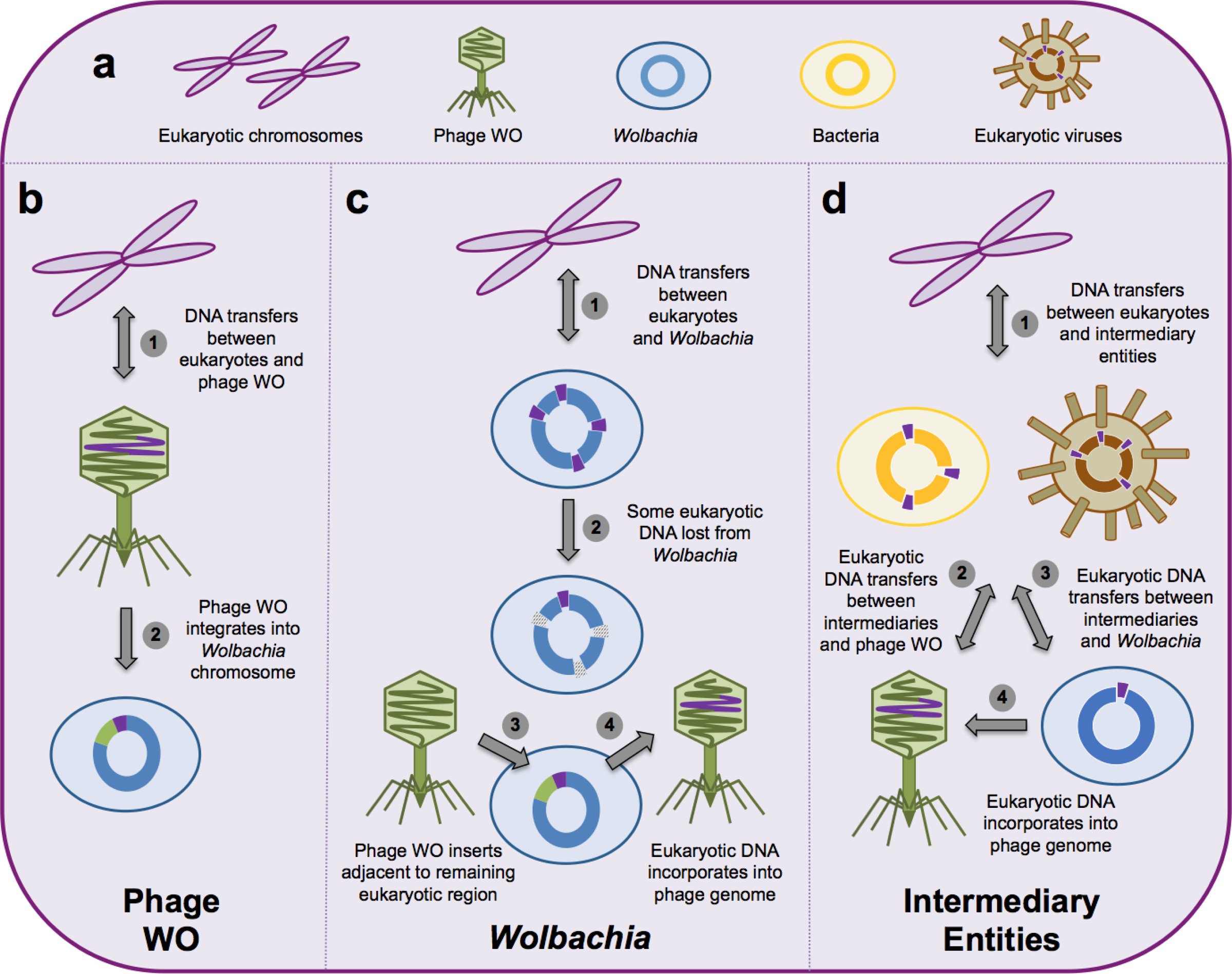 Figure 6
Peer Reviews
No public reviews on file for this paper yet. If you reviewed it on a platform where reviews are public (OpenReview, ICLR, NeurIPS, ICML), you can paste yours below so the community can read it here.
Videos
No videos yet. Explain this paper in a talk, walkthrough, or lecture? Add one.
Taxonomy
TopicsInsect symbiosis and bacterial influences · Bacteriophages and microbial interactions · CRISPR and Genetic Engineering
Introduction
Viruses are the most abundant and diverse biological entities in the biosphere^1,2^. Infecting organisms across the tree of life, they associate with every ecosystem on the planet. They are generally classified into polythetic groups according to ecological niche and mode of replication^3,4^. While any cellular domain can be infected by a virus, no extant virus is known to traverse more than one domain^5,6^. This domain-specific ecology of viruses underpins the current taxonomic paradigm of trifurcating viruses into eukaryotic, archaeal and bacterial categories, along with recent reappraisals of whether viruses constitute a fourth domain of life^7,8^. As a result of this domain-specific ecology, viruses often integrate host genes via specific highways of lateral gene transfer. Eukaryotic viruses tend to hijack genes directly from their eukaryotic hosts to evade, manipulate and counter-strike anti-viral immune responses^9,10^, with the exception of some giant viruses that appear to acquire genes from all domains of life^11^. Bacterial viruses, or bacteriophages (phages), integrate genetic material from their bacterial hosts including toxin^12^, photosynthesis^13^ and pigment biosynthesis genes^14^ that contribute to the fitness of their bacterial host. To date, however, there is no archetypal case of phage particles harboring genomes with eukaryotic DNA.
While all viruses are specific to one of the three domains of life, some bacteriophages target obligate intracellular bacteria of eukaryotic cells. For instance, phage WO infects the obligate intracellular alpha-proteobacteria Wolbachia, which in turn infect an estimated 40% of the most speciose group of animals worldwide - arthropods (as well as filarial nematodes). Wolbachia cause a range of host reproductive pathologies^15,16^, primarily infect the cells of host reproductive tissues, exist in Golgi-derived vesicles within the eukaryotic cytoplasm, and are enclosed by a bacterial cell membrane and one or more eukaryotic-derived membranes^17,18^. Nearly all sequenced Wolbachia genomes, with the exception of those acting as obligate mutualists, harbor prophage WO^19–21^. The prophage WO encode conserved structural modules (e.g., head, tail, baseplate) and exhibit Caudovirales morphology in electron micrographs of purified phages^20,22–25^. Electron microscopy and quantitative analyses indicate that prophages undergo a lytic phase capable of rupturing bacterial and eukaryotic cell membranes, and phage WO occurs in the extracellular matrix of arthropod gonads^23,26^. Therefore, phage WO appears to uniquely contend with the cellular exit, entry and defense mechanisms of two separate domains of life. WO is also a promising tool for genome editing of Wolbachia that has thus far been refractory to genetic modifcation.
Here we assemble the sequenced genomes of phage WO particles, resolve the bacteriophage attachment and bacterial integration sites, report a eukaryotic association module in bacteriophages, and discuss lateral gene transfers between eukaryotes and bacteriophages.
Results
Phage WO genomes reveal a eukaryotic association module
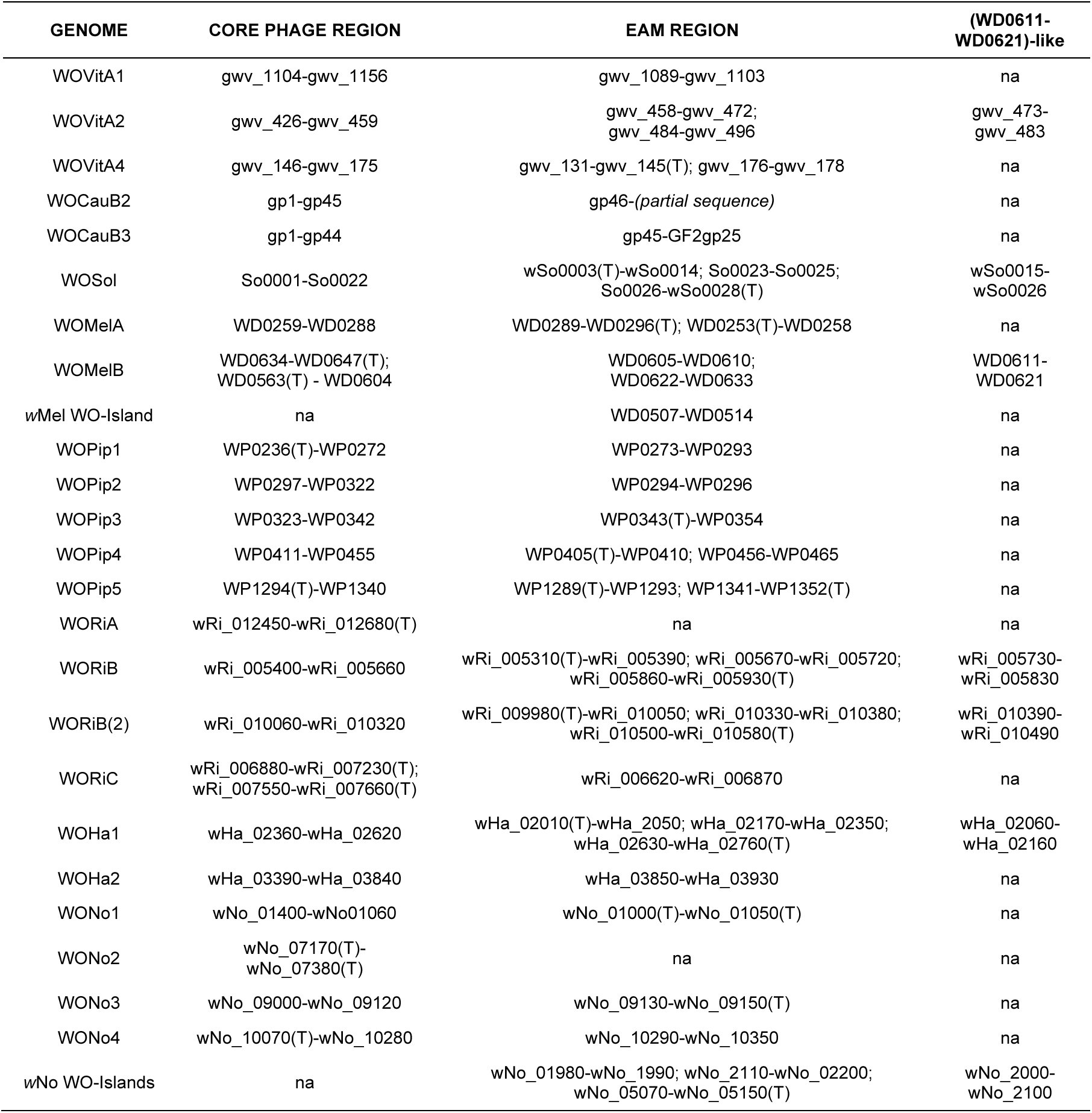
Here we report the metagenomic analysis of phage WO particles from wVitA-infected Nasonia giraulti wasps and wCauB-infected Ephestia kuehniella moths (the w-prefix indicates specific Wolbachia strain and WO-prefix indicates phage haplotype; see Supplementary Table 1 for a complete list). We identify the phage attachment sites and insertion regions and show from fully sequenced genomes that WO harbors all formerly described phage genetic modules (lysogeny, baseplate, head, replication, virulence, tail and patatin-like phospholipase^27^) as well as a new group of genes with atypical protein domains indicative of eukaryotic interaction. We collectively group these genes, which include the second largest gene in bacteriophages to date, into a ‘Eukaryotic Association Module’ (EAM; Fig. 1, white box). The EAM features genes that (i) encode protein domains and cleavage sites central to eukaryotic functions, (ii) occur in phage and metazoan hosts, (iii) are among the largest genes in phage genomes (up to 14,256 bp) and (iv) are absent from mutualistic, phage-free genomes such as the bedbug-infecting wCle and filarial nematode-infecting wBm and wOo. They occur in all complete prophage WO haplotypes (Supplementary Table 2).
Phage WO genomes harbor a Eukaryotic Association Module (EAM) The complete phage WO genome for (a) WOVitA1 was sequenced directly from purified viral particles using high throughput, metagenomic sequencing. The prophage (b) WOVitA1, (c) WOCauB3 and (d) WOCauB2 genomes were reannotated based on sequencing reads obtained from purified particles; complete genomes of WOCauB3 and WOCauB2 were not obtained. Each genome consists of a bacteriophage-like region (recombinase to patatin) and EAM highlighted in white. Gray slash marks indicate illustrative continuation of the genome. Dark blue dots indicate the discovery of the attL andattR sites of the prophage, which adjoin in the packaged WO genome to form attP. Numbers above the open reading frames indicate locus tags. Scale bar, 5,000 base pairs.
To verify the newly discovered EAM in the phage genome, we identified the terminal prophage WO genes and Sanger sequenced amplicons from an independent sample of phage WOVitA1 (Fig. 1a) across the linear phage attP site (hypothetical protein gwv_1089 to recombinase, Supplementary Fig. 1). Next, using the newly identified attR and attL sites, we extrapolated the bacterial attB site in WOVitA1, which is a noncoding, repetitive sequence in Wolbachia from Nasonia wasps (Supplementary Fig. 1e). The full length of the completely assembled, linear WOVitA1 genome is 65,653 bp, which is 23,531 bp larger than the previous prophage WO annotation. Similarly, we identified the new terminal ends of the WOCauB3 prophage [23,099 bp (51%) larger than original estimate of 45,078 bp], extending the previous observation that the end of the genome is beyond the patatin gene^25^, along with internal localization of the EAM genes by Sanger sequencing its attP site [Domain of Unknown Function (DUF)2426 to recombinase]. While we were not able to assemble a complete contig for WOCauB2, it is more than 6,854 bp larger than the original estimate of 43,016 bp, includes multiple ankyrin repeat genes homologous to those in WOVitA1, and, like many other prophage haplotypes (e.g., WORiC, WOVitA2, WOSuziC), integrates directly into Wolbachia’s magnesium chelatase (chlI) gene.
The EAM is enriched with eukaryotic-like domains
We then analyzed each phage WO protein domain for homology and surrounding peptide architecture. Unlike the single domain architecture of phage WO’s structural genes, EAM genes are highly polymorphic and encompass fusions of both eukaryotic and bacterial protein domains. By extending the analysis to include homologous prophage regions from all sequenced Wolbachia chromosomes, ten types of protein domains with putative eukaryotic functions were uncovered spanning four predicted functions: (i) toxins, (ii) host-microbe interactions, (iii) host cell suicide, and (iv) secretion of proteins through the cell membrane (Fig. 2). Notably, over half of these domain types [6/10; latrotoxin C-terminal domain (CTD), PRANC, NACHT, SecA, gwv_1093 N-terminal domain (NTD), Octomom-NTD] share greater amino acid homology to eukaryotic invertebrates than to bacteria in GenBank. Among this subset with eukaryotic sequence homology, the protein domains are almost exclusively found in the prophage EAM region (N=17) versus the Wolbachia chromosome (N=2). In the latter case, the two chromosomal latrotoxin-CTD domains (wNo_10650 and wHa_05390) are flanked by phage-associated genes and transposases, indicating a likely phage WO origin and subsequent genomic rearrangement. This pattern differs from other EAM protein domains with bacterial homology, which are equally dispersed in phage WO (N=19) and the Wolbachia chromosome (N=18) (Fig. 2, Fisher’s Exact Test, p = 0.0072). The difference importantly indicates that the eukaryotic-like protein domains are highly enriched in the EAM, suggesting a near exclusive role in phage WO biology.
Eukaryotic-like EAM genes are enriched in prophage WO regions EAM genes with (a) eukaryotic homology are most likely to be associated with prophage WO while those with (b) bacterial homology are both phage-associated and found scattered throughout the Wolbachia chromosome. () The two chromosomal latrotoxinCTD domains (wNo_10650 and wHa_05390) are located within phage-associated genes and transposases, indicating a potential genomic rearrangement. (†) SecA represents one ‘domain type’ but is listed separately because phage WO contains two different homologs (i.e., wHa_3920 and wHa_3930). Putative functional categories are: anti-eukaryotic toxins (orange); host-microbe interactions (green); host cell suicide (blue); secretion of virulence factors (pink); and unknown (black). Octomom refers to WD0513 of the wMel genome.*
The black widow latrotoxin-CTD
Latrotoxin-CTD is the most prevalent eukaryotic domain in prophage WO. Originally described for its major role in the venom of widow spiders (Latrodectus species), latrotoxins act extracellularly to cause the formation of ion-permeable membrane pores in their vertebrate or invertebrate victims. The CTD, specifically, is only associated with the latrotoxin precursor molecule (protoxin) and could possibly act intracellularly to facilitate disintegration of the spider’s toxin-producing cells^28^. While latrotoxins are generally considered exclusive to spiders, CTDhomologs in Wolbachia, Rickettsiella grylli^28^, and a transcriptome from a Wolbachia-infected stink bug^29^ have been reported. Here, phylogenetic analysis implies that the latrotoxin-CTD horizontally transferred between widow spiders and phage WO (Fig. 3). Reciprocal search queries using homologous spider and phage CTDs return the same BLASTP hits shown in Fig. 3. Notably, phage WO CTD sequences have the highest amino acid similarity to black widow spider homologs that target invertebrates, which are the primary hosts of Wolbachia. While convergent evolution could explain amino acid sequence similarities of the latrotoxin-CTD in black widows and Wolbachia, these two taxa occur in overlapping ecological niches (Wolbachia are known to infect spiders of the family Theridiidae) in which gene transfers are likely to happen^30^. We also confirmed the presence of Wolbachia in three independent Latrodectus geometricus samples by amplifying Wolbachia 16S rDNA and wsp membrane protein genes. The transfer event was apparently followed by a relatively more recent transfer from phage WO back to animals in the Aedes aegypti genome, where the region is located between genes of mosquito origin [fibrinogen-related protein (AAEL004156) and GalE3 (AAEL004196)].
Latrotoxin-CTD phylogeny supports lateral genetic transfers (a) Phylogeny of phage WO latrotoxin-CTD protein domains and their eukaryotic homologs was constructed by Bayesian analysis of 74 amino acids using the JTT model of evolution. Consensus support values are shown at the nodes. Comparative protein architecture shows that spider venom (b) vertebrate-specific alpha-latrotoxins and (c) invertebrate-specific alpha- and delta-latrotoxins are highly conserved, whereas (d) phage WO are not. Bolded nomenclature in (d) denotes the specific phage WO haplotype (listed as WO). Genome locus tags are listed in parentheses. Predicted furin cleavage sites, listed in Supplementary Table 3, are illustrated with gray triangles. () A second L. hesperus sequence represents a recently-described downstream paralog with unknown toxin activity32. (†) wNo_10650 is located within phage-associated genes and transposases, indicating a potential genomic rearrangement of a phage region. (‡) Architecture is not shown for sequences on incomplete contigs (WOBol1-b, WOAlbB, WODi, WOPipMol, WOVitB) because complete peptide information and specific phage association are unknown. Scale bar, 1,000 amino acids.*
Toxin activation by eukaryotic furin cleavage
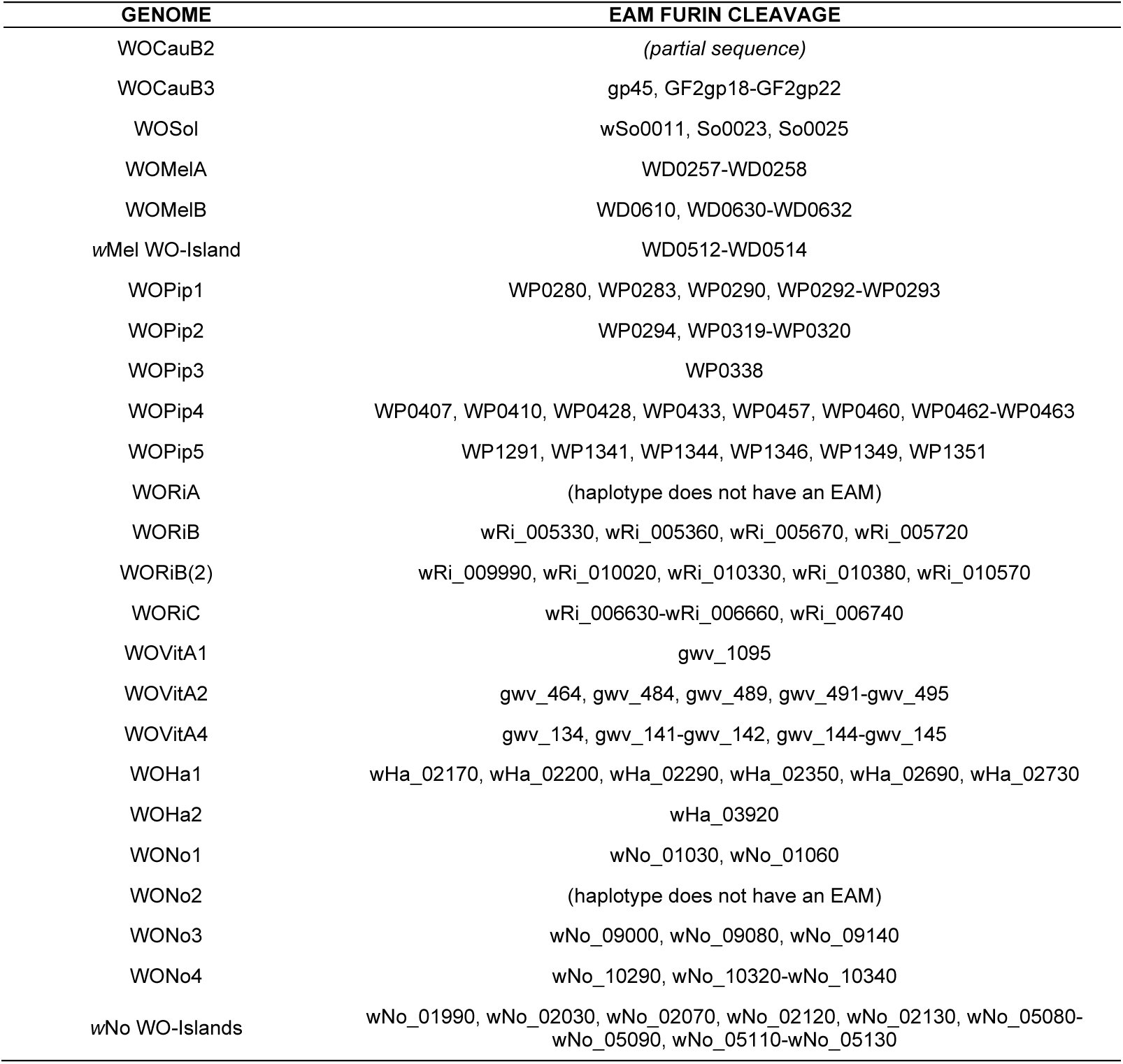
Latrotoxin-CTD is universally located at the 3’-terminal ends of both conserved spider latrotoxin genes^31^ and enormous, polymorphic, and eukaryotic-like phage WO genes (up to 14,256 bp). There is a high incidence of eukaryotic furin cleavage sites that immediately precede the latrotoxin-CTD. In spiders, cleavage at these sites by the eukaryotic furin protease in the trans-Golgi network or extracellular matrix is required for latrotoxin activation before the toxin exerts its effects upon the victim. We show that all prophage WO EAMs contain at least one site for eukaryotic furin cleavage (Supplementary Table 3), and the proportion of all EAM genes with predicted furin cleavage sites (25%) is two-fold greater than that of the genes in the core phage genome (11%, Fisher’s Exact Test, p < 0.0001), defined as the conserved bacteriophage region from recombinase to patatin. In regards to the phage WO latrotoxin-CTD, its preferential localization in prophage WO genomes versus the rest of the Wolbachia chromosome, conservation of eukaryotic furin cleaveage sites, large eukaryotic-like length, homology to invertbrate-specific toxins, and reduced divergence relative to the spider venom homologs is consistent with a eukaryotic origin and post-translational processing by furin peptidases.
Pox protein Repeats of ANkyrin C terminus (PRANC)
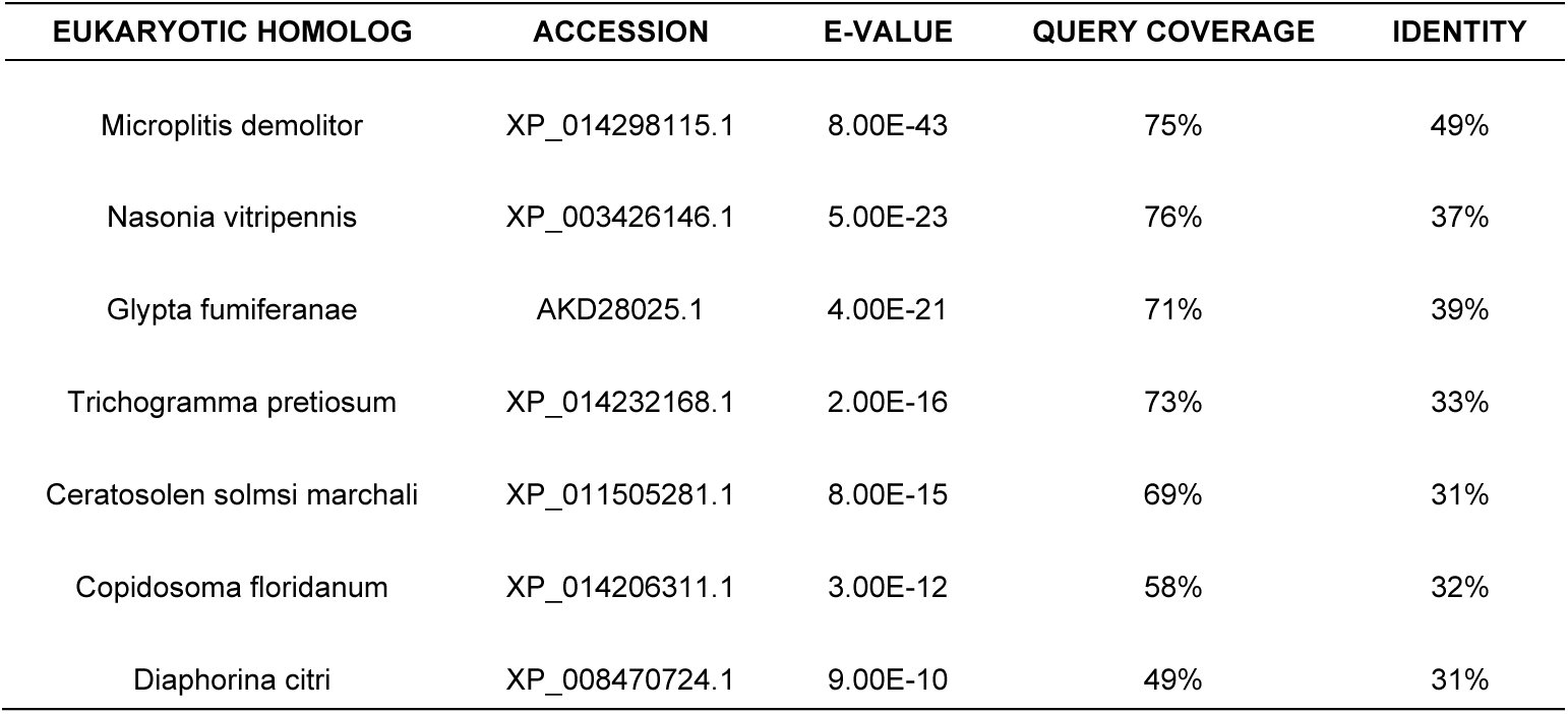
Domains central to modifying animal proteins are also abundant in the phage WO EAM. The PRANC domain in the WOVitA1 genome (gwv_1092) shares protein sequence homology with corresponding PRANC domains in multiple parasitic wasp hosts (Supplementary Table 4) and their eukaryotic viruses. Reciprocal BLASTP searches retrieve the same best hits and support previous findings that this protein domain horizontally transferred between eukaryotic viruses, animals, and Proteobacteria^33^. The discovery here of the eukaryotic-like PRANC domain in phage WO parallels its presence in the Poxviridae virus family, in which it functions in evasion of eukaryotic immune responses via modification of host ubiquitination. PRANC is related to amino acid sequences in F-box proteins, which are eukaryotic proteins involved in protein degradation. The PRANC domain also occurs in vaccina virus, ectromelia virus, cowpox virus and Orf virus and can regulate NF-κB signalling pathway to inhibit transcription of inflammatory cytokines^34^.
Conserved ankyrin and TetratricoPeptide Repeat (TPR) protein
Adjacent to the PRANC-encoding gene in WOVitA1’s EAM is an ankyrin and TPR-containing gwv_1093. Ankyrin repeats and TPRs mediate a broad range of protein-protein interactions (apoptosis, cell signaling, inflammatory response, etc.) within eukaryotic cells and are commonly associated with effector proteins of certain intracellular pathogens^35,36^. In Wolbachia, ankyrins within the core phage genome have been associated with reproductive manipulation of the insect host^37,38^. While generally rare in viral genomes (Supplementary Fig. 2 and 3), these repeat regions occur in all prophage WO haplotypes from sequenced Wolbachia genomes (N=23). Phylogenetic analysis using reciprocal BLASTP hits (Fig. 4) shows that the N-terminus sequences of the TPR-containing gwv_1093 are embedded within a diverse set of homologs from many athropod lineages (Fig. 4b), with the most recent transfer putatively occurring between phage WO and Solenopsis invicta (Fig. 4c). In this species, the gene is located between ant genes bicaudal D and rho guanine nucleotide exchange factor 11. As S. invicta can naturally harbor Wolbachia^39^, either a gene transfer event occurred between these ecologically-associated taxa or the S. invicta homolog could be an assembly artifact. This assembly was based on samples from a region rarely infected with Wolbachia (Y Wurm, personal communication, April 2016) and there are no other Wolbachia/prophage WO homologs in the S. invicta genome; therefore, the latter explanation seems unlikely. Moreover, other gwv_1093 homologs are from insect genome sequences of uninfected strains, i.e., N. vitripennis, and thus they can not be derived by an assembly artifact. Based on parsimony, the transfer event appears to have occurred from arthopod to phage WO since the arthropod taxa comprise a more diverse set of lineages. However, the reverse is plausible as transfers from Wolbachia to their arthropod hosts are common^40–42^.
Conserved TPR and anyrin proteins support lateral genetic transfer (a) A BLASTP query of WOVitA1’s gwv_1093 N-terminus reveals homologs in moquitoes, ants, beetles, a mealybug, a solitary bee and one obligate intracellular gammaproteobacteria. Bayesian phylogenetic trees were constructed based on (b) a 137-aa alignment of all homologs with E-value less than e-40 using the LG+G model of evolution. (c) To resolve taxa closest to phage WO, trees were reconstructed based on a 627-aa alignment of all homologs with an E-value of 0 using the JTT+I+G model of evolution. Isoforms were removed from each alignment. Both trees are unrooted. Consensus support values are shown at the nodes. Chromosomal neighborhood analyses of available animal genome sequences indicate that animal homologs to the phage WO protein are on contigs with other animal genes. Scale bar, 1,000 amino acids.
NACHT
Another instance of genetic transfer involves the programmed cell death (PCD) domain, NACHT (Fig. 5). Eukaryotic NACHT-containing proteins are typically engaged in PCD by acting as pathogen-sensors and signal transduction molecules of the innate immune system^43^. The polymorphic prophage WO homolog encodes ankyrin repeats and a latrotoxin-CTD directly downstream from the conserved NTPase domain (Fig. 5a). NACHT domains have been identified in animals, fungi and bacteria^44^ and phylogenetic patterns indicate multiple instances of horizontal transfer^45^. A NACHT-containing peptide was recently discovered in the Clostridium difficile-infecting phage phiCDHM1^46^. In contrast to prophage WO, it is bacterial in both amino acid homology and protein architecture. While all BLASTP and reciprocal BLASTP queries of the phiCDHM1 NACHT domain yield only bacterial homologs, BLASTP searches of the prophage WO NACHT domain yield only animal homologs, and reciprocal BLASTP searches of these yield only hits to prophage WO and other animals. Similar to the phylogeny of the N-terminus of the TPR-containing gwv_1093, this single NACHT domain sequence in prophage WO is embedded within a more diverse set of homologs in arthropods (Fig. 5b,c). Phylogenetic analyses place the prophage WO variants adjacent to a divergent Bombyx mori sequence, though these variants have slightly closer total homology to Culex quiquefasciatus mosquitoes that harbor Wolbachia with related prophage WO variants.
Phylogeny and protein architecture of the cell death domain, NACHT (a) A BLASTP query of prophage WO’s NACHT region reveals homologs throughout arthropods and crustaceans. (b) Bayesian phylogenetic trees were constructed based on a 271-aa alignment of all homologs with E-value less than e-15 and coverage greater than 70% using the cpREV+G model of evolution. To resolve taxa closest to prophage WO, all Daphnia sequences were removed from the alignment and clusters of highly divergent residues (i.e., 5 or more sequential residues with less than 15% pairwise identity) were trimmed. Trees were reconstructed based on this 262-aa alignment using the LG+G model of evolution. Consensus support values are shown at the nodes. Both trees are unrooted. Chromosomal neighborhood analyses of available animal genome sequences indicate that animal homologs to the prophage WO protein are on contigs with other animal genes. Scale bar, 1,000 amino acids.
Discussion
Metagenomic analysis of the complete genome from phage WO particles reveals all formerly described phage genetic modules (lysogeny, baseplate, head, replication, virulence, tail and patatin-like phospholipase^27^) as well as a new group of genes that we collectively group into a eukaryotic associatoin module (EAM). Some of these genes (i) encode protein domains and cleavage sites central to eukaryotic functions, (ii) occur in both phage and metazoan hosts, (iii) comprise the second largest phage gene to date (14,256 bp) and (iv) are absent from mutualistic, phage-free genomes of Wolbachia. Together, these genes increase the phage WO genome size by roughly 50% and include ten types of protein domains with four predicted eukaryotic functions: toxins, host-microbe interactions, host cell suicide, and secretion of proteins through the cell membrane. Notably, over half of these domain types share greater amino acid homology to eukaryotic invertebrates than to bacteria in GenBank. Among this subset with eukaryotic sequence homology, the protein domains are almost exclusively found in the phage EAM. An EAM has never before been reported in bacteriophage genomes, to our knowledge, possibly because phages of obligate intracellular bacteria occupy a unique eukaryotic-enclosed niche and are relatively understudied.
The presence of eukaryotic protein domains in bacteriophage genomes is of special note as they curiously mirror eukaryotic genes in large eukaryotic viruses that aid in viral mimicry and manipulation of host processes^47,48^. In phage WO, these animal protein domains are central to anti-eukaryotic functions including the black widow latrotoxin, programmed cell death (NACHT), immune evasion (PRANC), and protein-protein interactions.
Bacteriophage WO frequently transfer between Wolbachia coinfections in the same animal host^49,50^ and to the host genome as part of large transfers of the Wolbachia chromosome^40,41^. We previously reported that phage WO in Wolbachia of Nasonia vitripennis were also capable of transferring adjacent, flanking, non-phage genes in the process of exchange between coinfections^51^. For two of these flanking genes, sequence evidence indicated that Wolbachia genomes may be able to receive eukaryotic DNA^42,52,53^. However, the nature of these lateral genetic transfers remained to be elucidated as these regions were not previously known to be part of the packaged phage genome until now. Here, we demonstrate that genes with eukaryotic homology are constituents of phage WO and its EAM, and they either retain conservation of eukaryotic furin cleaveage sites and a large eukaryotic-like length (i.e., latrotoxin-CTD), or they exhibit markedly reduced or no diversity relative to the arthropod homologs as the WO sequences exist as single or a few representatives (NACHT and TPR-containing proteins). Moreover, WO protein domains with eukaryotic homology are highly enriched in the EAM over WO protein domains with bacterial homology. Based on this work, we suspect that systematic surveys of phage genomes in intimate host-associated bacteria may uncover a broad range of eukaryotic-like protein domains involved in phage lifecycle adaptations and phage-eukaryote interactions. Of particular note is the reported association between phage WO genes, specifically ankyrins, transcriptional regulators and the Ulp1 operon, and Wolbachia’s ability to manipulate host reproduction^37,38,54–56^.
The mechanisms by which eukaryotic protein domains are exchanged with phage WO are unknown and could follow at least three models (Fig. 6). First, animal genetic material could directly transfer to and from WO genomes during phage particle propagation in the cytoplasm of animal cells (Fig. 6b) or during packaging inside Wolbachia cells that are lysing and exposed to the eukaryotic cytoplasmic environment. Packaging of eukaryotic host RNAs, for instance, occur in the virions of herpesvirus^57^ and cytomegalovirus^58^. Second, genes may transfer between animal genomes and the Wolbachia chromosome and then to prophage WO. For this scenario to be plausible, animal genetic material transferred in random locations in the Wolbachia genome would have to be preferentially lost in non-phage associated locations from the Wolbachia chromosome (Fig. 6c) because domains with eukaryotic homology are highly enriched in the phage/prophage WO EAM versus the rest of the chromosome (Fig. 2). Third, DNA may transfer first between animal genomes and intermediary entities, such as eukaryotic viruses or other obligate intracellular bacteria, and then to phage WO and/or Wolbachia (Fib. 6d). In fact, the PRANC-domain (described in gwv_1092) was named for its discovery in and association with eukaryotic Pox viruses. Finally, once DNA is incorporated into a prophage genome, it is susceptible to recombination with other phage WO haplotypes located in the same Wolbachia chromosome and can transfer from one haplotype to another.
Models of lateral DNA transfer between eukaryotes and bacteriophages (a) The eukaryotic cell can harbor multiple microbes capable of horizontal gene transfer. Genetic transfers between eukaryotes and bacteriophages can, in theory, occur (b) directly between eukaryotic chromosomes and phage genomes; (c) indirectly between eukaryotic and Wolbachia chromosomes; or (d) indirectly between eukaryotic chromosomes and intermediary entities, such as eukaryotic viruses and other intracellular bacteria.
Alternatively, these protein domains could originate in the phage and be particularly prone to transfer, maintenance, and spread in their recipient arthropod genomes (Fig. 6b). For this scenario to be plausible, it would have to imply that phage genetic material independently and repeatedly transfers to athropods and spreads through the host population, which would subsequently be followed by loss of these phage genes or recombination with other non-transferred phage genetic material so that the eukaryotic sequence varation clusters seperately from the phage WO sequence(s). While each mode of transfer is possible, the eukaryotic length of these genes, presence of furin protease domains, and enrichment in the phage WO EAM provides evidence for their eukaryotic origin.
Why are these protein domains present in the EAM of bacteriophage WO? Some phages of obligate intracellular bacteria may have to overcome two major challenges not encountered by the well-studied phages of free-living bacteria. First, they are contained within both bacterial and eukaryotic membranes, posing an enigmatic “two-fold cell challenge”. They may not only have to breach peptidoglycan and permeabilize bacterial membranes, but they may also have to exit (and enter) across the eukaryotic membrane(s) that directly encapsulates the bacteria. Second, like their bacterial hosts, they must survive the internal cellular environment of the animal host, including the innate immune response and autophagy, while searching for phage-susceptible bacteria. Phage WO can dwell in the eukaryotic cytoplasm and extracellular matrix that they encounter upon bacterial lysis^26^, raising the likelihood of direct interaction with host membranes and intracellular biology. In this context, EAM protein domains are prime candidates to aid in functions including cell lysis (latrotoxin-CTD), manipulation of programmed cell death (NACHT and NB-ARC), host ubiquitination (OTU and Ulp1), insecticidal toxicity (ABC toxin) and interaction with host proteins (ankryin repeats and TPRs). Rather than simply act as virulence factors to benefit their bacterial host, their massive proportion of genomic real estate (up to 60% of the prophage genome, Supplementary Fig. 4) implies that they may be necessary to phage biology and likely have a direct impact on phage propagation. The concept of phage-mediated ecosystem modification as an alternative to bacterial virulence is not new^59^ but, much like the biology of phage WO, is relatively understudied.
Phage WO is not the only virus described within obligate intracellular bacteria. Chlamydiomicroviridae infect obligate intracellular bacteria, yet still do not directly contend with the eukaryotic membrane. Rather, they attach to dormant chlamydial cells (i.e., reticulate bodies) and enter via phagocytosis or endocytosis of the bacteria^60^. The phages then alter development of their bacterial host, which leads to disintegration of the chlamydial inclusion and subsequent lysis of the eukaryotic host cell^61,62^. The nature of phage WO’s lifestyle, on the other hand, may require a distinct interaction with multiple membranes and immune responses because lytic activity of phage WO has been associated with typical bacterial cell defects including degraded bacterial DNA, a detached inner membrane, and exit of the phage particles from inside Wolbachia and its host cell into the extracellular matrix of the reproductive tissues^26^. Bacteriophages of free-living bacteria also regularly colonize eukaryotic environments, particularly those associated with mucosal surfaces^63^. They, however, do not infect or traverse the eukaryotic membrane and are still within the genomic boundaries of the bacterial virosphere.
Temperate dsDNA phages also occur in facultative symbionts of aphids^64^ and tsetse flies^65^. While Wolbachia has never successfully been cultured outside of host cells^66^, these facultative symbionts can replicate both intra- and extracellularly (JW Brandt, personal communication, July 2015) suggesting that their phages are not constrained by the same two-fold cell challenge. In addition, their phages encode a traditional lytic cassette (holin and lysozyme) that correlates with the need to deal only with bacterial membranes. In some cases, the phages harbor bacterial-derived toxins that target eukaryotic cells^67^, and these function mutualistically in aphids by arresting development of parasitoid wasp larvae^64^. Furthermore, unlike phage WO that is stably maintained in the lab, these phages are readily lost in the absence of parasitoids during laboratory rearing, presumably due to the cost of their toxins^68^.
In addition to providing new insights into the evolution of bacteriophages and showing phage WO genomes to be far more complex than previously described, the findings here reveal evidence for gene sharing between metazoan hosts and phages of obligate intracellular bacteria. We suggest that the putative acquistion and retooling of intact eukaryotic domains in phage WO is analgous to the commandeering of host genes by eukaryotic viruses. Whether lateral genetic transfers between metazoans and bacteriophages are common in the symbiotic virosphere remains to be determined.
Methods
Insect and bacterial strains
The transfected line of the Mediterranean flour moth Ephestia kuehniella harboring Wolbachia strain wCauB was obtained with the help of Takema Fukatsu and Tetsuhiko Sasaki^22^. Moths were maintained at 24°C and 70% humidity on a diet consisting of wheat bran, glycerol and dried yeast (20:2:1 w/w). The introgressed line of the parasitoid wasp Nasonia giraulti harboring Wolbachia strain w VitA, termed IntG12.1, was previously derived by repeatedly backcrossing N. vitripennis (strain 12.1) females to N. giraulti males for nine generations^69^. The strain was incubated at 25°C using the flesh fly Sarcophaga bullata as host.
Phage particle purification
Phage particles were isolated according to Fujii et al^22^ with modifications. Approximately 4 g of adult insects were homogenized in 29.6 ml cold SM buffer (50mM Tris-HCl, pH 7.5, 0.1 M NaCl, 10mM MgSO_4_. 7H_2_0, and 0.1% (w/v) gelatin). NaCl and RNase A were added to a final concentration of 1M and 1µg/ml, respectively. The homogenate was incubated on a shaker at 4°C for 1 h and then centrifuged at 13,000g for 10 min at 4°C. Polyethylene glycol (PEG) 6000 was added to a final concentration of 10% to precipitate phage particles, incubated at 4°C for 1 hr with gentle shaking and centrifuged at 13,000g for 10 min. The pellet was resuspended in 5 ml TM buffer (50 mM Tris-HCl, pH 7.5, 10 mM MgCl_2_. 6H_2_O) and mixed with an equal volume chloroform. The suspension was centrifuged at 3,000g to remove PEG and the aqueous phase was filtered through a 0.22 µm filter to remove bacterial cells. The suspension was centrifuged at 60,000g for 1 h at 4°C to collect phage particles. The pellet was suspended in 10 µl TM buffer.
Phage DNA extraction and metagenomic sequencing
The phage suspension was treated with RQ1 RNase-Free DNase (Promega) for 30 min at 37°C, followed by heat inactivation for 10 min at 65°C, to remove host DNA contamination. Phage DNA was extracted from the suspension using the QIAamp MinElute Virus Spin Kit (Qiagen) and amplified using the REPLI-g Mini Kit (Qiagen). Following amplification, paired-end DNA libraries were prepared according to manufacturer’s (Illumina) instructions and samples were sequenced with an Illumina HiSeq 2000 (2×100-nt read length).
Bioinformatics and statistics
Metagenomic sequences (reads) were trimmed, paired and assembled into contigs using the CLC Assembler (CLC bio) with bubble size = 50, insertion and deletion cost = 3, mismatch cost = 2, length fraction = 0.6, minimum contig size = 130, similarity = 0.5, minimum distance = 90 and maximum distance = 200. Contigs were compared to the GenBank non-redundant database using NCBI’s BLASTN (http://blast.ncbi.nlm.nih.gov/Blast.cgi) and those with similarity to phage WO and/or Wolbachia(E-value <10^-10^) were manually annotated using Geneious (Biomatters Ltd.). Individual reads were mapped to reference sequences using Geneious. Open reading frame (ORF) homology searches were performed to determine putative function using NCBI’s BLASTP (http://blast.ncbi.nlm.nih.gov/Blast.cgi) and Wellcome Trust Sanger Institute’s pfam database (http://pfam.sanger.ac.uk). Coiled coil domains were predicted with EMBL’s Simple Modular Architecture Research Tool (SMART, http://smart.emblheidelberg.de). Furin cleavage sites were identified using PiTou (http://www.nuolan.net/reference.html). The number of genes with and without furin cleavage sites was analyzed with respect to phage-region using Fisher’s Exact Test (GraphPad Software). Phylogenetic trees were built using the Bayes plugin in Geneious and model selection for each Bayes analysis was estimated using ProtTest^70^.
Confirmation of phage WO terminal genes
Genomic DNA was extracted from wVitA-infected N. vitripennis (strain 12.1) and wCauB-infected E. kuehniella individuals using the Gentra Puregene Tissue Kit (Qiagen). Primers were designed for both WOVitA1 and WOCauB3 attP sites, respectively: VitA1_attF (5’- CGA AGA ACC AGC ACA GGG TGG-3’), VitA1_attR (5’- GCT GGA AGA GGG CAT CTG CAT C-3’), CauB3_attF (5’- TCG TGA CTG CCC TAT TGC TGC T – 3’) and CauB3_attR (5’- ATG CGG CCA AAG CTG GGT GT – 3’). Amplification was performed in a Veriti thermal cycler (Applied Biosystems) using GoTaq green master mix (Promega) under the following conditions: 94°C for 2 min; 35 cycles of 94°C for 30 s, 53°C for 30 s, 72°C for 1 min; and a final elongation cycle of 72°C for 10 min. PCR products were sequenced via Sanger sequencing (Genewiz, Inc).
Data availability
The phage WOVitA1 genome assembly reported in this paper has been deposited in NCBI under accession number KX522565. The N. vitripennis viral metagenome sequences have been deposited in the SRA under accession number SRR3560636 and BioProject PRJNA321548. The wCauB-infected E. kuehniella viral metagenome sequences have been deposited in the SRA under accession number SRR3536639 and BioProject PRJNA321549.
Data referenced in this study are available in NCBI with accession codes AE017196 (wMel), AM999887 (wPip), CTEH00000000 (wPipMol), ABZA00000000 (wPipJHB) CP001391 (wRi), CAOU00000000 (wSuzi), AMZJ00000000 (wDi), AAGB01000001 (wAna), CAGB00000000 (wAlbB), CAOH00000000 (wBol1-b), JYPC00000000 (wOb), CP003884 (wHa), CP003883 (wNo), LK055284 (wAu), AP013028 (wCle), HE660029 (wOo), PRJNA213627 (wVitA), AB478515 (WOCauB2), AB478516 (WOCauB3), KC955252 (WOSol), HQ906665 and HQ906666 (WOVitB).
The reference list from the paper itself. Each links out to its DOI / PubMed record.
- 1Edwards, R.A. & Rohwer, F. Viral metagenomics. Nat Rev Microbiol 3, 504–10 (2005).
- 2Hendrix, R.W., Smith, M.C., Burns, R.N., Ford, M.E. & Hatfull, G.F. Evolutionary relationships among diverse bacteriophages and prophages: all the world’s a phage. Proc Natl Acad Sci U S A 96, 2192–7 (1999).
- 3Brussow, H. The not so universal tree of life or the place of viruses in the living world. Philos Trans R Soc Lond B Biol Sci 364, 2263–74 (2009).
- 4King, A.M.Q., Adams, M.J., Lefkowitz, E.J. & Carstens, E.B. Virus taxonomy: classification and nomenclature of viruses: Ninth Report of the International Committee on Taxonomy of Viruses., 1327 (Elsevier, San Diego, 2012).
- 5Nasir, A., Forterre, P., Kim, K.M. & Caetano-Anolles, G. The distribution and impact of viral lineages in domains of life. Front Microbiol 5, 194 (2014).
- 6Prangishvili, D., Forterre, P. & Garrett, R.A. Viruses of the Archaea: a unifying view. Nat Rev Microbiol 4, 837–48 (2006).
- 7Forterre, P. Giant viruses: conflicts in revisiting the virus concept. Intervirology 53, 362–78 (2010).
- 8Raoult, D. TRUC or the need for a new microbial classification. Intervirology 56, 349–53 (2013).