OptoDyCE: Automated System for High-Throughput All-Optical Dynamic Cardiac Electrophysiology
Aleksandra Klimas, Jinzhu Yu, Christina M. Ambrosi, John C. Williams, Harold Bien, Emilia Entcheva

TL;DR
OptoDyCE is a high-throughput system using light to study heart cell electrophysiology, enabling fast and detailed drug testing.
Contribution
OptoDyCE introduces a fully automated, all-optical system for dynamic cardiac electrophysiology at high throughput.
Findings
OptoDyCE enables testing of over 600 multicellular samples or compounds per hour.
The system provides high-content data on drug effects over time, space, and doses.
Optical actuation is validated using optogenetic drivers in rat and human cardiomyocytes.
Abstract
The improvement of preclinical cardiotoxicity testing, the discovery of new ion-channel-targeted drugs, and the phenotyping and use of stem-cell-derived cardiomyocytes and other biologics all necessitate high-throughput (HT), cellular-level electrophysiological interrogation tools. Optical techniques for actuation and sensing provide instant parallelism, enabling contactless dynamic HT testing of cells and small-tissue constructs, not affordable by other means. Here, we consider, computationally and experimentally, the limits of all-optical electrophysiology when applied to drug testing, then implement and validate OptoDyCE, a fully automated system for all-optical cardiac electrophysiology. We validate optical actuation by virally introducing optogenetic drivers in (rat and human) cardiomyocytes or through the modular use of dedicated light-sensitive somatic “spark” cells. We show that…
Genes, proteins, chemicals, diseases, species, mutations and cell lines named across the full text — each resolved to its canonical identifier and authoritative record.
Click any figure to enlarge with its caption.
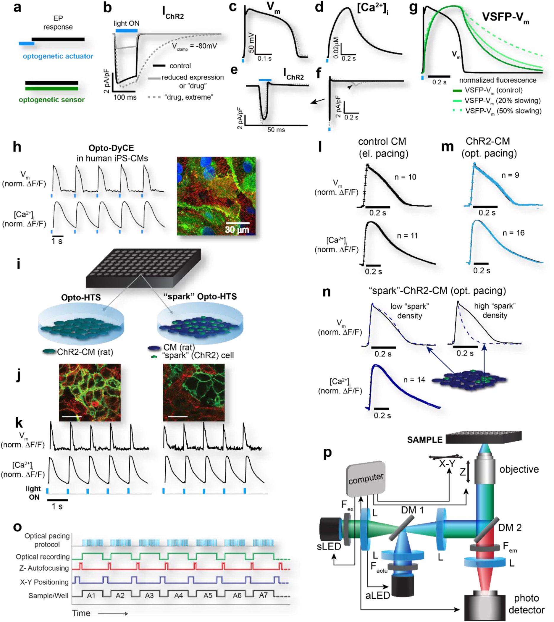 Figure 1
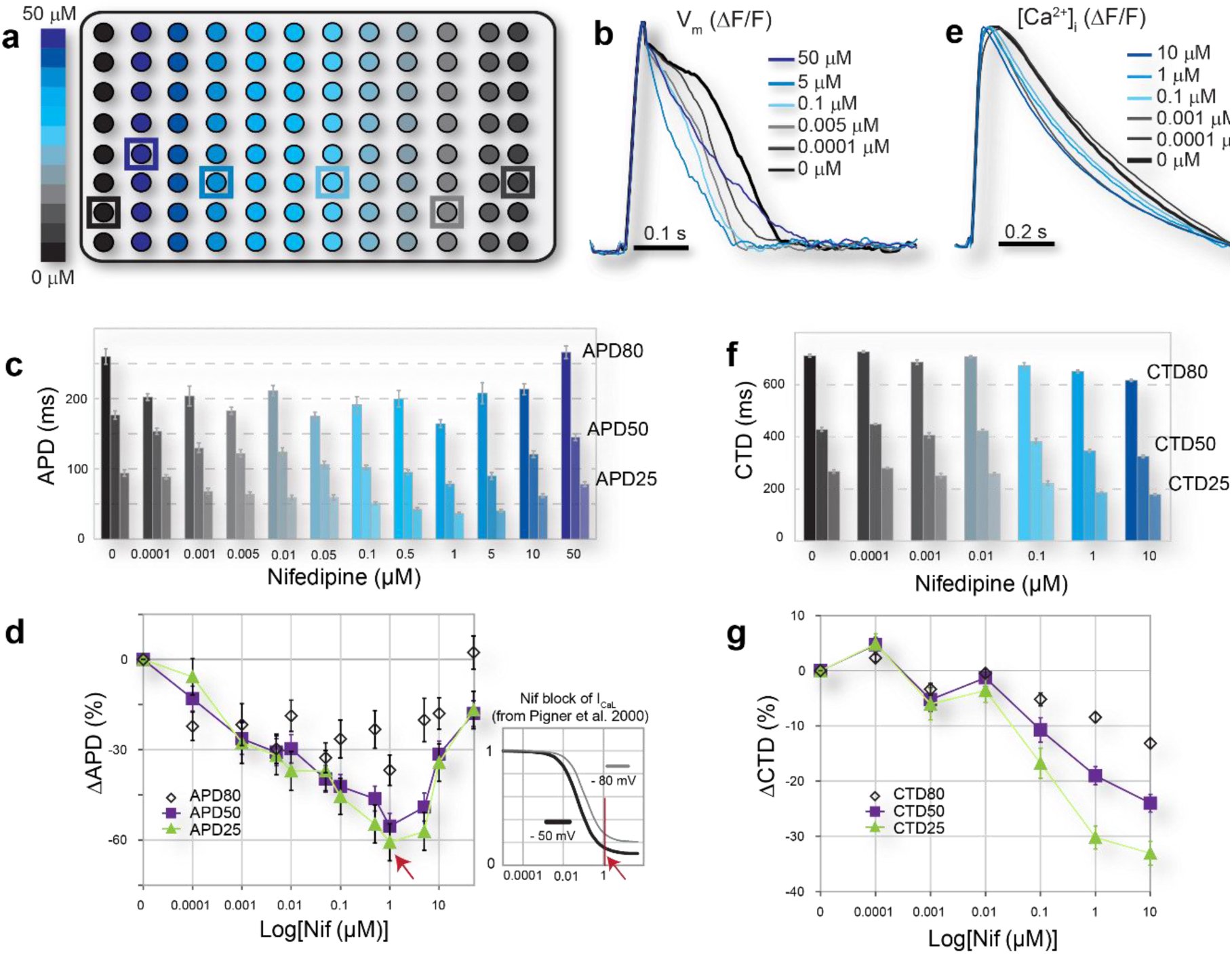 Figure 2
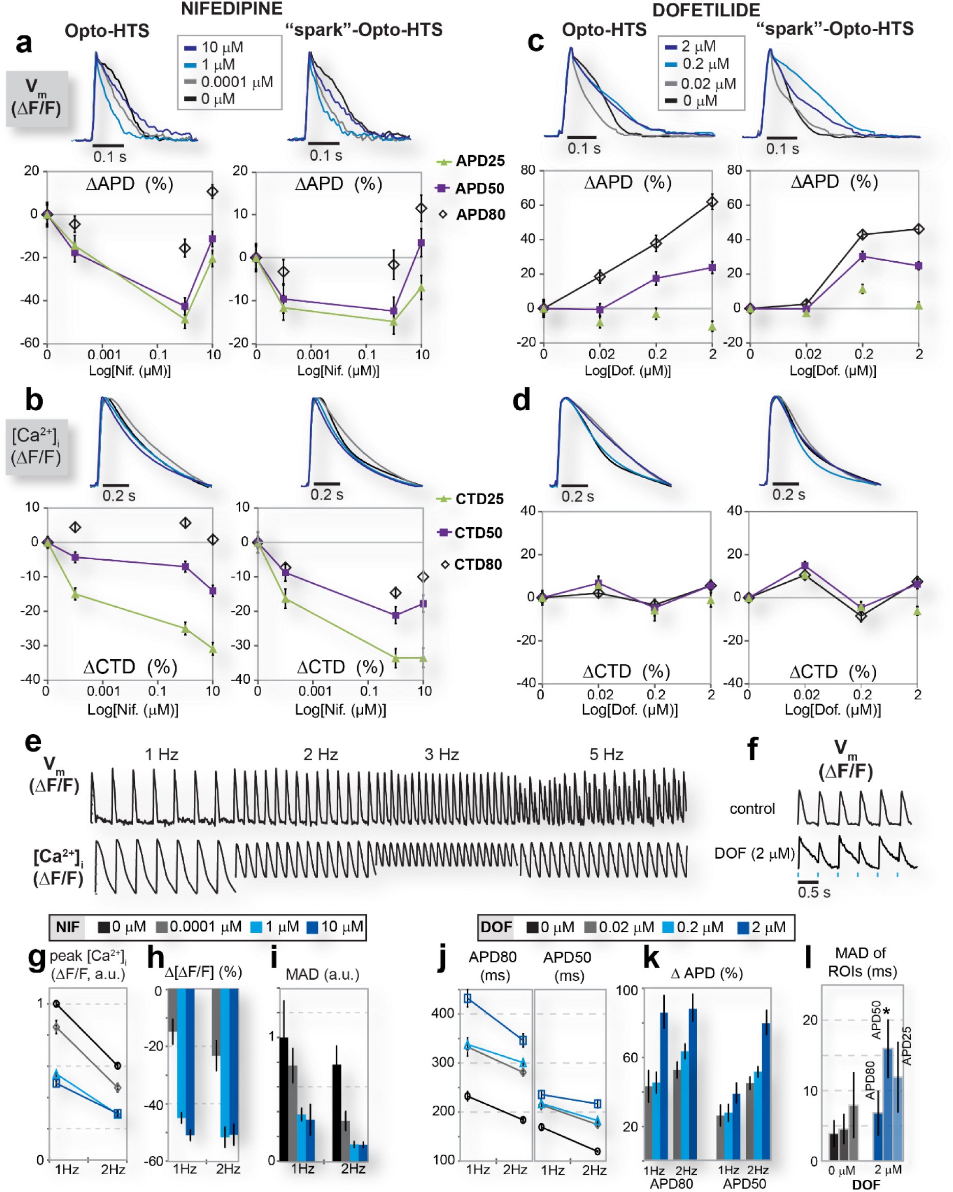 Figure 3
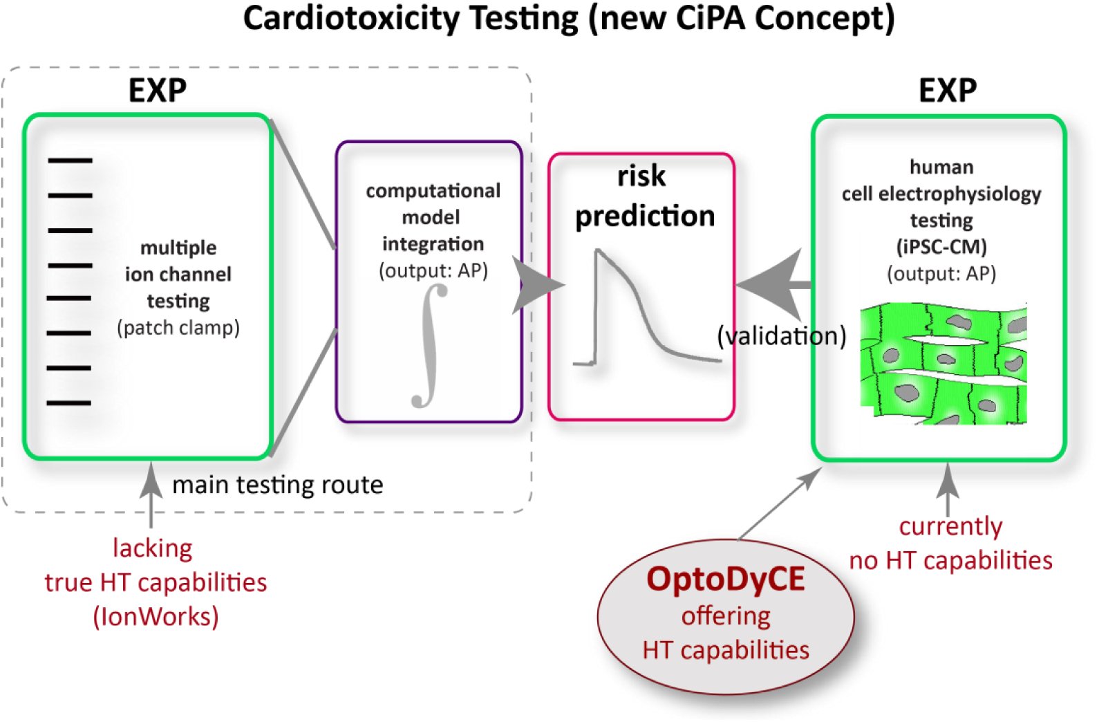 Figure 4
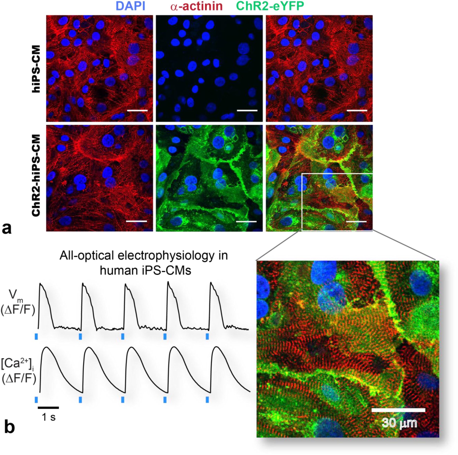 Figure 5
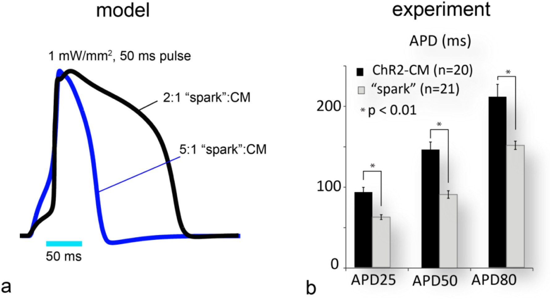 Figure 6
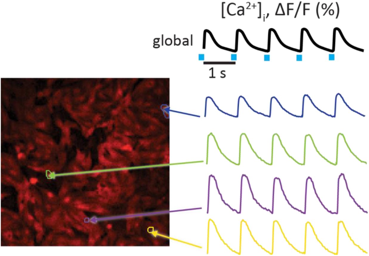 Figure 7
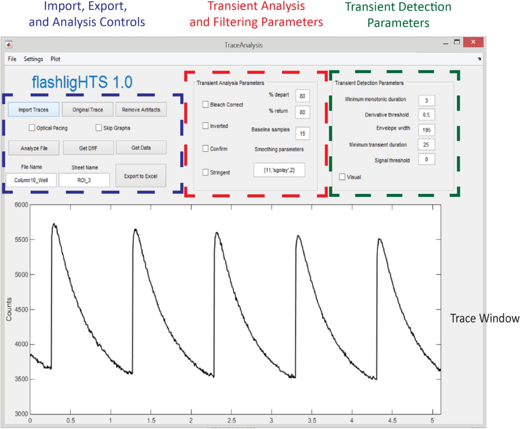 Figure 8
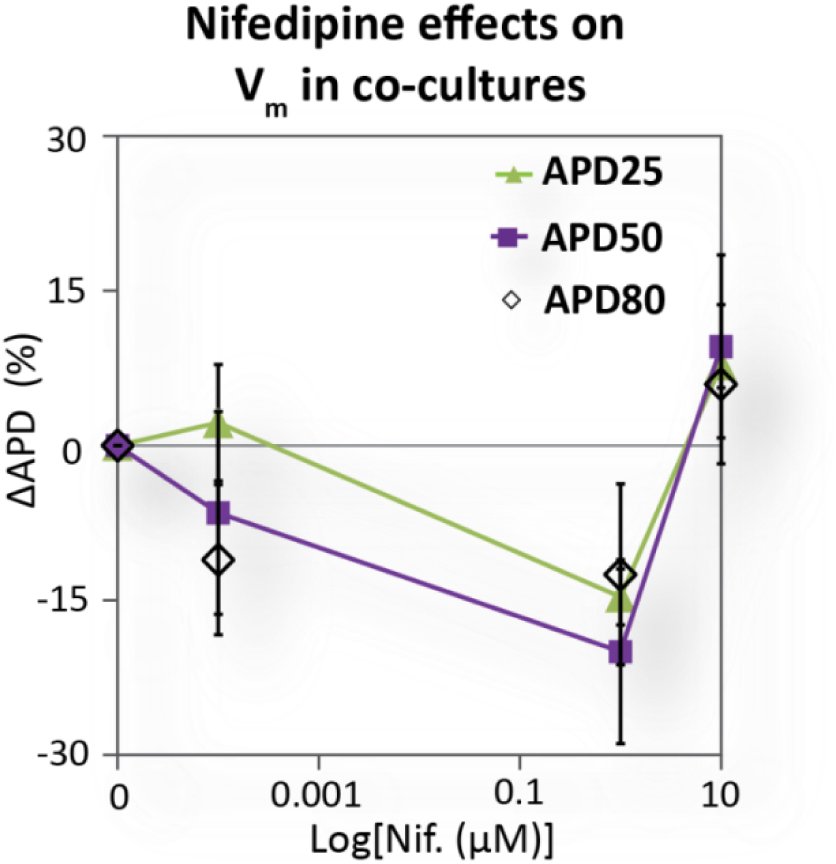 Figure 9
 Figure 10
Peer Reviews
No public reviews on file for this paper yet. If you reviewed it on a platform where reviews are public (OpenReview, ICLR, NeurIPS, ICML), you can paste yours below so the community can read it here.
Videos
No videos yet. Explain this paper in a talk, walkthrough, or lecture? Add one.
Taxonomy
TopicsPhotoreceptor and optogenetics research · Neuroscience and Neural Engineering · Receptor Mechanisms and Signaling
The development of new drugs is a lengthy, inefficient process;^1^ in the United States, <0.05% of all compounds undergoing preclinical tests become marketed drugs, and <30% of compounds evaluated in expensive clinical trials make it to market.^2^ Perhaps most costly, and with greatest negative societal impact, is the withdrawal of drugs from the market after they have been approved. In the last two decades, over 30% of such withdrawals have been due to cardiac toxicity (pro-arrhythmic effects mediated by cardiac ion channels).^2^ International regulatory agreements mandate the testing of all new drugs for cardiac liability, including drug-induced long QT interval (LQT) and risk for development of life-threatening arrhythmias, such as Torsade de Pointes (TdP).^3^ This mandated preclinical testing specifically concerns a drug’s action as a potential blocker of the hERG K^+^ channel, which supplies one of the main repolarizing currents in cardiomyocytes. However, it has recently been recognized that a drug’s pro-arrhythmic effect, i.e. “torsadogenicity”, is often shaped by its action on multiple ion channels;^3-7^ therefore, an integrative (cell-level or multicellular) view is essential, and current regulations need to be revisited (see Supplementary Fig. 1 about the CiPA initiative, Comprehensive In Vitro Pro-arrhythmia Assay^3^). Computational efforts are underway^5,7^ to integrate multi-channel data obtained in recombinant expression systems to predict the action of a drug on the human cardiac action potential (see Supplementary Fig. 1). Recently, great strides in the optimization and production-scaling of human patient-derived cardiomyocytes (induced pluripotent stem cell derived, iPSC-CMs) promise to provide an alternative, more direct and relevant experimental testbed for cardiotoxicity screening.^8, 9^ However, there are currently no high-throughput solutions (ability to screen >10,000 compounds a day) for performing robust cardiomyocyte electrophysiological testing.
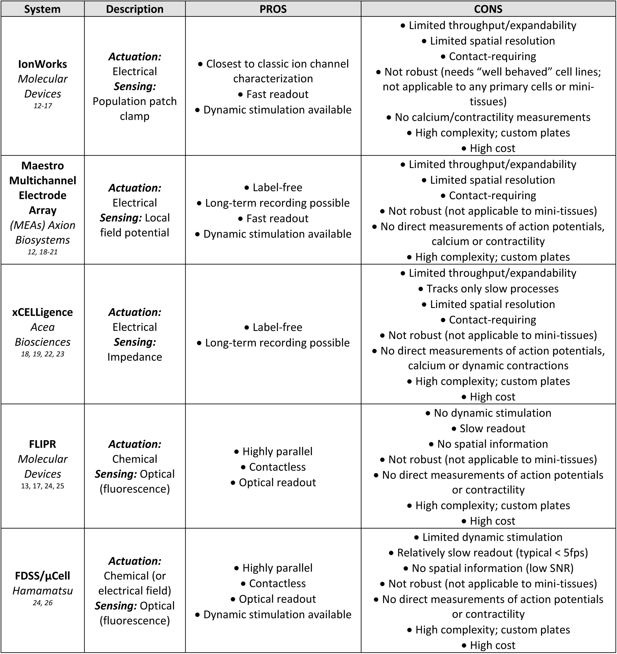
Classic electrophysiology involves physical contact and therefore is inherently very low throughput (manual). New technical developments towards increased throughput^10, 11^ include the automated planar patch, IonWorks by Molecular Devices, at the single-channel level; the Fluorometric Imaging Plate Reader (FLIPR) by Molecular Devices; Multichannel Electrode Arrays (MEAs) by Axion Biosystems; impedance-based assays with xCELLigence by Acea Biosciences; and the kinetic plate reader FDSS/μCell by Hamamatsu for cellular measurements (see Supplementary Table 1 for a detailed comparison). The following limitations of these systems motivate the need for further developments towards HT cell-level electrophysiology: 1) requirements for contact in IonWorks, MEAs, or xCELLigence prevent scaling to the HT-level – a non-contact modality is a must; 2) lack of electrophysiologically-relevant fast readout (e.g. optical sensing in FLIPR makes it highly parallel, but still unable to track fast action potentials); 3) inability for dynamic actuation and frequency-response testing (e.g. FLIPR), which is quite relevant in drug-induced cardiotoxicity;^12^ 4) cell-type restrictions: more phenotypic outputs, such as iPSC-CMs or primary CMs as testbeds, are desirable instead of the currently-employed recombinant expression systems, but handling limitations present challenges (e.g. in IonWorks, a proper seal can only be formed with “well-behaved” cell lines^10^); 5) none of the current methods can characterize tissue-level/multicellular effects, even though arrhythmias are inherently spatiotemporal phenomena.
An all-optical electrophysiology approach^13-15^ can address these limitations and facilitate HT-level testing through built-in parallelism. New fast optogenetic tools for actuation^16-18^ and sensing^15, 19, 20^ offer attractive solutions for manipulating and observing multiple cells optically, but their limitations need to be considered carefully in the context of drug screening (Fig. 1a-g). Both actuators and sensors contain essential elements of ion channel proteins, making them susceptible to the drugs being tested. However, the action of a fast optogenetic actuator, e.g. Channelrhodopsin-2 (ChR2), in cardiomyocytes can be viewed as “time-detached” from the electrophysiological (EP) response, Fig. 1a, hence mostly benign. Computationally, we show that for brief light pulses, even dramatic drug effects on the ChR2 current amplitude and/or kinetics are practically inconsequential for the optically-triggered action potentials (APs) and calcium transients (CTs), if light irradiances are maintained at supra-threshold levels (Fig. 1b-f, details in Supplement). In contrast, an optogenetic sensor, e.g. VSFP2.3,^21^ is fully “temporally-convolved” with the EP response (Fig. 1a), and even a mild drug action on the sensor can profoundly alter the EP readout (see computational predictions, Fig. 1g); the same applies to other voltage^15, 20^ or calcium (GCaMP) optogenetic sensors, regardless of their kinetics. While great for long-term monitoring,^20^ channel-based optogenetic sensors may not be ideal for acute drug-testing applications due to such potential direct interference; instead, classic synthetic optical dyes (for voltage and calcium) may be more suitable, as they are already used in industrial applications.
Here, we present OptoDyCE, the first fully automated platform for all-optical dynamic interrogation of cardiomyocyte electrophysiology, including human iPSC-CMs (Fig. 1h, Supplementary Fig. 2), with applicability to drug testing. We demonstrate the HT capabilities of OptoDyCE using multicellular samples in 96-well format by combining optogenetic actuation (via ChR2) with simultaneous optical sensing of voltage or intracellular calcium by synthetic red-shifted dyes (di-4-ANBDQBS and Rhod-4AM, respectively), illustrated in Fig. 1h-p. Contactless optical pacing reliably triggers voltage (V_m_) and calcium ([Ca^2+^]i) signals (Fig. 1h, k). We impart the ability for optical pacing via one of two quick and efficient transduction methods (within 48 hours prior to experimentation) to yield: 1) OptoHTS: using direct adenoviral gene delivery (in human, ChR2-hiPSC-CM, or neonatal rat ventricular cardiomyocytes, ChR2-CM);^13, 22, 23^ or 2) sOptoHTS: “sprinkling” of dedicated light-sensitive (“spark”) cells on top of samples of non-transduced cardiomyocytes, a version of our “tandem-cell-unit” concept,^24^ (Fig. 1i-k, see Supplement).
We validate OptoHTS by comparing AP and CT morphology of optically-stimulated ChR2-CM samples and electrically-paced non-transduced CM samples, confirming optogenetic pacing is a suitable alternative to electrical stimulation for drug testing purposes (Fig. 1l-m), as predicted computationally.^22, 25^ The sOptoHTS provides a more attractive modular method of light sensitization, as a bank of generic “spark” cells (light-sensitized somatic cells) can be used in conjunction with a variety of non-modified experimental cardiac systems. However, caution should be applied regarding the geometry of “spark” cell distribution, since loading effects of higher “spark”-cell concentrations can locally shorten the AP (Fig. 1n, Supplementary Fig. 3), having minimal effects on CT morphology. Proper “spark”-cell delivery (for example, a localized pacing site) can easily address the issue.
A fully automated high-throughput version of OptoDyCE in 96-well format is demonstrated here using an optical setup custom-built around an inverted microscope, an automation protocol, and custom-developed software for semi-automated analysis (Fig. 1o-p, Supplementary Fig. 4-5, details in Supplement). The simplicity of our implementation makes it easy to adopt; compared to a prior report on all-optical electrophysiology in neurons,^15^ we use low-power LED light sources and standard equipment.
To our knowledge, this is the first scalable automated all-optical platform for cardiac electrophysiology that can meet the HT standard (see considerations in Supplementary Fig. 4). With robotic dispensing, the 96-well format can be instantly upgraded to 384-well or other standard plate formats, with very simple reprogramming. The current implementation has built-in parallelism within a well, interrogating hundreds of cells simultaneously (Supplementary Fig. 4), but relies on serial traversing of the wells; a macroscopic version (see^26^ or in FDSS/μCell) of OptoDyCE with larger field-of-view can further increase throughput by order(s) of magnitude.
Even in the current proof-of-concept implementation of OptoDyCE, dynamic drug-dose testing can be performed on a 96-well platform (over 30,000 single-cell readouts, multi-beat pacing protocol) in less than 10 minutes (Fig. 2a, Supplementary Fig. 4). As an illustration, nifedipine, a class-IV antiarrhythmic agent, was applied in 12 doses (0 to 50μM) to 96 ChR2-CM samples (Fig. 2a). Using optical pacing at 1Hz, we quantified the dose-response to nifedipine in terms of AP duration (APD) and CT duration (CTD) (Fig. 2b-g). Expected APD shortening (Fig. 2b-d) and CTD shortening (Fig. 2e-g) was observed, especially at the plateau phase (APD25/CTD25 and APD50/CTD50), due to nifedipine blocking the inward L-type calcium current, I_CaL_. Nifedipine caused CTD to monotonically decrease up to 10 μM (Fig. 2f,g). In contrast, after maximum APD shortening at around 1 μM, corresponding to maximum block of I_CaL_ reached at that concentration^27^ (Fig. 2d inset), the APD response to nifedipine reversed its direction, as seen clinically.^6^ This was likely due to indirect (voltage-mediated) or non-specific action on other ion channels, partially countering the I_CaL_ block (Fig. 2d). Note that the benefits of our in vitro HT platform are in the ability to quickly and finely probe many concentrations and to help determine the “therapeutic window”, i.e. the window for which a drug is both effective (has the desired action) and safe. Clinically, the drug-metabolizing action of the cytochrome P450 enzymes can amplify or suppress the effect of a drug which can can result in a lower or higher apparent drug dose (as in some failed drugs^2^); our data can be used to judge the “room for error” in the therapeutic window for a drug.
Seeking validation for sOptoHTS, we further compared the dose-dependent effects of nifedipine and of dofetilide using the two methods, OptoHTS vs. sOptoHTS (Fig. 3). Dofetilide, a class-III anti-arrhythmic agent and intended hERG channel blocker, has a known risk for drug-induced LQT and TdP due to its APD-prolonging action.^6^ sOptoHTS was able to successfully track the drug-dose dependent effects on APD and CTD, similar to OptoHTS (Fig. 3a-d). With proper tuning of the “spark” cell distribution, this simple and truly modular approach provided by sOptoHTS can be reliably applied to HT electrophysiological drug testing.
Electrophysiological responses are frequency-dependent, therefore passive observation of spontaneous activity^20^ is generally insufficient in drug testing. Unlike most currently-employed systems (Supplementary Table 1), our platform allows for active dynamic interrogation, such as robust pacing protocols that can reveal V_m_ and [Ca^2+^]i frequency response (restitution) and temporal instabilities (Fig. 3e). For example, a consistent generation of voltage instabilities known as alternans can be captured at 2Hz optical pacing in the presence of 2 μM dofetilide due to drug-induced APD prolongation (Fig. 3f). Restitution and temporal or spatial variability (assessed by median absolute deviation (MAD), see Supplement) can be quantified as function of drug dose (Fig. 3g-l). These are directly relevant to the “torsadogenecity” of a drug, providing a much more complete assessment than traditional (single-channel block) testing or current state-of-the-art assays (Supplementary Table 1). Our dynamic testing data reveal that nifedipine action on peak calcium (% change) is dose-dependent (p<0.05) but frequency-independent (Fig. 3g-h). Furthermore, nifedipine reduces temporal variability of peak calcium (assessed by MAD), and this reduction is augmented by higher-frequency pacing (Fig. 3i). For dofetilide, we found enhanced relative APD50 prolongation at higher frequency, which is opposite to purported reverse-use dependence (Fig. 3j-k). Furthermore, because of the ability to study multicellular samples, spatial variability can be quantified as a function of drug dose by analysing individual cells or regions of interest within the same sample/well (Fig. 3l, see also Supplementary Fig. 4). For example, we found that dofetilide at 2 μM increases spatial variability in APD (i.e. increases dispersion of repolarization -- a known pro-arrhythmic factor), compared to control during 1Hz pacing (p<0.05 for APD50). Most of the electrophysiological results presented in Figs. 2-3 can be corroborated by published work, albeit certainly not in an all-encompassing study. We chose to conduct our demonstration with these known clinically-used drugs so that the emphasis of our study can be the illustration of usability and the potential impact of our methodology rather than the electrophysiological insights per se.
These results also illustrate the high-content data that can be obtained with OptoDyCE, that allows the quantification of a drug’s pro-arrhythmic risk in a much more comprehensive way than with any of the current platforms. The contactless nature of interrogation in OptoDyCE makes it especially versatile and applicable to not only single cells and cell monolayers, but also to small-thickness 3D engineered tissues (within the optical penetration depth). By offering a currently missing option for automated HT cardiomyocyte electrophysiology, OptoDyCE can profoundly impact developments concerning human iPSC-CMs^8, 9^ (see Supplementary Fig. 1) by allowing for combinatorial optimization of factors involved in cell maturation, phenotype selection, and tissue engineering. In turn, the utilization of these new optimized human (potentially patient-specific) experimental models, in conjunction with our HT testing platform, has the potential to dramatically improve pre-clinical drug testing, reduce cost, reduce animal use, and increase a therapy’s likelihood of clinical success.
The reference list from the paper itself. Each links out to its DOI / PubMed record.
- 1Reichert, J.M. Trends in development and approval times for new therapeutics in the United States. Nat Rev Drug Discov 2, 695–702 (2003).
- 2Preziosi, P. Science, pharmacoeconomics and ethics in drug R&D: a sustainable future scenario? Nat Rev Drug Discov 3, 521–526 (2004).
- 3Fermini, B. et al. A New Perspective in the Field of Cardiac Safety Testing through the Comprehensive In Vitro Proarrhythmia Assay Paradigm. J Biomol Screen (2015).
- 4Bowlby, M.R., Peri, R., Zhang, H. & Dunlop, J. h ERG (KCNH 2 or Kv 11.1) K+ channels: screening for cardiac arrhythmia risk. Current drug metabolism 9, 965–970 (2008).
- 5Mirams, G.R. et al. Simulation of multiple ion channel block provides improved early prediction of compounds’ clinical torsadogenic risk. Cardiovasc Res 91, 53–61 (2011).
- 6Redfern, W.S. et al. Relationships between preclinical cardiac electrophysiology, clinical QT interval prolongation and torsade de pointes for a broad range of drugs: evidence for a provisional safety margin in drug development. Cardiovasc Res 58, 32–45 (2003).
- 7Rodriguez, B. et al. The systems biology approach to drug development: application to toxicity assessment of cardiac drugs. Clin Pharmacol Ther 88, 130–134 (2010).
- 8Bellin, M., Marchetto, M.C., Gage, F.H. & Mummery, C.L. Induced pluripotent stem cells: the new patient? Nat Rev Mol Cell Biol 13, 713–726 (2012).